Abstract
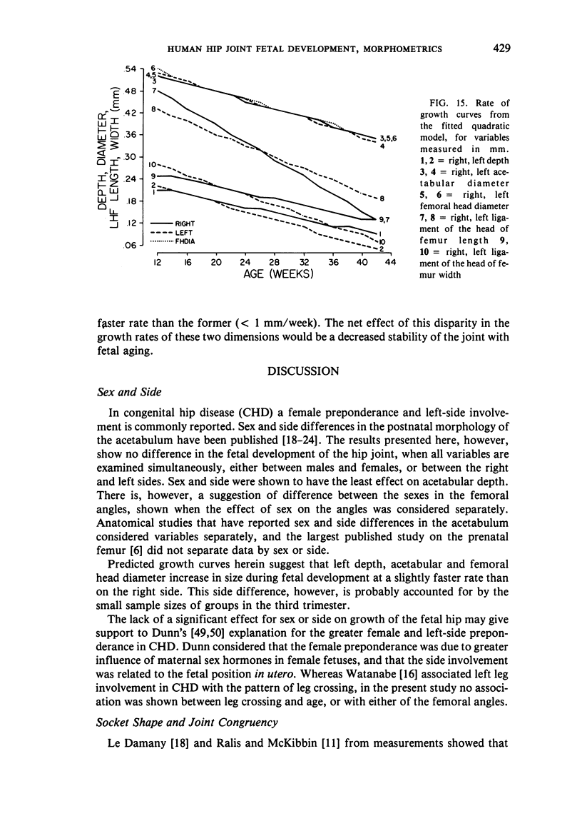
Hip joints (280) from 140 human fetuses, obtained from abortions and deaths in the perinatal period, were studied. The fetuses ranged from 8.7 to 40 cm in crown-rump length and are believed to be between 12 and 42 weeks in age. The joints were dissected, morphology inspected, and measurements taken of the depth and diameter of the acetabulum, the diameter of the femoral head, length and width of the ligament of the head, the neck-shaft, and torsion angles of the proximal femur. Regression models were fitted to determine which would best predict the growth pattern. Multivariate analysis of variance showed no significant differences between males and females or between the right and left sides. Acetabular depth was shown to be the slowest-growing hip variable, increasing less than fourfold in the period studied. Acetabular indices less than 50 percent indicate a shallow socket at term. Femoral head and acetabular diameter demonstrated a strong relationship (r = 0.860) and in many joints the femoral head diameter exceeded that of the acetabulum. Considerable variability was demonstrated in both femoral angles. The femoral angles showed only low correlation with the other hip variables. These observations indicate that soft tissue structures about the joint must play an important role in neonatal joint stability. The explanation of greater female and left side involvement in congenital hip disease must lie in factors other than growth changes of hip dimensions. Neither angle appears to be a useful indicator of normal joint development.
Full text
PDF

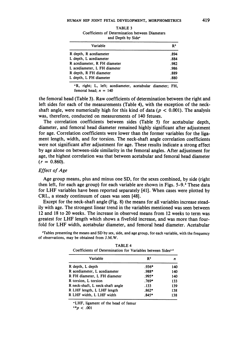
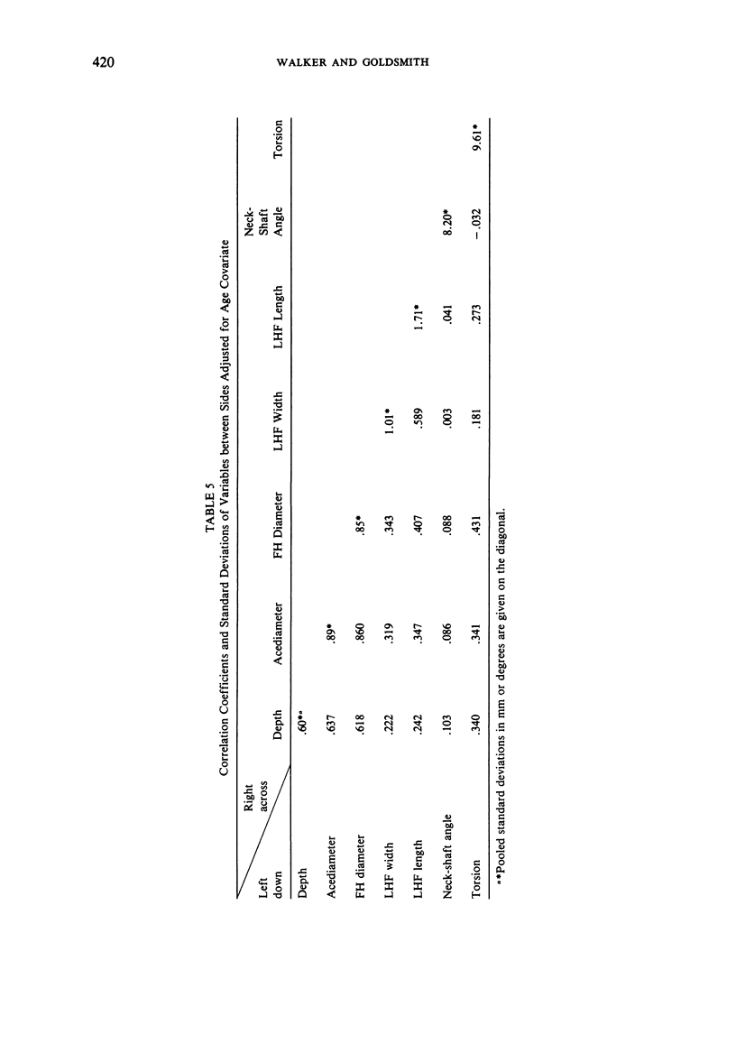

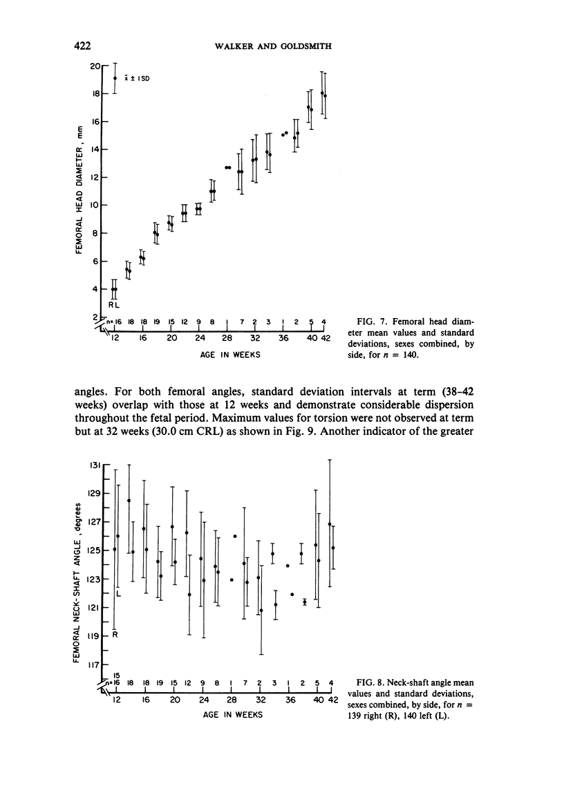
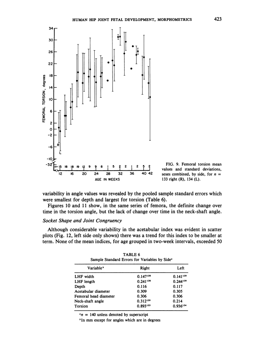



Images in this article
Selected References
These references are in PubMed. This may not be the complete list of references from this article.
- ANDERSEN H. Histochemical studies of the development of the human hip joint. Acta Anat (Basel) 1962;48:258–292. doi: 10.1159/000141844. [DOI] [PubMed] [Google Scholar]
- Blowers D. H., Elson R., Korley E. An investigation of the sphericity of the human femoral head. Med Biol Eng. 1972 Nov;10(6):762–775. doi: 10.1007/BF02477387. [DOI] [PubMed] [Google Scholar]
- Browne D. Congenital Deformities of Mechanical Origin: (Section for the Study of Disease in Children). Proc R Soc Med. 1936 Sep;29(11):1409–1431. [PMC free article] [PubMed] [Google Scholar]
- CAFFEY J., AMES R., SILVERMAN W. A., RYDER C. T., HOUGH G. Contradiction of the congenital dysplasia-predislocation hypothesis of congenital dislocation of the hip through a study of the normal variation in acetabular angles at successive periods in infancy. Pediatrics. 1956 May;17(5):632–641. [PubMed] [Google Scholar]
- CHEYNEL J., HUET R. Anatomie comparée de la hanche du nouveau-né blanc et noir; le problème de la luxation congénitale. Rev Chir Orthop Reparatrice Appar Mot. 1952 Jul-Sep;38(3-4):279–286. [PubMed] [Google Scholar]
- Crelin E. S. An experimental study of hip stability in human newborn cadavers. Yale J Biol Med. 1976 May;49(2):109–121. [PMC free article] [PubMed] [Google Scholar]
- DUNLAP K., SHANDS A. R., Jr, HOLLISTER L. C., Jr, GAUL J. S., Jr, STREIT H. A. A new method for determination of torsion of the femur. J Bone Joint Surg Am. 1953 Apr;35-A(2):289–311. [PubMed] [Google Scholar]
- Dunn P. M. Congenital postural deformities. Br Med Bull. 1976 Jan;32(1):71–76. doi: 10.1093/oxfordjournals.bmb.a071327. [DOI] [PubMed] [Google Scholar]
- Dunn P. M. Morphology and pathology of hip-joint at birth. Arch Dis Child. 1969 Feb;44(233):135–135. doi: 10.1136/adc.44.233.135. [DOI] [PMC free article] [PubMed] [Google Scholar]
- Dunn P. M. The anatomy and pathology of congenital dislocation of the hip. Clin Orthop Relat Res. 1976 Sep;(119):23–27. [PubMed] [Google Scholar]
- FELTS W. J. The prenatal development of the human femur. Am J Anat. 1954 Jan;94(1):1–44. doi: 10.1002/aja.1000940102. [DOI] [PubMed] [Google Scholar]
- GARDNER E., GRAY D. J. Prenatal development of the human hip joint. Am J Anat. 1950 Sep;87(2):163–211. doi: 10.1002/aja.1000870202. [DOI] [PubMed] [Google Scholar]
- GARDNER E., GRAY D. J. Prenatal development of the human hip joint. Am J Anat. 1950 Sep;87(2):163–211. doi: 10.1002/aja.1000870202. [DOI] [PubMed] [Google Scholar]
- Hadziselimović H., Sećerov D. Appearance of the upper end of the femur in infants under normal conditions and in cases of dislocation. (X-ray-anatomical studies). Acta Anat (Basel) 1968;70(4):509–523. [PubMed] [Google Scholar]
- Hammond B. T., Charnley J. The sphericity of the femoral head. Med Biol Eng. 1967 Sep;5(5):445–453. doi: 10.1007/BF02479138. [DOI] [PubMed] [Google Scholar]
- Harris N. H. Acetabular growth potential in congenital dislocation of the hip and some factors upon which it may depend. Clin Orthop Relat Res. 1976 Sep;(119):99–106. [PubMed] [Google Scholar]
- Hughes J. R. Acetabular dysplasia in congenital dislocation of the hip. Proc R Soc Med. 1974 Nov;67(11):1178–1180. [PMC free article] [PubMed] [Google Scholar]
- Humphry The Angle of the Neck with the Shaft of the Femur at Different Periods of Life and under Different Circumstances. J Anat Physiol. 1889 Jan;23(Pt 2):273–282. [PMC free article] [PubMed] [Google Scholar]
- James D. K., Dryburgh E. H., Chiswick M. L. Foot length--a new and potentially useful measurement in the neonate. Arch Dis Child. 1979 Mar;54(3):226–230. doi: 10.1136/adc.54.3.226. [DOI] [PMC free article] [PubMed] [Google Scholar]
- KINGSLEY P. C., OLMSTED K. L. A study to determine the angle of anteversion of the neck of the femur. J Bone Joint Surg Am. 1948 Jul;30A(3):745–751. [PubMed] [Google Scholar]
- LAURENSON R. D. DEVELOPMENT OF THE ACETABULAR ROOF IN THE FETAL HIP; AN ARTHROGRAPHIC AND HISTOLOGICAL STUDY. J Bone Joint Surg Am. 1965 Jul;47:975–983. [PubMed] [Google Scholar]
- MELLBIN T. The children of Swedish nomad Lapps. A study of their health, growth and development. Acta Paediatr Suppl. 1962 Jan;131:1–97. [PubMed] [Google Scholar]
- McKibbin B. Anatomical factors in the stability of the hip joint in the newborn. J Bone Joint Surg Br. 1970 Feb;52(1):148–159. [PubMed] [Google Scholar]
- Robinson D. C., De Buse P. J. Dislocatable hip in Ugandan newborn infants. East Afr Med J. 1970 Jul;47(7):395–397. [PubMed] [Google Scholar]
- Skirving A. P., Scadden W. J. The African neonatal hip and its immunity from congenital dislocation. J Bone Joint Surg Br. 1979 Aug;61-B(3):339–341. doi: 10.1302/0301-620X.61B3.479257. [DOI] [PubMed] [Google Scholar]
- Strayer L. M. The Embryology of the Human Hip Joint. Yale J Biol Med. 1943 Oct;16(1):13–26.6. [PMC free article] [PubMed] [Google Scholar]
- Trotter M., Peterson R. R. Weight of bone during the fetal period. Growth. 1969 Jun;33(2):167–184. [PubMed] [Google Scholar]
- Walker J. M. Growth characteristics of the fetal ligament of the head of femur: significance in congenital hip disease. Yale J Biol Med. 1980 Jul-Aug;53(4):307–316. [PMC free article] [PubMed] [Google Scholar]
- Walker J. M. Morphological variants in the human fetal hip joint. Their significance in congenital hip disease. J Bone Joint Surg Am. 1980 Oct;62(7):1073–1082. [PubMed] [Google Scholar]
- Watanabe R. S. Embryology of the human hip. Clin Orthop Relat Res. 1974 Jan-Feb;(98):8–26. doi: 10.1097/00003086-197401000-00003. [DOI] [PubMed] [Google Scholar]
- Wilkinson J. A. Breech malposition and intra-uterine dislocations. Proc R Soc Med. 1966 Nov;59(11 Pt 1):1106–1108. [PMC free article] [PubMed] [Google Scholar]
- Wosko I. Obserwacje makroskopowe i mikroskopowe dachu panewki w biodrach normalnych. Chir Narzadow Ruchu Ortop Pol. 1974;39(6):753–763. [PubMed] [Google Scholar]
- Wynne-Davies R. Acetabular dysplasia and familial joint laxity: two etiological factors in congenital dislocation of the hip. A review of 589 patients and their families. J Bone Joint Surg Br. 1970 Nov;52(4):704–716. [PubMed] [Google Scholar]


