Abstract
The eIF4F cap-binding complex mediates the initiation of cellular mRNA translation. eIF4F is composed of eIF4E, which binds to the mRNA cap, eIF4G, which indirectly links the mRNA cap with the 43S pre-initiation complex, and eIF4A, which is a helicase necessary for initiation. Viral nucleocapsid proteins (N) function in both genome replication and RNA encapsidation. Surprisingly, we find that hantavirus N has multiple intrinsic activities that mimic and substitute for each of the three peptides of the cap-binding complex thereby enhancing the translation of viral mRNA. N binds with high affinity to the mRNA cap replacing eIF4E. N binds directly to the 43S pre-initiation complex facilitating loading of ribosomes onto capped mRNA functionally replacing eIF4G. Finally, N obviates the requirement for the helicase, eIF4A. The expression of a multifaceted viral protein that functionally supplants the cellular cap-binding complex is a unique strategy for viral mRNA translation initiation. The ability of N to directly mediate translation initiation would ensure the efficient translation of viral mRNA.
Keywords: IRES, mRNA, nucleocapsid, RNA virus, translation
Introduction
Members of the hantavirus genus of the family Bunyaviridae are enveloped viruses harbouring three negative-sense, single-stranded genomic RNA molecules (Schmaljohn, 1996). The nucleocapsid peptide (N) has a vital function in hantavirus replication. Multiple studies show that N recognizes viral RNA (vRNA) with specificity indicative of its function during encapsidation (Gott et al, 1993; Severson et al, 1999, 2001; Osborne and Elliott, 2000; Jonsson et al, 2001; Jonsson and Schmaljohn, 2001; Mir and Panganiban, 2006). Each of the three genome segments form pseudocircular structures through a short imperfect ‘panhandle' composed of hydrogen-bonded nucleotides from the 5′ and 3′ termini (Pettersson and von Bonsdorff, 1975; Obijeski et al, 1976; Raju and Kolakofsky, 1989). The terminal panhandle is both necessary and sufficient for high-affinity binding by N (Mir and Panganiban, 2004b, 2005). N also functions in viral genome replication, as complementary in vitro and in vivo studies indicate that N, from diverse negative-sense RNA viruses, serves in vRNA replication working in coordinated manner with the viral polymerase or through interaction with template RNA (Bridgen and Elliott, 1996; Blakqori et al, 2003; Pinschewer et al, 2003; Kohl et al, 2004; Ikegami et al, 2005). Although hantavirus replication is exclusively cytoplasmic, generation of viral mRNA uses an orthomyxovirus-like cap-snatching mechanism yielding mRNAs with 5′ m7G caps derived from cellular mRNAs (Dunn et al, 1995; Garcin et al, 1995; Hutchinson et al, 1996).
The vast majority of eukaryotic mRNA translation is m7G cap dependent. Translation initiation involves the recognition of capped mRNA by a set of initiation factors (components of eIF4F cap-binding complex) (Dever, 1999; Gingras et al, 1999; Richter and Sonenberg, 2005). This heterotrimeric complex includes eIF4E, which directly binds to the mRNA cap (von der Haar et al, 2004; Richter and Sonenberg, 2005), and eIF4A, which is a DEAD box RNA helicase (Rogers et al, 2002; Hernandez and Vazquez-Pianzola, 2005). The third component of the eIF4 complex is eIF4G, a peptide that interacts with both eIF4E and eIF4A (Mader et al, 1995; Hentze, 1997; Dever, 1999). In addition, eIF4G interacts with eIF3 (Hinnebusch, 2006) to bridge the mRNA–eIF4 cap-binding complex and the 43S ‘pre-initiation complex.' The 43S complex is composed of the 40S small ribosomal subunit, initiator methionine transfer RNA, eIF2 and GTP (Hershey and Merrick, 2000). Scanning by this large set of proteins then proceeds from the capped 5′ end of the mRNA in a process that may require the helicase activity of eIF4A (Rogers et al, 2002; Hernandez and Vazquez-Pianzola, 2005). When an AUG start codon in optimal context is encountered the 60S large ribosomal subunit and additional factors are recruited and translation begins (Kozak, 1991, 1992).
Typically, viruses use this cellular machinery for translation of their mRNAs, and most have capped mRNAs. However, the picornaviruses, some flaviviruses, a few additional viruses contain a cis-acting internal ribosomal entry sites (IRESs) to enable cap-independent ribosomal entry at a site in the mRNA immediately proximal to the start codon (Hellen and Sarnow, 2001; Jang, 2006). Along the same lines, poxviruses contain a cis-acting poly A sequence in their 5′ leader that facilitates association of the pre-initiation complex with viral mRNA (Shirokikh and Spirin, 2008). In the course of experiments to examine hantavirus nucleocapsid (N) protein function, we noted that the expression of N in cells appeared to surprisingly result in increased expression of heterologous indicator mRNAs. Here, we describe this phenomenon in detail. N can replace the activities of eIF4F to mediate mRNA translation. In particular, N binds with high affinity to the capped 5′ end of viral mRNAs, an activity that mimics that of eIF4E. N substitutes for the standard requirement for the bridging peptide, eIF4G, by directly recruiting the 43S pre-initiation complex to the 5′ mRNA cap. Finally, N replaces the helicase, eIF4A, in the cap-binding complex. Thus, this viral strategy is the functional complement to that of an IRES. N supplants the eIF4F complex in trans, whereas an IRES replaces cap-dependent translation in cis.
Results
N facilitates translation of capped mRNA
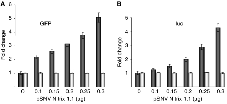
Co-expression of N with various reporter mRNAs yielded unexpected evidence, consistent with the idea that the steady-state expression of reporter proteins was augmented by N (data not shown). To examine this apparent N-dependent increase in protein expression, we co-transfected HeLa cells with increasing amounts of a plasmid that expresses Sin nombre hantavirus (SNV) N (or an empty expression vector) and a constant amount of a reporter plasmid expressing either green fluorescent protein (GFP) or luciferase (luc) mRNA. At 36 h after transfection, cells were harvested, and GFP expression was quantified by flow cytometry and luc was measured using a quantitative enzymatic assay. We observed a concomitant increase of about five-fold in both GFP expression and luc expression with increasing amounts of N expression plasmid (Figure 1A and B). Quantitative RT–PCR (real-time PCR) with primers corresponding to a segment in the centre of the mRNA indicated that N does not detectibly affect intracellular amounts of either GFP or luciferase mRNA (Figure 1A and B), suggesting that N augments expression at the translational level.
Figure 1.
N increases the expression of reporter proteins. HeLa cells were transfected with a constant amount of reporter plasmid and increasing amounts of a plasmid expressing hantavirus N. Evaluation of N expression on western blots with anti-N antibody indicated that N expression increased along with increasing amounts of plasmid, as expected (not shown). At 36 h after transfection, cells were harvested and GFP or luc expression was quantified, by flow cytometry or enzymatically, respectively (dark bars). (A) Expression of GFP is shown. (B) luc as a function of increasing N is shown. Steady-state GFP mRNA and luc mRNA were quantified using ‘real-time' RT–PCR with primers specific for a segment in the centre of each reporter RNA. In both (A, B), the results of this latter analysis are depicted with light bars.
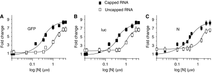
We next used rabbit reticulocyte extracts to carry out in vitro translation reactions with reporter RNA containing or lacking a 5′ m7G cap. When increasing amounts of bacterially expressed purified N, were added translation of each of the three reporter mRNAs was enhanced. This effect of N was significantly more efficient and manifested at a lower N concentration when the reporter mRNA contained a 5′ m7G cap (Figure 2A–C). It should be noted that the expression of N from the mRNA in Figure 2C was used merely as a reporter, analogous to GFP or luc, and did not significantly contribute to the amount of N in the reaction.
Figure 2.
N augments the translational expression of capped mRNA. We examined the effect of increasing N on the translational expression of three reporter mRNAs containing or lacking a 5′ cap using rabbit reticulocyte lysates. Capped and uncapped mRNA encoding GFP, luc or N were translated in vitro in the presence of 35S-methionine and increasing amounts of N in (A–C), respectively. Translation products were then electrophoresed on SDS polyacrylamide gels, and the amount of translation product was quantified by phosphorimage analysis. Translation of capped and uncapped RNA is depicted with filled and open squares, respectively. The amount of labelled protein synthesized with capped RNA in reactions lacking N was normalized to 1 (this cannot be indicated on the log scale). In the absence of N, expression of the indicator proteins was slightly higher with uncapped than capped RNA. This is consistent with earlier observations (Svitkin et al, 1996). Thus, the amount of expression at the lower concentrations of N is equivalent to background levels of expression from capped and uncapped RNA for each of the indicator RNAs.
N binds to 5′ caps and mediates preferential translation of viral mRNA
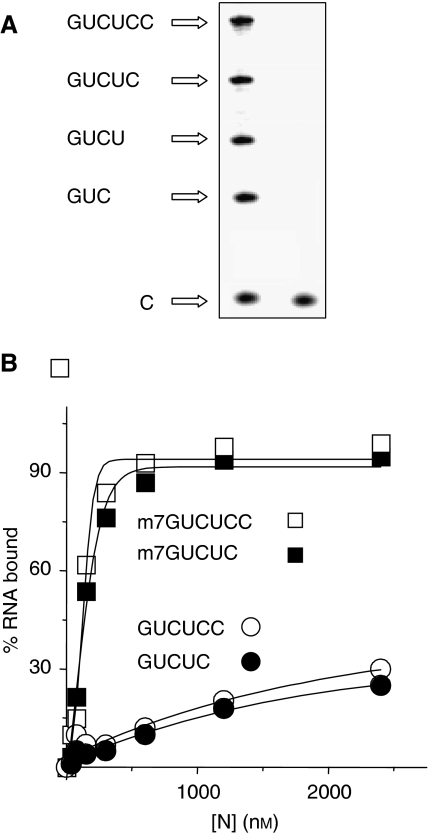
As N preferentially enhanced the translation of capped mRNAs, we next asked whether N interacts with the 5′ end of capped RNAs. We synthesized radioactively labelled capped and uncapped RNAs, 3–6 nt in length (Figure 3A) and carried out filter binding studies with each of these short RNAs to assess binding by N. This indicated that N binds to capped but not uncapped penta- and hexanucleotide RNA at a Kd of 120–130 nM (Figure 3B). However, there was no detectible binding with either capped or uncapped tri- and tetranucleotide RNA (Supplementary Figure S1). We also examined interaction between N and free cap using fluorescence spectroscopic analysis. The interaction of N with free cap is at least three orders of magnitude weaker than that observed for N with capped penta- or hexanucleotide RNA (Supplementary Figure S2). These data suggest that translation enhanced by N is superior for capped mRNAs owing simply to the ability of N to bind to capped 5′ ends.
Figure 3.
N binds to capped oligoribonucleotides. (A) Short radioactively labelled capped and uncapped RNAs were synthesized using T7 RNA polymerase and α-32P-CTP. As the third nucleotide of transcription products arising from the T7 promoter is the first C residue of the RNA, only molecules 3 nt long or greater were labelled. These short RNAs were separated on, and recovered from, a high percentage denaturing polyacrylamide gel. The image depicts such a gel. The leftward lane displays the series of short oligoribonucleotides arising from transcription. The rightward lane contains unincorporated CTP as a migration control. (B) Each RNA was incubated with increasing concentrations of purified N, and association of RNA with N was quantified by filter binding. Binding of N with capped (m7GUCUCC) or uncapped (GUCUCC) are indicated with open squares and circles, respectively, and with m7GUCUC and GUCUC with closed squares and circles, respectively. Binding experiments carried out with both capped and uncapped RNAs less than 5 nt in length exhibited negligible binding with N. For example, binding with the 4-nt long RNA, GUCU, in capped and uncapped form is shown in Supplementary Figure S1.
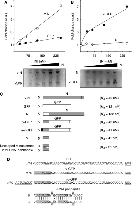
Hantaviruses do not abrogate general cellular mRNA translation. Nonetheless, if N enhances translation, viral mRNA might be preferentially translated relative to non-viral mRNA. The previously described ‘reporter RNA' encoding N (Figure 2C) contained the N gene but lacked this viral 5′ non-coding region, and N-mediated translation of this RNA was similar to that of the GFP and luc reporter mRNAs. As with all bunyavirus, the 5′ ends of hantavirus mRNAs contain approximately 10 non-viral nucleotides that arise from cap-snatching. The 5′ non-coding region from hantavirus S segment mRNA is 44 nt in length, not including non-viral nucleotides. We carried out a competitive assay to examine the translation of an mRNA containing the viral 5′ non-coding sequences relative to a second reporter (GFP) containing a non-viral leader of equal length. Equimolar amounts of these two capped RNAs were added together to reticulocyte extracts with increasing amounts of purified N. N-mediated enhancement of translation was superior for the viral mRNA when compared with the GFP RNA, yielding an increase in viral mRNA expression of about seven-fold (Figure 4A). We generated two additional mRNAs in which the 44 nt leader regions were interchanged between the two reporter genes. Increasing amounts of N resulted in preferential expression of GFP from the chimaeric mRNA containing the capped viral 5′ leader (Figure 4B). Thus, the translation of non-viral mRNA can be facilitated by N (Figure 2), but in a competitive reaction containing both viral and non-viral mRNA translation of mRNA containing the 5′ non-coding sequences from the virus is robust when compared with mRNA harbouring a non-viral leader.
Figure 4.
N preferentially augments the translation of viral mRNA. (A) Equimolar amounts of capped mRNA containing the 5′ untranslated region from S segment mRNA and encoding N (v-N), and an mRNA containing a non-viral leader region and encoding GFP (GFP) were added together to reticulocyte extracts containing increasing concentrations of N as indicated. The concentration of each mRNA was approximately 45 nM. Labelled N and GFP were separated by PAGE and quantified by phosphorimage analysis (shown below the graph). Similar results were obtained in three separate experiments. In (B), the leader regions from the mRNAs of (A) were interchanged. Thus, one mRNA contained the 5′ viral UTR preceding the GFP gene (v-GFP), and a second mRNA contained the non-viral leader preceding the N gene (N). Translation and quantitation were as in (A). (C) Radioactively labelled capped RNAs were used in binding reactions with purified N and the binding affinity (Kd) was determined for each. Viral sequences are shown schematically in grey, whereas non-viral sequences are in white. n-v-GFP contains a 9 nt non-viral cap simulating cellular RNA derived from cap-snatching (shown in black). Note: The leader regions are not shown to scale relative to the N and GFP genes. The untranslated leaders, GFP gene, and N gene are 43, 798, and 1287 nt in length, respectively. (D) Comparison of the leader sequences from GFP, v-GFP, and n-v-GFP mRNA and minus strand S segment viral RNA. Nucleotides required for high-affinity binding to the vRNA panhandle are depicted with shading and include nucleotides from both the 5′ and 3′ termini (Mir and Panganiban, 2005). The 5′ terminal nucleotides of +strand mRNA required for binding by N is also indicated by shading. As the termini of the viral genome segments consist of imperfect inverted repeats, the 5′ sequences of both plus and minus strand viral RNA are similar. Nucleotide differences in the 5′ sequence of mRNA relative to the 5′ sequence of minus strand vRNA are indicated with bold lettering. Leader sequences of v-GFP and n-v-GFP. The 9-nt-long non-viral leader of n-v-GFP, and the start codon of the mRNAs are underlined.
RNA-binding assays of N for each of these capped RNAs indicated that RNA containing the 5′ leader region from viral mRNA interacted with N at significantly higher affinity than RNA with a non-viral leader (Figure 4C). Further, the viral leader region was sufficient for higher affinity binding by N. Preferential translation of viral mRNA, and high-affinity binding by N, is not diminished by the presence of capped non-viral nucleotides at the 5′ end, as would be present on bona fide viral mRNA (Figure 4C) (Mir and Panganiban, submitted).
N stably binds to the 43S pre-initiation complex and replaces eIF4G
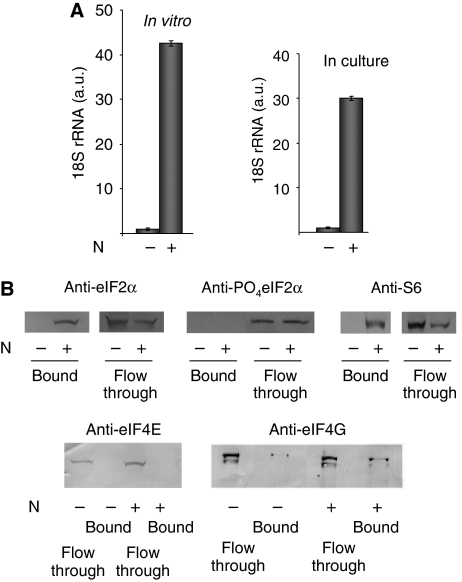
eIF4F binds to mRNA by way of eIF4E, and to the eIF3 and the 43S pre-initiation complex by way of eIF4G. To determine whether N may interact with the 43S pre-initiation complex, we added his6-tagged N to rabbit reticulocyte lysates, recovered N using Ni-NTA beads and used a quantitative assay for 18S rRNA to quantify 40S ribosomal subunits associated with N. These assays indicated that 18S rRNA was associated with N (Figure 5A), suggesting that N interacts with 40S eukaryotic ribosomal subunit. We carried out a similar experiment with lysates derived from 293 cells expressing his6-tagged N following transfection with an N expression construct. Again we observed that 18S rRNA was recovered with N on Ni-NTA beads, indicating that N interacts directly or indirectly with the 43S pre-initiation complex in vivo (Figure 5A).
Figure 5.
N interacts with the pre-initiation complex. (A) N was incubated with rabbit reticulocyte lysates and recovered with Ni-NTA beads. The bound material was eluted from the Ni-NTA, RNA was purified, and 18S rRNA was quantified using real-time RT–PCR. The leftward graph depicts the relative amount of 18S rRNA associated with Ni-NTA beads in the absence and presence of N. The rightward graph depicts an analogous experiment carried out with 293 cells that were transfected with either an N-expressing plasmid or its parental vector, as a negative control. N was recovered from the lysates of transfected cells using Ni-NTA and 18S rRNA that co-purified with N quantified by real-time RT–PCR. (B) A set of western blots to examine the association of peptide constituents of the 43S pre-initiation complex, and the eIF4F cap-binding complex, that co-purify with N. N was expressed by transfection, isolated from the lysates of these cells using Ni-NTA columns, bound material was recovered and subjected to western blot analysis with primary antibodies as indicated. Peptides that co-purify with N (bound), or that flow through the column are indicated.
In the unphosphorylated form, eIF2α is a functional component of the 43S pre-initiation complex, whereas phosphorylated eIF2α is not associated with the pre-initiation complex. We determined whether eIF2α co-purified with his6-tagged N from lysates of transfected cells on Ni-NTA columns using western blot analysis with antibody specific for the unphosphorylated and phosphorylated forms of eIF2α. This indicated that unphosphorylated eIF2α but not phosphorylated eIF2α was associated with N (Figure 5B). Moreover, western blot analysis indicated that S6 ribosomal protein also co-purified with N (Figure 5B). These data again suggest that N interacts with the 43S pre-initiation complex. In contrast, components of the eIF4 cap-binding complex, eIF4E and eIF4G, did not co-purify with N in parallel western blots (Figure 5B). Thus, association of the 43S pre-initiation complex with N does not require the eIF4F cap-binding complex.
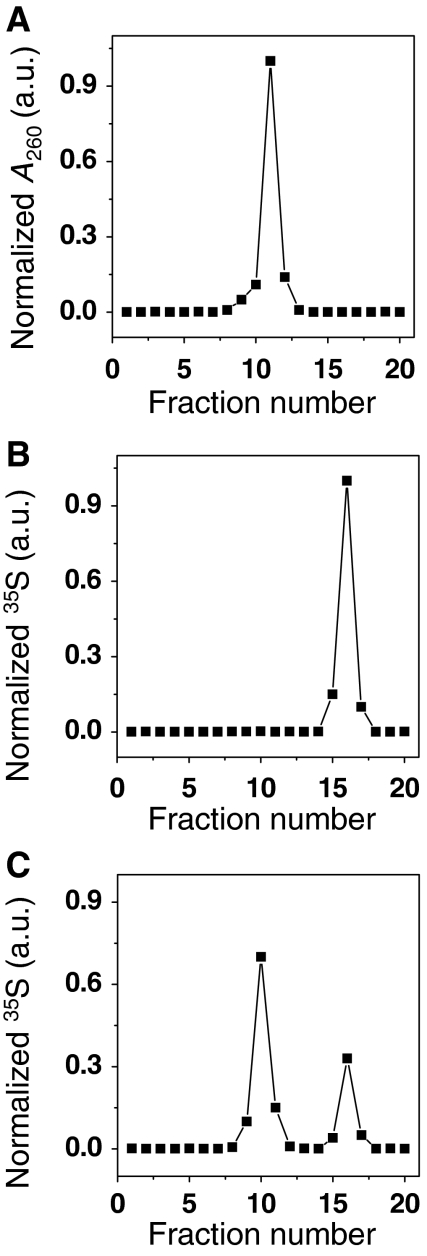
To verify that interaction between N and the 43S pre-initiation complex was mediated by way of direct interaction rather than indirectly through an mRNA bridge, we dissociated ribosomes into large and small subunits by incubation with puromycin, purified 40S small ribosomal subunits by sucrose gradient centrifugation, and asked whether N could interact directly with purified 40S subunits. The purified 40S subunits were resedimented, detected by monitoring optical density and yielded a sedimentation profile indicative of a homogeneous 40S preparation (Figure 6A). We synthesized 35S-labelled N in reticulocyte extracts, and purified the labelled protein by denaturation, recovery on Ni-NTA columns, and renaturation. Sedimentation analysis of this purified 35S-labelled N protein indicated that N migrated to a distinct position high in the gradient (Figure 6B). Significantly, incubation of N with purified 40S subunits resulted in co-migration of N with small subunits indicative of interaction between N and the 40S subunit (Figure 6C). A radioactively labelled control protein (GFP) did not interact with 40S ribosomal subunits and remained near the top of the gradient (data not shown). These data indicate that N binds directly to a component of the small ribosomal subunit and that association is not through an mRNA bridge. However, the data do not unequivocally distinguish between whether N interacts directly with the 40S subunit or with residual eIF3 bound to the 40S subunit.
Figure 6.
N binds directly to the small ribosomal subunit. 40S small ribosomal subunits were prepared by incubation of ribosomes in the presence of puromycin and purified from large ribosomal subunits and mRNA. (A) Purified 40S subunits were then resedimented on a sucrose gradient. (B) N protein was expressed by in vitro translation in the presence of 35S-methionine, purified from the translation mixture by denaturation with urea, recovery on Ni-NTA beads, renaturation by dialysis and sedimented in parallel with 40S subunits. (C) N was incubated with excess purified 40S subunits prior to sedimentation. Leftward fractions correspond to those from the bottom of the gradient.
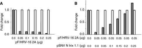
We next asked whether N is likely to functionally replace eIF4G. Some members of the picornavirus family shut off host mRNA translation through proteolytic cleavage of eIF4G, a process mediated by the viral 2A protease (Etchison et al, 1982; Liebig et al, 1993; Haghighat et al, 1996). We co-transfected cells with a plasmid expressing GFP reporter mRNA along with increasing amounts of a plasmid that expresses the 2A protease of human rhinovirus 16 (HRV-16). Although the 2A cleavage products of eIF4G sometimes retain residual activity (Ali et al, 2001), as expected, the presence of this plasmid expressing 2A protease dramatically reduced translational expression of the GFP reporter mRNA (Figure 7A). Significantly, co-expression of N overcame this translational inhibition resulting from the 2A-expressing plasmid (Figure 7B). Thus, N-mediated translation initiation appears to take place under conditions where eIF4G is proteolytically inactivated. Taken together, all these data are consistent with a simple model where N supplants eIF4G in bridging to the 43S pre-initiation complex.
Figure 7.
N replaces eIF4G. (A) HeLa cells were co-transfected with a plasmid expressing reporter GFP, along with increasing amounts of pF/HRV-16 2A, which expresses the 2A protease of HRV-16. GFP expression was quantified using flow cytometry as in Figure 1 (dark bars) and GFP mRNA was quantified using real-time PCR (light bars). (B) Cells were transfected with a constant amount of GFP expression plasmid, a constant amount of 2A expression plasmid (0.05 μg) sufficient for significantly reducing translation of the reporter gene, and increasing amounts of an N expression plasmid. GFP expression (dark bars) and steady-state GFP mRNA (light bars) were quantified as in (A). In the experiments of (A, B), the total amount of DNA used in the transfections was held constant by the addition of parental vector.
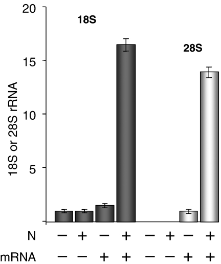
We used a ribosome-loading assay to see whether N can facilitate loading of small ribosomal subunits onto the 5′ end of mRNA. A 415-nt-long mRNA containing a 15-nt-long poly A tail and 200-nt-long non-coding 5′ leader sequence was incubated in rabbit reticulocyte lysates in the presence or absence of N. This mRNA was then purified from the reaction mixture by virtue of its poly A tail using poly-dT sepharose beads. Ribosomes loaded onto the isolated mRNA were then quantified by measuring 18S and 28S rRNA associated with the isolated mRNA. We observed a significant increase in both 18S and 28S rRNA when both N and mRNA were present in rabbit reticulocyte lysates indicative of enhanced ribosome loading onto the isolated mRNA (Figure 8). As the recruitment of the 43S pre-initiation complex to the 5′ end of mRNAs is conventionally considered to be the rate-limiting step for translation, these data suggest that N increases the rate of recruitment of the 43S pre-initiation complex onto mRNAs.
Figure 8.
N promotes ribosome loading. A synthetic mRNA containing 3′ poly A was incubated in reticulocyte lysates to allow translation. The synthetic polyadenylated RNA was recovered from the translation mixture using oligo dT beads. Ribosomes associated with the polyadenylated RNA were quantified by real-time RT–PCR with primer sets specific for 18 and 28S rRNA.
Cellular mRNA is circularized through association between eIF4G in the eIF4F cap-binding complex at the 5′ end and poly A-binding protein (PABP) at the 3′ end leading to more efficient translation (Tarun and Sachs, 1996; Gray et al, 2000). As eIF4G appeared to be dispensable for N-mediated translation initiation, we asked whether N may interact with PABP to effect circularization. We carried out co-precipitation experiments in which his6-tagged N from lysates of transfected cells was recovered on Ni-NTA beads and the recovered material was analysed by western blot analysis with anti-PABP antibody. On the basis of this approach, there was not detectible stable association between N and PABP (Supplementary Figure S3).
N replaces eIF4A
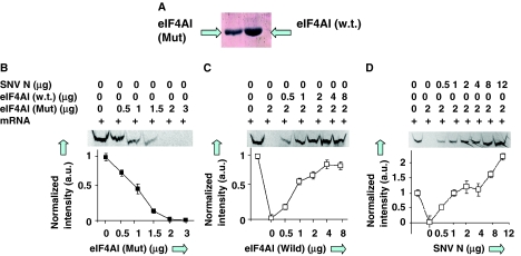
We next wanted to see whether N replaces the activity of the third constituent of the eIF4F complex, eIF4A. eIF4A is a DEAD box RNA helicase required for eIF4F function during cap binding and has been postulated to function in the scanning of the pre-initiation complex to the AUG start codon (Rogers et al, 2002; Hernandez and Vazquez-Pianzola, 2005). In comparison, N has an intrinsic ATP-independent activity that facilitates transient RNA duplex dissociation (Mir and Panganiban, 2006). eIF4A migrates on and off the eIF4F complex, where it functions in concert with eIF4E and eIF4G, and this interconversion between the complexed and free forms of eIF4A appears to be necessary for eIF4A function. We used a dominant-negative mutant of eIF4A defective in cycling through eIF4F complex, which dramatically inhibits translation in rabbit reticulocyte lysates, to see whether N mediates translation initiation in an eIF4F-independent manner (Pause et al, 1994). Wild-type and dominant-negative eIF4A were expressed in bacteria and purified (Figure 9A). Consistent with published characterization of this dominant-negative eIF4A protein (Pause et al, 1994), 2 μg of the dominant-negative mutant protein inhibited translation of a reporter mRNA in rabbit reticulocyte lysates by about 98% (Figure 9B), and translation could be significantly rescued by the addition of 2–4 μg of wild-type eIF4A when the dominant-negative protein was present (Figure 9C). Notably, translation inhibited by dominant-negative eIF4A could also be completely overcome by adding 1.5 μg of N protein to the translation reaction (Figure 9D). These data indicate that that N functionally substitutes for eIF4A, and together with the earlier presented data indicate that N functionally substitutes for the entire eIF4F cap-binding complex.
Figure 9.
N functionally replaces eIF4A. (A) Bacterially expressed and purified wild-type and mutant eIF4A were used in in vitro translation reactions containing luciferase mRNA. (B) Effect of dominant-negative mutant eIF4A on translation. Translation was quantified by SDS–PAGE followed by phosphorimage analysis of radioactively labelled luc. (C) Similar reactions were carried out in the presence of fixed amount (2 μg) of mutant eIF4A and increasing amounts of wild-type eIF4A. (D) Translation reactions in the presence of 2 μg of mutant eIF-4A and increasing amounts of N.
Discussion
N as a translation initiation factor
N-mediated translation initiation is a viral strategy that is the complement to the use of an IRES. The latter tactic is employed by the picornaviruses and some flaviviruses. Although an IRES is a cis-acting element that functionally supplants the requirement for cap-dependent translation, N is a trans-acting element that replaces eIF4F.
It is likely that N mediates initiation through a simple mechanism. There is no overt similarity between N and the eIF4F components. Nonetheless, the three-dimensional structure of cellular cap-binding peptides human eIF4E includes two W residues (W52 and W102) that hold the guanine residue of a cap-analogue through stacking interactions. An acidic residue (E103) further stabilizes this association (Marcotrigiano et al, 1997; Matsuo et al, 1997). There may be weak alignment of this region with a segment of N of identical length (a.a. 119–166) containing appropriately spaced W119 and Y165E166 residues. Binding of eIF4G with the eIF3 and the 43S pre-initiation complex is mediated by a portion of the central region of eIF4G (Korneeva et al, 2000; Schutz et al, 2008). ClustalW comparison of N with this region of eIF4G indicates weak alignment with the amino-terminus of N. These regions of both peptides contain antiparallel alpha-helices, in coiled-coil (N) or in HEAT (eIF4G) configuration (Marcotrigiano et al, 2001; Boudko et al, 2007). However, interaction of N with 40S subunits is likely mediated through a domain dissimilar to that responsible for eIF3 recognition by eIF4G. Finally, there is no apparent similarity between eIF4A and N. This may be expected as eIF4A is an ATP-dependent DEAD box helicase, N is an ATP-independent RNA chaperone.
N augments translation of both viral and non-viral mRNA. However, viral mRNA is recognized at higher affinity by N, and translation of viral mRNA is more robust in competitive in vitro translation reactions with non-viral mRNA. mRNA from all minus strand segmented RNA viruses is initiated with nucleotides acquired from the 5′ ends of cellular mRNA by cap snatching. Nonetheless, the motif in viral mRNA preferentially recognized by N is situated in the viral 5′ UTR and not affected by the presence of a short non-viral cap. Mutational analysis of the viral UTR indicates that the motif recognized by N for preferential translation is less than 10 nt in length (Figure 4D) (Mir and Panganiban, submitted). In contrast, the motif in minus strand vRNA recognized at high affinity, which ostensibly initiates genome encapsidation, is the predominantly double-stranded viral panhandle formed by the juxtaposition of the 5′ and 3′ vRNA termini (Figure 4D) (Mir and Panganiban, 2004). The panhandle is recognized at higher affinity than viral mRNA. During replication, viral mRNA synthesis precedes and overlaps with genome replication. It will be of interest to see common RNA-binding domains of N function in both processes.
It is likely that the general strategy of encoding a trans-acting factor to ensure efficient viral translation is not restricted to hantaviruses. It is probable that the N peptides of members of the other genera of the bunyavirus family, and perhaps the members of diverse families of segmented and non-segmented minus strand RNA viruses, also supplant the eIF4F complex. Several observations hint that RNA viruses may use this general scheme. Subgenomic Sindbis virus mRNA is translated when eIF4G is inactivated (Castello et al, 2006). Vesicular stomatitis virus impairs eIF4E function through dephosphorylation but sustain translation of its own mRNAs (Connor and Lyles, 2002). Similarly, influenza mRNA translation can occur when eIF4E is impaired (Burgui et al, 2007). Expression of Sendai virus N is required for the expression of a reporter gene from a viral vector (Wiegand et al, 2007). This could be due to an effect on transcription, as suggested by the authors, but their data are also consistent with a positive role of SeV N in translation.
Implications for viral transcription
The bunyaviruses are unique among the negative-stranded RNA viruses in that transcription requires concomitant translation of the nascent viral mRNA (Bellocq and Kolakofsky, 1987; Barr, 2007). Coupling of transcription with translation appears to be necessary for successful RNA elongation by the RdRp through spurious premature transcription termination signals. Mechanistically, this may occur as ribosomes trailing the RdRp block the formation of higher order structures in the nascent RNA that function as inappropriate termination sites. N may promote more efficient loading of ribosomes onto nascent viral mRNA leading to higher ribosome density and ensuring mRNA elongation.
Apparent lack of circularization of hantavirus mRNA
Circularization of cellular mRNA mediated by the interaction between eIF4G and PABP enhances translation efficiency (Tarun and Sachs, 1996; Gray et al, 2000). However, circularization is probably not required for efficient N-mediated translation of viral mRNA, or else occurs through unidentified factors. Initiation can take place independently of eIF4F and we were unable to detect association between N and PABP. Moreover, of the three hantavirus mRNAs only that encoding the viral envelope protein is polyadenylated, whereas mRNA encoding N and the RdRp are not (Hutchinson et al, 1996).
It is worthwhile to contrast N-mediated translation initiation with viral peptides that associate with eIF4F. The potyviruses, a set of plant viruses related to the picornaviruses, encode a VPg that is attached to the 5′ end of the genome and that also operationally associates with eIF4E, potentially enabling functional circularization of the genome (Kang et al, 2005). Rotavirus NSP3A and Alfalfa mosaic virus (AMV) coat protein associate with both the 3′ end of their respective genomes and with eIF4G (Piron et al, 1998; Bol, 2005). This association with the eIF4F complex also enables genome circularization as the 3′ termini of these viral genomes are not polyadenylated. Thus, NSP3A and AMV coat function as surrogates for PABP. For these viruses, interaction with the eIF4F complex is required for efficient replication.
There are a remarkable number of activities and functions associated with hantavirus N. These include its structural role as the capsid protein, its role as the principle player mediating the encapsidation of vRNA, its role as an RNA chaperone capable of reconfiguring the higher order structure of RNA, its role in genome replication in coordination with the viral polymerase, and now an unexpected role as a translational initiation factor with multiple complementary activities dedicated to that function. At the same time, N-mediated translation of viral mRNA can probably be considered to be a narrowly focused function ensuring efficient production of the viral peptides. In this regard, translation initiation by N would not be accompanied by the elegant and subtle regulatory capacity of the multicomponent cellular translational complex.
Materials and methods
Plasmids
pSNV N TriEx 1.1 expresses N containing a C-terminal histidine tag both in vivo and in vitro (Mir et al, 2006). pEGF-P (Promega) and pGL3 plasmid (Clone Tech) were used for the expression of GFP and luciferase (luc), respectively. pF/HRV-16 2A, which expresses human rhinovirus 16 2A protease from the EMCV IRES at high level, was kindly provided by Yury Bochkov, Alex Aminev, and Ann Palmenberg (Bochkov and Palmenberg, 2006).
Flow cytometry
All transfections were carried out in triplicate in six-well plates. Flow cytometry was carried out using a FACScan (BD Biosciences), obtaining 10 000 gated events for each sample. The fluorescence of the gated cells was quantified and a histogram was generated to display the distribution of fluorescence intensity in the cell population. The mean fluorescence value of positive and negative events was calculated.
Ribosome-loading assay
RNA molecules generated were 415 nt long, containing a 200 nt non-coding sequence at the 5′ end, followed by an AUG and 200 additional nucleotides followed by a 15-nt-long poly A tail. Here, 5 μg of this mRNA was added to 20 μl in vitro translation reactions with or without N and incubated at 30°C for 15 min. RNA was recovered with 20 μl of oligotex (poly-dT beads) and reverse transcribed using random primers as described in the ‘Real-time PCR' section. Then, 2 μl of the resulting cDNA was used for the quantitation of 18S and 28S rRNA using appropriate primers with the standard curve method as in the Real-time PCR section.
Preparation of mRNA substrates for in vitro T7 transcription reactions
PCR products containing T7 promoters were gel purified and used as a template in T7 transcription reactions with Ribomax T7 (Promega). Following synthesis, template DNA was degraded with DNase I, the RNA was purified by RNAeasy (Qiagen), and stored in 10 μl aliquots at −70°C. RNA molecules with a terminal 5′ m7G cap were synthesized by the incorporation of m7G cap analogue in the transcription reactions. Short RNA molecules, 3–6 nt long, were synthesized from a 100-nt-long DNA template containing a terminal T7 promoter. 5′ caps were incorporated in the short transcripts by adding m7G cap analogue to the T7 reaction mixtures. Transcription reactions lacked ATP to terminate the reaction after the incorporation of first 6 nt in the transcript resulting in the synthesis of oligoribonucleotides from 3–6 nt in length (5′-GUCUCC). Reaction mixtures were fractionated on denaturing 18% polyacrylamide gels containing urea. This resulted in a ladder composed of RNAs 3, 4, 5 and 6 nt in length. RNA was recovered from gel slices and stored in 10 μl aliquots at −70°C.
In vitro translation
Nuclease-treated rabbit reticulocyte lysates were used for the translation of mRNA in the presence and absence of supplemented, bacterially expressed nucleocapsid protein. Translation reactions were carried out in 50 μl containing 35 μl of rabbit reticulocyte lysate, 1 μl amino-acid mixture minus methionine (1 mM), 1 μl 35S-methionine (1175 Ci/mmol), 2 μl RNase inhibitor (40 μg/μl), 4 μl mRNA in water (250 ng/μl), and 7 μl of RNase-free water. The final RNA concentration in the reactions was approximately 90 nM. Reaction mixtures were incubated at 30°C for 30 min. Samples were electrophoresed on 10% SDS gels and quantified with a phosphorimager.
Real-time PCR
RNA was isolated from cells, or from rabbit reticulocyte lysates, using ‘RNAeasy' (Qiagen) and treated with RNase-free DNase I. RNA was reverse transcribed using Mo-MLV reverse transcriptase and random primers in a total volume of 50 μl. An absolute standard curve was used for real-time PCR, using a ABI prism 7700 (Applied Biosystems) using a 150-nt-long sequence of the GFP, or luciferase, gene. Primer validation was carried out following the manufacturer's protocol (Applied Biosystems). Real-time PCR reactions were carried out in 20 μl, including 10 μl ribogreen mastermix (Applied Biosystems), 2 μl of template, 3.6 μl of each forward and reverse primer, and 0.8 μl water. Each reaction was carried out in triplicate.
RNA filter binding
All binding reactions were carried out in RNA-binding buffer (Mir and Panganiban, 2004) at a constant concentration of RNA (1 pM) with increasing concentration of N protein. Reaction mixtures were incubated at room temperature for 30–45 min and filtered through nitrocellulose membranes under vacuum. The amount of RNA retained on the filter by virtue of N was measured using a scintillation counter. Data points were fit to a hyperbolic equation using the program Origin 6 (Microcal). The apparent dissociation constant (Kd) corresponded to the concentration of N protein required to obtain the half saturation in the binding profile.
‘Pull-down' experiments with Ni-NTA
His-tagged N from reticulocyte lysates was recovered on, and eluted from, Ni-NTA columns (Qiagen) in 50 μl of elution buffer (50 mM NaH2PO4, 300 mM NaCl, and 500 mM imidazole). The eluted sample (25 μl) was used for the purification of total RNA. RNA was reverse transcribed and analysed by real-time PCR to measure 18S or 28S rRNA. His-tagged N was recovered from HeLa and 293 cells with Ni-NTA columns. Columns were washed with 600 μl of wash buffer and bound material was eluted with elution buffer as described above. Total RNA purification and real-time PCR studies were carried out as described above. Pooled fractions were also analysed for the presence of translation initiation factors (eIF4E, eIF4G, unphosphorylated and phosphorylated eIF2α, and ribosomal protein S6) using the corresponding antibodies (Cell Signaling Technology). Here, 50% of the eluted and 10% of the flowthrough material, respectively, were used for the detection of ribosomal proteins or other initiation factors involved in translation by western blot analysis.
40S ribosomal subunit preparations
40S ribosomal subunits were purified using a standard protocol (Pestova et al, 1996). Briefly, rabbit reticulocyte lysates were diluted 10-fold in the presence of 1 mM dithiothreitol (DTT), and centrifuged for 4 h at 100 000 g using a fixed angle NVT 90 rotor. The resulting pellet was resuspended in 5 ml of buffer A (0.25 M sucrose, 0.05 M Tris–HCl (pH 7.5), 1 mM DTT, 6 mM MgCl2, and 0.1 mM EDTA), followed by the addition of 0.5 M KCl with continuous stirring on ice for 30 min. The mixture was centrifuged for 2 h at 180 000 g using a NVT 90 rotor. The pellet was dissolved in a small volume of buffer A, layered over a sucrose cushion (1.0 M sucrose, 0.5 M KCl, 0.02 M Tris–HCl (pH 7.5), 2 mM MgCl2, and 0.1 mM EDTA) and centrifuged at 275 000 g for 3 h. The pellet, containing 80S ribosomes, was resuspended in a small volume of buffer A and further diluted 10-fold in buffer B (0.5 M KCl, 0.05 M Hepes (pH 7.5), 2 mM MgCl2, and 1 mM puromycin). The mixture was incubated for 10 min on ice followed by further incubation for 10 min at 37°C and 5 min on ice. The solution was layered onto a 5–20% sucrose gradient prepared in buffer C (0.5 M KCl, 0.05 M Hepes (pH 7.5), 5 mM MgCl2, 1 mM DTT, and 0.1 mM EDTA) and centrifuged at 50 000 r.p.m. for 3 h. Here, 0.25-ml fractions were collected and monitored by checking their absorbance at 280 nm. Two peaks corresponding to 40S and 60S subunits were detected. The fractions containing 40S subunit were pooled and concentrated by further centrifugation at 70 000 r.p.m. for 10 h. The purified 40S subunit pellet was resuspended in subunit storage buffer (0.05 M Tris–HCl (pH 7.5), 0.25 M sucrose, 1 mM DTT, 0.1 mM EDTA, 10 mM KCl, and 1 mM MgCl2) and stored at −80°C.
Interaction of 40S subunits with N
mRNA encoding N was translated in rabbit reticulocyte lysates and labelled with 35S-met. Labelled N was purified under denaturing conditions using Ni-NTA affinity column. Purified N was renatured, concentrated, and sedimented on a 10–30% sucrose gradient prepared in 0.05 M Hepes (pH 7.5), 1 mM DTT, 0.1 mM EDTA, 10 mM KCl, and 1 mM MgCl2. N was incubated with purified 40S ribosomal subunits at 37°C for 1 h prior to fractionation on 10–30% sucrose gradients.
Expression and purification of wild-type and mutant eIF4AI
Plasmids expressing wild-type eIF4A and R362Q eIF4A were generously provided by Nahum Sonenberg and Colin Lister (Pause et al, 1994). Following expression in Escherichia coli BL21 cells, recovery of protein from sonicated cells, and ammonium sulphate fractionation, the peptides were fractionated on a DEAE sephacel column using a 0.1–0.5 M KCl gradient. eIF4A elutes from the column at about 0.2 M KCl. Fractions containing eIF4A were identified by western blot analysis with anti-eIF4A antibodies (provided by the Sonenberg lab). Pooled fractions were diluted with buffer A (1:1 dilution) and fractionated on Hi-Trap blue columns (Pharmacia) using a 0.1–2 M KCl gradient. Fractions containing eIF4A were dialysed and fractionated on a mono Q 5/5 column using sequential gradients of 120–160 mM KCl and160–200 mM KCl. eIF4A elutes from the column between 180 and 190 mM KCl. Fractions containing eIF4A were pooled, concentrated, and used in translation experiments.
Supplementary Material
Supplementary Figure S1
Supplementary Figure S2
Supplementary Figure S3
Supplementary Data
Acknowledgments
We thank Dave Bear for advice and assistance in preparing wild-type and mutant eIF4A. This study was supported by research grant R01AI074011 from the NIH.
References
- Ali IK, McKendrick L, Morley SJ, Jackson RJ (2001) Truncated initiation factor eIF4G lacking an eIF4E binding site can support capped mRNA translation. EMBO J 20: 4233–4242 [DOI] [PMC free article] [PubMed] [Google Scholar]
- Barr JN (2007) Bunyavirus mRNA synthesis is coupled to translation to prevent premature transcription termination. RNA 13: 731–736 [DOI] [PMC free article] [PubMed] [Google Scholar]
- Bellocq C, Kolakofsky D (1987) Translational requirement for La Crosse virus S-mRNA synthesis: a possible mechanism. J Virol 61: 3960–3967 [DOI] [PMC free article] [PubMed] [Google Scholar]
- Blakqori G, Kochs G, Haller O, Weber F (2003) Functional L polymerase of La Crosse virus allows in vivo reconstitution of recombinant nucleocapsids. J Gen Virol 84 (Part 5): 1207–1214 [DOI] [PubMed] [Google Scholar]
- Bochkov YA, Palmenberg AC (2006) Translational efficiency of EMCV IRES in bicistronic vectors is dependent upon IRES sequence and gene location. Biotechniques 41: 283–284, 286, 288 passim [DOI] [PubMed] [Google Scholar]
- Bol JF (2005) Replication of alfamo- and ilarviruses: role of the coat protein. Annu Rev Phytopathol 43: 39–62 [DOI] [PubMed] [Google Scholar]
- Boudko SP, Kuhn RJ, Rossmann MG (2007) The coiled-coil domain structure of the Sin Nombre virus nucleocapsid protein. J Mol Biol 366: 1538–1544 [DOI] [PMC free article] [PubMed] [Google Scholar]
- Bridgen A, Elliott RM (1996) Rescue of a segmented negative-strand RNA virus entirely from cloned complementary DNAs. Proc Natl Acad Sci USA 93: 15400–15404 [DOI] [PMC free article] [PubMed] [Google Scholar]
- Burgui I, Yanguez E, Sonenberg N, Nieto A (2007) Influenza virus mRNA translation revisited: is the eIF4E cap-binding factor required for viral mRNA translation? J Virol 81: 12427–12438 [DOI] [PMC free article] [PubMed] [Google Scholar]
- Castello A, Sanz MA, Molina S, Carrasco L (2006) Translation of Sindbis virus 26S mRNA does not require intact eukaryotic initiation factor 4G. J Mol Biol 355: 942–956 [DOI] [PubMed] [Google Scholar]
- Connor JH, Lyles DS (2002) Vesicular stomatitis virus infection alters the eIF4F translation initiation complex and causes dephosphorylation of the eIF4E binding protein 4E-BP1. J Virol 76: 10177–10187 [DOI] [PMC free article] [PubMed] [Google Scholar]
- Dever TE (1999) Translation initiation: adept at adapting. Trends Biochem Sci 24: 398–403 [DOI] [PubMed] [Google Scholar]
- Dunn EF, Pritlove DC, Jin H, Elliott RM (1995) Transcription of a recombinant bunyavirus RNA template by transiently expressed bunyavirus proteins. Virology 211: 133–143 [DOI] [PubMed] [Google Scholar]
- Etchison D, Milburn SC, Edery I, Sonenberg N, Hershey JW (1982) Inhibition of HeLa cell protein synthesis following poliovirus infection correlates with the proteolysis of a 220,000-dalton polypeptide associated with eucaryotic initiation factor 3 and a cap binding protein complex. J Biol Chem 257: 14806–14810 [PubMed] [Google Scholar]
- Garcin D, Lezzi M, Dobbs M, Elliott RM, Schmaljohn C, Kang CY, Kolakofsky D (1995) The 5′ ends of Hantaan virus (Bunyaviridae) RNAs suggest a prime-and-realign mechanism for the initiation of RNA synthesis. J Virol 69: 5754–5762 [DOI] [PMC free article] [PubMed] [Google Scholar]
- Gingras AC, Raught B, Sonenberg N (1999) eIF4 initiation factors: effectors of mRNA recruitment to ribosomes and regulators of translation. Annu Rev Biochem 68: 913–963 [DOI] [PubMed] [Google Scholar]
- Gott P, Stohwasser R, Schnitzler P, Darai G, Bautz EK (1993) RNA binding of recombinant nucleocapsid proteins of hantaviruses. Virology 194: 332–337 [DOI] [PubMed] [Google Scholar]
- Gray NK, Coller JM, Dickson KS, Wickens M (2000) Multiple portions of poly(A)-binding protein stimulate translation in vivo. EMBO J 19: 4723–4733 [DOI] [PMC free article] [PubMed] [Google Scholar]
- Haghighat A, Svitkin Y, Novoa I, Kuechler E, Skern T, Sonenberg N (1996) The eIF4G–eIF4E complex is the target for direct cleavage by the rhinovirus 2A proteinase. J Virol 70: 8444–8450 [DOI] [PMC free article] [PubMed] [Google Scholar]
- Hellen CU, Sarnow P (2001) Internal ribosome entry sites in eukaryotic mRNA molecules. Genes Dev 15: 1593–1612 [DOI] [PubMed] [Google Scholar]
- Hentze MW (1997) eIF4G: a multipurpose ribosome adapter? Science 275: 500–501 [DOI] [PubMed] [Google Scholar]
- Hernandez G, Vazquez-Pianzola P (2005) Functional diversity of the eukaryotic translation initiation factors belonging to eIF4 families. Mech Dev 122: 865–876 [DOI] [PubMed] [Google Scholar]
- Hershey JWB, Merrick WC (2000) Pathway and mechanism of initiation of protein synthesis. In Translational Control of Gene Expression, Sonenberg N, Hershey JWB, Mathews MB (eds), pp 33–88. Cold Spring Harbor, NY: Cold Spring Harobor Press [Google Scholar]
- Hinnebusch AG (2006) eIF3: a versatile scaffold for translation initiation complexes. Trends Biochem Sci 31: 553–562 [DOI] [PubMed] [Google Scholar]
- Hutchinson KL, Peters CJ, Nichol ST (1996) Sin Nombre virus mRNA synthesis. Virology 224: 139–149 [DOI] [PubMed] [Google Scholar]
- Ikegami T, Peters CJ, Makino S (2005) Rift valley fever virus nonstructural protein NSs promotes viral RNA replication and transcription in a minigenome system. J Virol 79: 5606–5615 [DOI] [PMC free article] [PubMed] [Google Scholar]
- Jang SK (2006) Internal initiation: IRES elements of picornaviruses and hepatitis C virus. Virus Res 119: 2–15 [DOI] [PubMed] [Google Scholar]
- Jonsson CB, Gallegos J, Ferro P, Severson W, Xu X, Schmaljohn CS, Fero P (2001) Purification and characterization of the Sin Nombre virus nucleocapsid protein expressed in Escherichia coli. Protein Expr Purif 23: 134–141 [DOI] [PubMed] [Google Scholar]
- Jonsson CB, Schmaljohn CS (2001) Replication of hantaviruses. Curr Top Microbiol Immunol 256: 15–32 [DOI] [PubMed] [Google Scholar]
- Kang BC, Yeam I, Frantz JD, Murphy JF, Jahn MM (2005) The pvr1 locus in capsicum encodes a translation initiation factor eIF4E that interacts with tobacco etch virus VPg. Plant J 42: 392–405 [DOI] [PubMed] [Google Scholar]
- Kohl A, Hart TJ, Noonan C, Royall E, Roberts LO, Elliott RM (2004) A bunyamwera virus minireplicon system in mosquito cells. J Virol 78: 5679–5685 [DOI] [PMC free article] [PubMed] [Google Scholar]
- Korneeva NL, Lamphear BJ, Hennigan FL, Rhoads RE (2000) Mutually cooperative binding of eukaryotic translation initiation factor (eIF) 3 and eIF4A to human eIF4G-1. J Biol Chem 275: 41369–41376 [DOI] [PubMed] [Google Scholar]
- Kozak M (1991) Structural features in eukaryotic mRNAs that modulate the initiation of translation. J Biol Chem 266: 19867–19870 [PubMed] [Google Scholar]
- Kozak M (1992) Regulation of translation in eukaryotic systems. Annu Rev Cell Biol 8: 197–225 [DOI] [PubMed] [Google Scholar]
- Liebig HD, Ziegler E, Yan R, Hartmuth K, Klump H, Kowalski H, Blaas D, Sommergruber W, Frasel L, Lamphear B (1993) Purification of two picornaviral 2A proteinases: interaction with eIF-4 gamma and influence on in vitro translation. Biochemistry 32: 7581–7588 [DOI] [PubMed] [Google Scholar]
- Mader S, Lee H, Pause A, Sonenberg N (1995) The translation initiation factor eIF-4E binds to a common motif shared by the translation factor eIF-4 gamma and the translational repressors 4E-binding proteins. Mol Cell Biol 15: 4990–4997 [DOI] [PMC free article] [PubMed] [Google Scholar]
- Marcotrigiano J, Gingras AC, Sonenberg N, Burley SK (1997) Cocrystal structure of the messenger RNA 5′ cap-binding protein (eIF4E) bound to 7-methyl-GDP. Cell 89: 951–961 [DOI] [PubMed] [Google Scholar]
- Marcotrigiano J, Lomakin IB, Sonenberg N, Pestova TV, Hellen CU, Burley SK (2001) A conserved HEAT domain within eIF4G directs assembly of the translation initiation machinery. Mol Cell 7: 193–203 [DOI] [PubMed] [Google Scholar]
- Matsuo H, Li H, McGuire AM, Fletcher CM, Gingras AC, Sonenberg N, Wagner G (1997) Structure of translation factor eIF4E bound to m7GDP and interaction with 4E-binding protein. Nat Struct Biol 4: 717–724 [DOI] [PubMed] [Google Scholar]
- Mir MA, Brown B, Hjelle B, Duran WA, Panganiban AT (2006) Hantavirus N protein exhibits genus-specific recognition of the viral RNA panhandle. J Virol 80: 11283–11292 [DOI] [PMC free article] [PubMed] [Google Scholar]
- Mir MA, Panganiban AT (2004) Trimeric hantavirus nucleocapsid protein binds specifically to the viral RNA panhandle. J Virol 78: 8281–8288 [DOI] [PMC free article] [PubMed] [Google Scholar]
- Mir MA, Panganiban AT (2005) The hantavirus nucleocapsid protein recognizes specific features of the viral RNA panhandle and is altered in conformation upon RNA binding. J Virol 79: 1824–1835 [DOI] [PMC free article] [PubMed] [Google Scholar]
- Mir MA, Panganiban AT (2006) The bunyavirus nucleocapsid protein is an RNA chaperone: possible roles in viral RNA panhandle formation and genome replication. RNA 12: 272–282 [DOI] [PMC free article] [PubMed] [Google Scholar]
- Obijeski JF, Bishop DH, Murphy FA, Palmer EL (1976) Structural proteins of La Crosse virus. J Virol 19: 985–997 [DOI] [PMC free article] [PubMed] [Google Scholar]
- Osborne JC, Elliott RM (2000) RNA binding properties of bunyamwera virus nucleocapsid protein and selective binding to an element in the 5′ terminus of the negative-sense S segment. J Virol 74: 9946–9952 [DOI] [PMC free article] [PubMed] [Google Scholar]
- Pause A, Methot N, Svitkin Y, Merrick WC, Sonenberg N (1994) Dominant negative mutants of mammalian translation initiation factor eIF-4A define a critical role for eIF-4F in cap-dependent and cap-independent initiation of translation. EMBO J 13: 1205–1215 [DOI] [PMC free article] [PubMed] [Google Scholar]
- Pestova TV, Hellen CU, Shatsky IN (1996) Canonical eukaryotic initiation factors determine initiation of translation by internal ribosomal entry. Mol Cell Biol 16: 6859–6869 [DOI] [PMC free article] [PubMed] [Google Scholar]
- Pettersson RF, von Bonsdorff CH (1975) Ribonucleoproteins of Uukuniemi virus are circular. J Virol 15: 386–392 [DOI] [PMC free article] [PubMed] [Google Scholar]
- Pinschewer DD, Perez M, de la Torre JC (2003) Role of the virus nucleoprotein in the regulation of lymphocytic choriomeningitis virus transcription and RNA replication. J Virol 77: 3882–3887 [DOI] [PMC free article] [PubMed] [Google Scholar]
- Piron M, Vende P, Cohen J, Poncet D (1998) Rotavirus RNA-binding protein NSP3 interacts with eIF4GI and evicts the poly(A) binding protein from eIF4F. EMBO J 17: 5811–5821 [DOI] [PMC free article] [PubMed] [Google Scholar]
- Raju R, Kolakofsky D (1989) The ends of La Crosse virus genome and antigenome RNAs within nucleocapsids are base paired. J Virol 63: 122–128 [DOI] [PMC free article] [PubMed] [Google Scholar]
- Richter JD, Sonenberg N (2005) Regulation of cap-dependent translation by eIF4E inhibitory proteins. Nature 433: 477–480 [DOI] [PubMed] [Google Scholar]
- Rogers GW Jr, Komar AA, Merrick WC (2002) eIF4A: the godfather of the DEAD box helicases. Prog Nucleic Acid Res Mol Biol 72: 307–331 [DOI] [PubMed] [Google Scholar]
- Schmaljohn CM (1996) Molecular Biology of Hantaviruses. New York: Plenum Press [Google Scholar]
- Schutz P, Bumann M, Oberholzer AE, Bieniossek C, Trachsel H, Altmann M, Baumann U (2008) Crystal structure of the yeast eIF4A–eIF4G complex: an RNA-helicase controlled by protein–protein interactions. Proc Natl Acad Sci USA 105: 9564–9569 [DOI] [PMC free article] [PubMed] [Google Scholar]
- Severson W, Partin L, Schmaljohn CS, Jonsson CB (1999) Characterization of the Hantaan nucleocapsid protein–ribonucleic acid interaction. J Biol Chem 274: 33732–33739 [DOI] [PubMed] [Google Scholar]
- Severson WE, Xu X, Jonsson CB (2001) cis-Acting signals in encapsidation of Hantaan virus S-segment viral genomic RNA by its N protein. J Virol 75: 2646–2652 [DOI] [PMC free article] [PubMed] [Google Scholar]
- Shirokikh NE, Spirin AS (2008) Poly(A) leader of eukaryotic mRNA bypasses the dependence of translation on initiation factors. Proc Natl Acad Sci USA 105: 10738–10743 [DOI] [PMC free article] [PubMed] [Google Scholar]
- Svitkin YV, Ovchinnikov LP, Dreyfuss G, Sonenberg N (1996) General RNA binding proteins render translation cap dependent. EMBO J 15: 7147–7155 [PMC free article] [PubMed] [Google Scholar]
- Tarun SZ Jr, Sachs AB (1996) Association of the yeast poly(A) tail binding protein with translation initiation factor eIF-4G. EMBO J 15: 7168–7177 [PMC free article] [PubMed] [Google Scholar]
- von der Haar T, Gross JD, Wagner G, McCarthy JE (2004) The mRNA cap-binding protein eIF4E in post-transcriptional gene expression. Nat Struct Mol Biol 11: 503–511 [DOI] [PubMed] [Google Scholar]
- Wiegand MA, Bossow S, Schlecht S, Neubert WJ (2007) De novo synthesis of N and P proteins as a key step in Sendai virus gene expression. J Virol 81: 13835–13844 [DOI] [PMC free article] [PubMed] [Google Scholar]
Associated Data
This section collects any data citations, data availability statements, or supplementary materials included in this article.
Supplementary Materials
Supplementary Figure S1
Supplementary Figure S2
Supplementary Figure S3
Supplementary Data


