Abstract
The green fluorescent protein (GFP) has emerged as a powerful reporter molecule for monitoring gene expression, protein localization and protein-protein interaction. However, the detection of low concentrations of GFPs is limited by the weakness of the fluorescent signal and the low photostability. In this report, we observed the proximity of single GFPs to metallic silver nanoparticles increases its fluorescence intensity approximately 6-fold and decreases the decay time. Single protein molecules on the silvered surfaces emitted 10-fold more photons as compared to glass prior to photobleaching. The photostability of single GFP has increased to some extent. Accordingly, we observed longer duration time and suppressed blinking. The single-molecule lifetime histograms indicate the relatively heterogeneous distributions of protein mutants inside the structure.
Keywords: Green fluorescent protein (GFP), single molecule, metal-enhanced fluorescence, photostability, lifetime
Introduction
The green fluorescent proteins (GFPs) originated from the bioluminescent jellyfish Aequorea victoria, were discovered by Shimomura in the early 1960s(1). In the last few years, green fluorescent protein (GFP) has become one of the most widely used tools in molecular and cell biology(2–6). As a noninvasive fluorescent marker in living cells, GFP allows for numerous applications where it functions as a probe of gene expression, intercellular tracer or as a measure of protein-protein interactions. Furthermore, the mutation of the amino acid sequence of wild-type GFP has resulted in new fluorescent proteins like blue, cyan, or yellow-green GFP. The development of GFP fluorescent probes has also generated increased interest in single molecule spectroscopy(7–16). Single molecule studies of individual green fluorescent protein molecules have yielded the first example of single molecule optical switch at room temperature(11). GFP is a large intrinsically-fluorescent protein which consists of 238 amino acids. The fluorophore confers the typical green color and fluorescent group is contained within a compact barrel structure, which is influenced by the three dimensional structure in the immediate environment. Small changes to individual amino acids can yield large changes in photophysical behavior. Hereby single molecule spectroscopy of GFP can give insight into its photophysics and thereby contribute to the development of new mutants with improved properties. Additionally, single molecule imaging experiments have shown the principle possibility to detect individual GFP molecules(12, 17–19). To achieve the ultimate goal of detecting single GFP molecules in vivo, the experimental setup and conditions for imaging have to be optimized. Single-molecule experiments are performed by investigating molecules either diffusing in and out of the observation volume or fixed in a space by different immobilization procedures(14, 17, 18, 20–23).
Discovery of GFP constitutes an important improvement for living cell studies on submicron resolution allowing in vivo fluorescence labeling. Due to the fast photobleaching of the molecules and the resulting poor statistics, these experiments do not appear appropriate for studies of dynamic processes of the different GFP mutants. In case where extremely low expression levels are of interest, the emission properties of single copies of GFP can be important. The short obtainable fluorescence time traces prohibit any resolution of heterogeneities between individual molecules. Typically only 105 photons are emitted before photobleaching. This remains to be a major problem. For the blinking effect, a reversible transition between the on state and another dark state with unknown identity has been suggested(11). This complex photophysical or photochemical behaviors makes the use of GFP as single-molecule fluorescent probe challenging.
We now know it is possible to modify the radiative decay rates of fluorophores by proximity of fluorophores to metallic particles such as silver colloids(24–30). The effects of metals on fluorescence have been subject to prior reports. Using intrinsic(24), visible(25, 26, 28, 29) and NIR(27) fluorophores we have shown that the incident light-induced fields around the metallic nanoparticles can result in locally enhanced excitation, and the excited state fluorophores can crease plasmons in the particles, resulting in increased emission from the system of fluorophores and particles. We have shown proximity of fluorophores to metallic surfaces can increase the total radiative decay rate(31). Fluorophores can become more photostable, less prone to optical saturation, have higher maximum emission rates, and dramatically decreased fluorescence lifetimes. Most of metal-enhanced fluorescence (MEF) effects were experimentally performed on ensemble samples and demonstrated with visible or NIR fluorophores. This is because it is widely believed that MEF cannot occur with proteins and that their emission would be quenched by the silver. Our single molecule studies of GFP immobilized on silvered surfaces (silver island films, SIFs) indicated that this is not necessarily the case and show that fluorescence characteristics of GFP change in terms of brightness, photo-stability, and lifetime. One effect that makes a substantial contribution to the enhancement is the through-space electromagnetic interactions among the optical fields, the nearby molecules and the electronic plasma resonances localized on the roughness features of the metal surface. The enhanced brightness highlights interesting applications in the design of fluorescent hybrid systems and in biological applications.
Materials and Methods
Samples were prepared by immobilizing the proteins in water-filled nanopores of polyacrylamide gels. The investigated Green Fluorescent Protein (rGFP) is a 27 kDa recombinant protein purified from E.Coli (Roche Diagnostics) with Ex/Em = 488/508 nm. Proteins were diluted at 1 mg/mL in phosphate-buffered saline (PBS) containing 1 mg/mL bovine serum albumin and embedded in nondenaturing polyacrylamide gels prepared in PBS buffer (polyacrylamide 15%, N,N'-methylenebisacrylamide 3%), the aqueous solution was well mixed and protein was included in the mixture to a final concentration of 1 nM. A 10μL aliquot was added to a precleaned glass coverslip and spincast at 4000 rpm for 30 seconds. Each sample consisted of that polymerized rapidly on the cover slip. The gel preparation provides pore sizes small enough for convenient immobilization of each protein molecule while maintaining its naturally fluorescent and native conformation. Silver island films (SIFs) were deposited on cleaned glass coverslips by reduction of silver nitrate as reported previously(31). The formed silver island films are greenish and non-continuous. Only one side of each slide was coated with SIF. The particles are typically 100–500 nm across and 70 nm high covering about 20% of the surface(32).
Single-molecule measurements were performed using a confocal microscopy system (MicroTime 200, Picoquant, Germany) with an excitation line at 470 nm. Narrow band clean-up filters ensured that no parasitic light reached the sample GFP fluorescence emission was separated from the excitation light by a filter set (dichroic mirror , Z476RDZ, Chroma; emission filter 535RDF45, Omega). Images were recorded by raster scanning (in a bidirectional fashion) the sample over the focused spot of the incident laser with a pixel integration of 0.6 ms. The excitation power into the microscope was maintained less than 0.1 μW. Time-dependent fluorescence data were collected with a dwell time of 50 ms. The fluorescence lifetimes of single molecules were measured by time-correlated single photon counting (TCSPC) with the TimeHarp 200 PCI-board (PicoQuant)
Results and Discussion
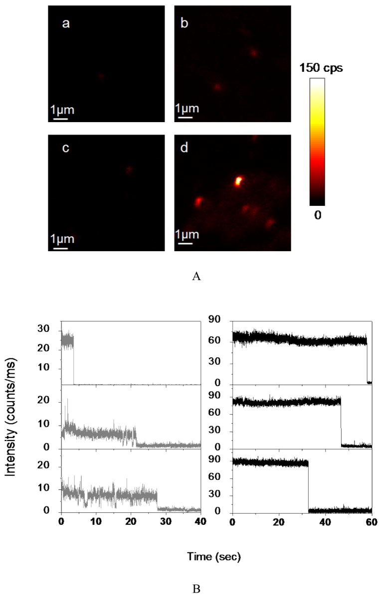
Figure 1A illustrates typical fluorescence images of the previously described sample, embedded in the gel and spincast on bare glass coverslips. Imaging of blank PAA gels on glass and SIF, respectively, using PBS instead of protein solution, show no fluorescence signals, confirming that the images in Figure 1(b, d) show specific GFP fluorescence. SIF film itself typically generates relatively weak scattering light, which were invisible in such images under similar circumstance. The representative images show well-defined fluorescent spots over background. The evidence that the observed spots are single GFP molecules is also based on some other arguments. One evidence is that the number of identified spots is in the proportional to the corresponding dilutions from the stock solution. Gel matrix does not alter the protein conformation and dynamics. It is worth noting that the fluorescence level of different single GFP molecules deposited on bare glass is remarkable similar. The proteins are assumed to be spatially confined in the gel and rotational diffusion is negligible. One can clearly observe that molecules emit nearly continuously when excited. Emission intensity of single GFP molecules on silvered varies. The observed heterogeneity of brightness is likely due to site-to-site variations in local electromagnetic field between the fluorophore and the metallic nanoparticle.
Figure 1.
(A) Typical 10×10 μm fluorescence images. The fluorescence intensity is displayed in a colorized scale, ranging from dark to light. a) PAA PBS gel spin-cast on a glass coverslip; b) GFP molecules in PAA gel spin-cast on a glass coverslip with incubation concentration of 0.5 nM; c) PAA PBS gel spin-cast on silver island film; d) GFP molecules in PAA gel spin-cast on silver island film with incubation concentration of 0.5 nM. (B) Representative time traces of single GFP molecules immobilized on a glass coverslip (left, gray lines) and silver island films (right, dark lines).
Furthermore, strong evidence for single molecule detection is also given by the time transients (Figure 1B). The time transients show sudden drop of fluorescence down to background level. This clear onestep photobleaching behavior corresponds to the typical behavior expected for a single molecule(8, 10, 12). In some cases, a switching “on/off” behavior is observed, the time trace clearly displays fast emission intensity fluctuations with several long “off” durations, which is related to internal photodynamcis of individual GFP molecules(11, 33). Intensity variations are also related to different characteristic local nanoenvironments of the probes, which could also affect their fluorescence lifetime(19). In contrast, it is well noticed that much higher and fairly constant emission rates are observed from the time profiles in the presence of silver nanostructure, which are generally more than 6-fold from those observed in the absence of SIF. “On/off” blinking appears to be significantly suppressed for GFP molecules deposited on SIF. In addition to increased emission rates on silver nanostructure, GFP molecules appear to emit for longer periods of time prior to photobleaching. As depicted in Figure 1B, the molecules display nearly constant emission intensity more than 30 seconds before undergoing irreversible photobleaching. The longer survival time occurs on a higher frequent basis for GFP near silvered surfaces compared to those on glass.
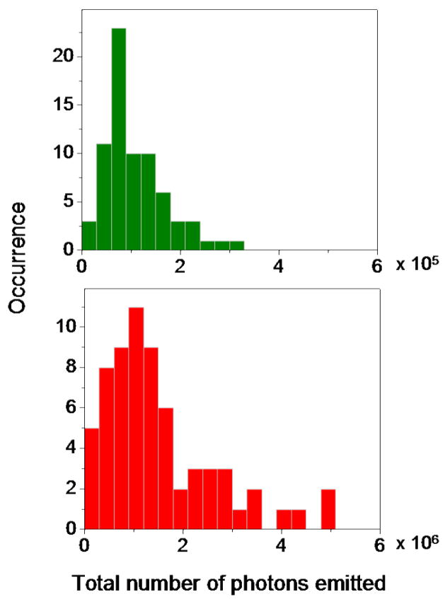
In comparison to GFP immobilized on bare glass coverslip, GFP on silvered surfaces show another clear advantage. The total number of photons emitted before photobleaching is particular interesting. Hence we observed more than 60 different GFP molecules on glass and the SIF until the proteins were photobleached. This was done by taking as many 10 x10 μm2 fluorescence images as necessary. These traces revealed the total number of photons observed for each fluorophore until it stopped emitting. The total number of detected photons was determined by integrating of individual single-molecule time transients as illustrated in Figure 1B. The histograms of these results are shown in Figure 2. It is obvious from these histograms that the protein molecules on glass are prone to fast photobleaching and emitting total photons in the range of 105. In the presence of SIF, proteins emit significantly more total photons of a maximum value of 5x10⁶ before photobleaching. The approximately 10-fold increase in total emitted photons on silvered surfaces suggest a possible increase in the fluorescence quantum yield of the protein molecules and also an improvement in photostability as frequently manifested by the reduced “on/off” blinking.
Figure 2.
Histograms of total number of photons detected before photobleaching. Top: GFP molecules on glass(green bars) ; Bottom: GFP molecules on silver island films (red bars).
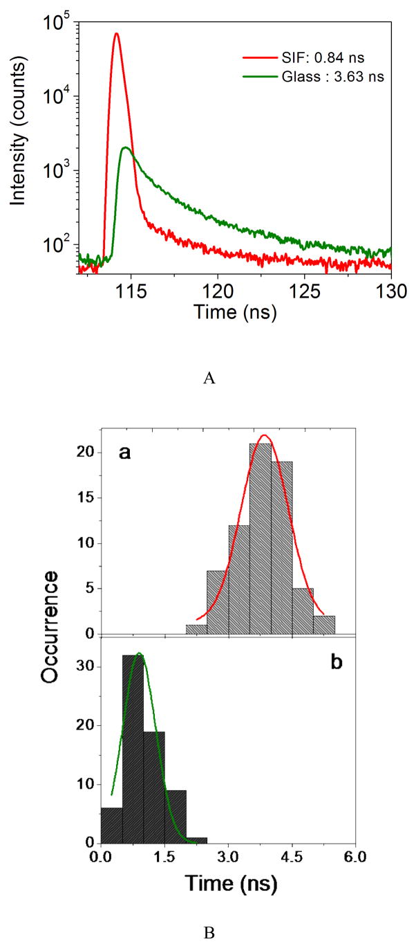
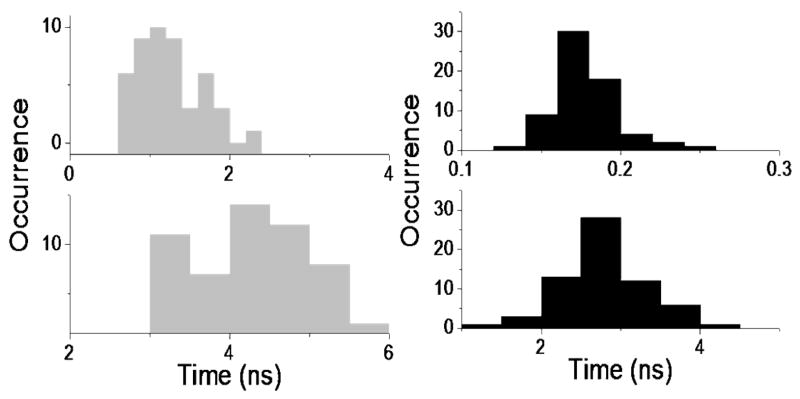
The single molecule fluorescence lifetime measurement is implemented using Time-Correlated Single Photon Counting (TCSPC) by plotting a histogram of time lags between the excitation pulses and the detected fluorescence photons. Fluorescence lifetime decay profiles are constructed by binning all of the arrived photons within a defined time-interval produces. The exponential fit to the observed decay profile gives the fluorescence lifetime. The average lifetime τav shown in the experiment is the amplitude-weighted averaged lifetime calculated from the fit result. In Figure 3A, the fluorescence decay of specific single GFP on silvered surfaces (red line) is compared with that of GFP on bare glass (green line). A bioexponential decay model best fits the GFP fluorescence decay deconvoleved from the instrument response function (IRF), as evaluated by the residuals and values of χ2. The analysis of the fluorescence signal of GFP on glass yields an averaged lifetime τ = 3.63 ns. The curve decays exponentially with two lifetime components of τ1 = 4.35 ns and τ2 = 1.34 ns, with relative amplitudes of 54% and 46%, respectively (χ2=1.031). The relative amplitudes of bioexponential fits to the GFP fluorescence decays are found to be relatively constant upon deposited on bare glass substrates, which suggest that most of the molecules adsorbed on glass are in a relatively homogeneous environment. Histograms of averaged lifetimes are illustrated in Figure 3B. The two lifetimes can be assigned to two different emitting species identified in the absorption spectrum(22, 34). In contrast, the fluorescence of protein deposited on SIF decay much faster and yields a much shorter averaged lifetime of 0.84 ns. Moreover, the intensity amplitudes differ considerably. Two lifetime components of τ1 = 3.12 ns and τ2 = 0.18 ns were obtained with relative amplitudes of 1.6% and 98.4 %, respectively (χ2=1.087). In the presence of SIF, we observed a predominant intensity contribution (> 90%) from the faster lifetime component (< 0.3 ns) for the investigated single molecules (Figure 5). Such detailed information related to the bioexponential fits of the decay curves can lead to the results presented in Figure 4. The histograms provide a means for evaluating the lifetime components of the single molecule data. On bare glass surfaces, the intensity decays of proteins can be seen from two major populations, in the ranges of decay time from 800ps to 2.3 ns and from 3 to 6 ns, respectively. Different kinds of ensemble spectroscopic measurements on GFP and some of its mutants have revealed that the protein system exhibits complex excited-state dynamics. The breadth of these distributions may also arise from variations in local polymer matrix environment, or they may result from variations in a number of other physical and chemical parameters associated with the nanopore environment in which each protein molecule is entrapped. Significant shifts to shorter values of the lifetime distributions are observed for the samples bound to SIF. In the presence of silver nanoparticles, the distributions of lifetime components become relatively narrow and symmetric; the intensity decay is dominated by a short decay time around 200 ps. The minor component is also observed around 2.4 ± 0.75 ns. It seems reasonable therefore to assume that the longer lifetime component arises from protein molecules exposed to the glass substrate and its contribution decreases dramatically where the surface becomes occupied by metallic nanostructure. Recent studies indicate that GFP tertiary structure resembles a barrel. It consists of 11 antiparallel β sheets and a single central α helix surrounded by the β sheets(3, 6). The chromophore resides in the center of the barrel, completely shielded from the external environment. The compact structure of GFP most likely contributes to the spectral properties of GFP. The pocket containing the fluorophore has a large number of charged residues in the immediate environment. The positive charge distribution forms a “rim” around the fluorophore inside the barrel(35).The interaction between these charges and the plasmon from the metallic nanostructure might induce a compression to the β cylinder structure near the chromophore. As a result, we observe relatively homogeneous lifetime component distributions in the presence of SIF. Furthermore, this more rigid environment would lead to an increased quantum yield and also to a protection of protein against bleaching.
Figure 3.
(A) Typical TCSPC decay curves of single GFP molecules embedded in gel on glass (green) and on silver island film (red). (B) Histograms of averaged lifetimes of single GFP molecules on (a) glass and (b) silver island films, the histograms were constructed from more than 60 single molecules, respectively.
Figure 4.
Histograms of lifetime components analyzed by bioexponential fitting to TCSPC decay curves. Left: on glass (gray bars); right: on silver island films (dark bars).
The strong energy transfer from the excited molecules to a nearby metallic nano-objective or an increase in the radiative decay rate of the fluorophore can dramatically shorten the lifetime of the excited state, leading to a fast de-excitation, which is consistent with our recorded data in this experiment. Considering the usual definition of lifetime (τ):
| (1) |
Where Γ is the radiative decay rate and knr Is the sum of the nonradiative decay rates. The expression for the radiative decay rate is in the form of(36):
| (2) |
Where n is the refractive index of the medium, f is a factor which relates the local electric field. It is assumed that the excited state decay rate is an intrinsic property of the molecule. Increased excitation rates will not affect Γ or knr. However, Equation (2) shows that it depends profoundly on the dielectric properties of the surrounding environment through f. Proximity to metal nanostructures could induce changes in local electromagnetic field. Unusual effects are expected and the increase in the radiative decay results in a decrease in lifetime. This effect increases the number of excitation cycles a molecule can survive until photobleaching. As a result, a dramatic increase in the number of photons is observed from a single fluorophore as described above.
In conclusion, GFP has found extensity use as a fluorophore in single molecule experiments for varying purposes. Single molecule imaging experiments have shown the principle possibility to detect individual GFP molecule. However, GFP photobleaches and blinks like most organic fluorophore. The intrinsic emission rate of a GFP molecule is not changed in most fluorescence experiments. The short obtainable fluorescence time traces prohibit any resolution of heterogeneities between individual molecules. This situation is changed near metal nanoparticles, which can increase the intrinsic radiative rates of nearby fluorophores. The metal particle fluorophore interaction is through-space, with maximum brightness enhancement factor more than 100-fold at distance of about 10nm(31). The increase in brightness is accompanied by a shortening of lifetimes and often by higher photostability. In this report, we illustrated the proximity of single GFPs to silver island films increases the fluorescence intensity approximately 6-fold. Single protein molecules on the silvered surfaces emitted 10-fold more photons as compared to glass prior to photobleaching. Additionally, we observed longer duration time and reduced blinking. The single-molecule lifetime histograms indicate the relatively heterogeneous distributions of protein mutants inside the protein. However, in addition to the shortened lifetimes, relatively homogeneous lifetime component distributions were identified in the presence of SIF. In closing, the ability to detect and track single labeled biomolecules within cells is severely limited by the brightness of the probes and their photostability. The detection of metal-surface enhanced fluorescence from GFP suggests the more extensive use of metallic nanostructures in imaging and single molecule detection.
Acknowledgments
The present work was supported by grants from the National Institutes of Health NHGRI (HG-002655) and NIBIB (EB-006521).
Footnotes
Publisher's Disclaimer: This is a PDF file of an unedited manuscript that has been accepted for publication. As a service to our customers we are providing this early version of the manuscript. The manuscript will undergo copyediting, typesetting, and review of the resulting proof before it is published in its final citable form. Please note that during the production process errors may be discovered which could affect the content, and all legal disclaimers that apply to the journal pertain.
References
- 1.Shimomura O, Johnson FH, Saga Y. Extraction, purification and properties of aequorin, a bioluminescent protein from the luminous hydromedusan, Aequorea. J Cell Comp Physiol. 1962;59:223–229. doi: 10.1002/jcp.1030590302. [DOI] [PubMed] [Google Scholar]
- 2.Shimomura O. The discovery of aequorin and green fluorescent protein. Journal of Microscopy-Oxford. 2005;217:3–15. doi: 10.1111/j.0022-2720.2005.01441.x. [DOI] [PubMed] [Google Scholar]
- 3.Tsien RY. The Green Fluorescent Protein. Annual Review of Biochemistry. 1998;67:509–544. doi: 10.1146/annurev.biochem.67.1.509. [DOI] [PubMed] [Google Scholar]
- 4.Schmid JA, Neumeier H. Evolutions in science triggered by green fluorescent protein (GFP) Chembiochem. 2005;6:1149. doi: 10.1002/cbic.200500029. [DOI] [PubMed] [Google Scholar]
- 5.March JC, Rao G, Bentley WE. Biotechnological applications of green fluorescent protein. Applied Microbiology and Biotechnology. 2003;62:303–315. doi: 10.1007/s00253-003-1339-y. [DOI] [PubMed] [Google Scholar]
- 6.Zimmer M. Green fluorescent protein (GFP): Applications, structure, and related photophysical behavior. Chemical Reviews. 2002;102:759–781. doi: 10.1021/cr010142r. [DOI] [PubMed] [Google Scholar]
- 7.Haupts U, Maiti S, Schwille P, Webb WW. Dynamics of fluorescence fluctuations in green fluorescent protein observed by fluorescence correlation spectroscopy. Proc Natl Acad Sci USA. 1998;95:13573–13578. doi: 10.1073/pnas.95.23.13573. [DOI] [PMC free article] [PubMed] [Google Scholar]
- 8.Weber MA, Stracke F, Meixner AJ. Dynamics of single dye molecules observed by confocal imaging and spectroscopy. Cytometry. 1999;36:217–223. [PubMed] [Google Scholar]
- 9.Cinelli RAG, Ferrari A, Pellegrini V, Tyagi M, Giacca M, Beltram F. The enhanced green fluorescent protein as a tool for the analysis of protein dynamics and localization: Local fluorescence study at the single-molecule level. Photochemistry and Photobiology. 2000;71:771–776. doi: 10.1562/0031-8655(2000)071<0771:tegfpa>2.0.co;2. [DOI] [PubMed] [Google Scholar]
- 10.Peterman EJG, Brasselet S, Moerner WE. The fluorescence dynamics of single molecules of green fluorescent protein. Journal of Physical Chemistry A. 1999;103:10553–10560. [Google Scholar]
- 11.Dickson RM, Cubitt AB, Tsien RY, Moerner WE. On/off blinking and switching behaviors of single molecules of green fluorescent protein. Nature. 1997;388:355–358. doi: 10.1038/41048. [DOI] [PubMed] [Google Scholar]
- 12.Moerner WE, Peterman EJG, Brasselet S, Kummer S, Dickson RM. Optical methods for exploring dynamics of single copies of green fluorescent protein. Cytometry. 1999;36:232–238. doi: 10.1002/(sici)1097-0320(19990701)36:3<232::aid-cyto13>3.0.co;2-l. [DOI] [PubMed] [Google Scholar]
- 13.Garcia-Parajo MF, Segers-Nolten GMJ, Veerman JA, Greve J, van Hulst NF. Real-time light-driven dynamics of the fluorescence emission in single green fluorescent protein molecules. Proceedings of the National Academy of Sciences of the United States of America. 2000;97:7237–7242. doi: 10.1073/pnas.97.13.7237. [DOI] [PMC free article] [PubMed] [Google Scholar]
- 14.Pierce DW, Vale RD. Single-molecule fluorescence detection of green fluorescence protein and application to single-protein dynamics. Methods in Cell Biology. 1999;58:49–73. doi: 10.1016/s0091-679x(08)61948-2. [DOI] [PubMed] [Google Scholar]
- 15.Moerner WE. Single-molecule optical spectroscopy of autofluorescent proteins. Journal of Chemical Physics. 2002;117:10925–10937. [Google Scholar]
- 16.Habuchi S, Cotlet M, Gronheid R, Dirix G, Michiels J, Vanderleyden J, Schryver FCD, Hofkens J. Single-molecule surface enhanced resonance raman spectroscopy of the enhanced green fluorescent protein. J Am Chem Soc. 2003;125:8446–8447. doi: 10.1021/ja0353311. [DOI] [PubMed] [Google Scholar]
- 17.Jung G, Wiehler J, Gohde W, Tittel J, Basche T, Steipe B, Brauchle C. Confocal microscopy of single molecules of the green fluorescent protein. Bioimaging. 1998;6:54–61. [Google Scholar]
- 18.Harms GS, Cognet L, Lommerse PHM, Blab GA, Schmidt T. Autofluorescent proteins in single-molecule research: Applications to live cell imaging microscopy. Biophysical Journal. 2001;80:2396–2408. doi: 10.1016/S0006-3495(01)76209-1. [DOI] [PMC free article] [PubMed] [Google Scholar]
- 19.Iino R, Koyama I, Kusumi A. Single molecule imaging of green fluorescent proteins in living cells: E-cadherin forms oligomers on the free cell surface. Biophysical Journal. 2001;80:2667–2677. doi: 10.1016/S0006-3495(01)76236-4. [DOI] [PMC free article] [PubMed] [Google Scholar]
- 20.Turner EH, Lauterbach K, Pugsley HR, Palmer VR, Dovichi NJ. Detection of green fluorescent protein in a single bacterium by capillary electrophoresis with laser-induced fluorescence. Analytical Chemistry. 2007;79:778–781. doi: 10.1021/ac061778r. [DOI] [PubMed] [Google Scholar]
- 21.Kohl T, Schwille P. Microscopy Techniques. 2005. Fluorescence correlation spectroscopy with autofluorescent proteins; pp. 107–142. [DOI] [PubMed] [Google Scholar]
- 22.Cotlet M, Hofkens J, Habuchi S, Dirix G, Van Guyse M, Michiels J, Vanderleyden J, De Schryver FC. Identification of different emitting species in the red fluorescent protein DsRed by means of ensemble and single-molecule spectroscopy. Proceedings of the National Academy of Sciences of the United States of America. 2001;98:14398–14403. doi: 10.1073/pnas.251532698. [DOI] [PMC free article] [PubMed] [Google Scholar]
- 23.Mashanov GI, Tacon D, Knight AE, Peckham M, Molloy JE. Visualizing single molecules inside living cells using total internal reflection fluorescence microscopy. Methods. 2003;29:142–152. doi: 10.1016/s1046-2023(02)00305-5. [DOI] [PubMed] [Google Scholar]
- 24.Lakowicz JR, Shen B, Gryczynski Z, D'Auria S, Gryczynski I. Intrinsic fluorescence from DNA can be enhanced by metallic particles. Biochemical and Biophysical Research Communications. 2001;286:875–879. doi: 10.1006/bbrc.2001.5445. [DOI] [PMC free article] [PubMed] [Google Scholar]
- 25.Malicka J, Gryczynski I, Geddes CD, Lakowicz JR. Metal-enhanced emission from indocyanine green: a new approach to in vivo imaging. Journal of Biomedical Optics. 2003;8:472–478. doi: 10.1117/1.1578643. [DOI] [PMC free article] [PubMed] [Google Scholar]
- 26.Mackowski S, Wormke S, Maier AJ, Brotosudarmo THP, Harutyunyan H, Hartschuh A, Govorov AO, Scheer H, Brauchle C. Metal-enhanced fluorescence of chlorophylls in single light-harvesting complexes. Nano Letters. 2008;8:558–564. doi: 10.1021/nl072854o. [DOI] [PubMed] [Google Scholar]
- 27.Anderson JP, Griffiths M, Boveia VR. Near-infrared fluorescence enhancement using silver island films. Plasmonics. 2006;1:103–110. [Google Scholar]
- 28.Fu Y, Zhang J, Lakowicz JR. Reduced blinking and long-lasting fluorescence of single fluorophores coupling to silver nanoparticles. Langmuir. 2008;24:3429–3433. doi: 10.1021/la702673p. [DOI] [PMC free article] [PubMed] [Google Scholar]
- 29.Fort E, Gresillon S. Surface enhanced fluorescence. Journal of Physics D-Applied Physics. 2008;41 [Google Scholar]
- 30.Zhang J, Fu Y, Lakowicz JR. Enhanced Forster resonance energy transfer (FRET) on a single metal particle. Journal of Physical Chemistry C. 2007;111:50–56. doi: 10.1021/jp062665e. [DOI] [PMC free article] [PubMed] [Google Scholar]
- 31.Fu Y, Lakowicz JR. Enhanced fluorescence of Cy5-labeled oligonucleotides near silver island films: A distance effect study using single molecule spectroscopy. Journal of Physical Chemistry B. 2006;110:22557–22562. doi: 10.1021/jp060402e. [DOI] [PMC free article] [PubMed] [Google Scholar]
- 32.Fu Y, Lakowicz JR. Enhanced fluorescence of Cy5-labeled DNA tethered to silver island films: Fluorescence images and time-resolved studies using single-molecule spectroscopy. Analytical Chemistry. 2006;78:6238–6245. doi: 10.1021/ac060586t. [DOI] [PMC free article] [PubMed] [Google Scholar]
- 33.Webb W, Helms V, McCammon JA, Langhoff PW. Shedding light on the dark and weakly fluorescent states of green fluorescent proteins. Proc Natl Acad Sci USA. 1999;96:6177–6182. doi: 10.1073/pnas.96.11.6177. [DOI] [PMC free article] [PubMed] [Google Scholar]
- 34.Heikal AA, Hess ST, Webb WW. Multiphoton molecular spectroscopy and excited-state dynamics of enhanced green fluorescent protein (EGFP): acid-base specificity. Chemical Physics. 2001;274:37–55. [Google Scholar]
- 35.Diaspro A, Krol S, Campanini B, Cannone F, Chirico G. Enhanced Green Fluorescent Protein (GFP) fluorescence after polyelectrolyte caging. Optics Express. 2006;14:9815–9824. doi: 10.1364/oe.14.009815. [DOI] [PubMed] [Google Scholar]
- 36.Tomczak N, Vallee RAL, van Dijk E, Garcia-Parajo M, Kuipers L, van Hulst NF, Vancso GJ. Probing polymers with single fluorescent molecules. European Polymer Journal. 2004;40:1001–1011. [Google Scholar]


