Abstract
There is an association between expression of the MHC class I molecule HLA-B27 and protection following human infection with either HIV or HCV. In both cases, protection has been linked to HLA-B27 presentation of a single immunodominant viral peptide epitope to CD8+ T cells. If HIV mutates the HLA-B27–binding anchor of this epitope to escape the protective immune response, the result is a less-fit virus that requires additional compensatory clustered mutations. Here, we sought to determine whether the immunodominant HLA-B27–restricted HCV epitope was similarly constrained by analyzing the replication competence and immunogenicity of different escape mutants. Interestingly, in most HLA-B27–positive patients chronically infected with HCV, the escape mutations spared the HLA-B27–binding anchor. Instead, the escape mutations were clustered at other sites within the epitope and had only a modest impact on replication competence. Further analysis revealed that the cluster of mutations is required for efficient escape because a combination of mutations is needed to impair T cell recognition of the epitope. Artificially introduced mutations at the HLA-B27–binding anchors were found to be either completely cross-reactive or to lead to substantial loss of fitness. These results suggest that protection by HLA-B27 in HCV infection can be explained by the requirement to accumulate a cluster of mutations within the immunodominant epitope to escape T cell recognition.
Introduction
HCV, a positive-strand RNA virus of the Flaviviridae family, is a major cause of chronic liver disease. More than 170 million people worldwide are chronically infected and are at risk of developing complications such as liver cirrhosis and hepatocellular carcinoma (1). Growing evidence suggests an important role for virus-specific CD8+ T cells in the outcome of HCV infection (2). The onset of virus-specific CD8+ T cell responses in the peripheral blood and liver correlates with control of acute-phase viremia, and antibody-mediated depletion of the CD8+ T cell subset prolongs viral replication in chimpanzees (3–6). The central role of virus-specific CD8+ T cells is further supported by the finding that the HLA class I allele B27 (HLA-B27) is strongly associated with spontaneous viral clearance, which has been mechanistically linked to a dominant CD8+ T cell epitope (7–9). This epitope resides in the C-terminal region of the NS5B RNA-dependent RNA polymerase (RdRp) and spans residues 2,841–2,849 of the HCV polyprotein (residues 421–429 of the NS5B protein). Of note, HLA-B27 also has a protective role in HIV infection, indicated by its association with slow disease progression (10). The immune response to HIV in these patients is focused on an immunodominant gag epitope. This response occurs early in infection and coincides with a decline in viral load and subsequent long-term suppression (11–14). HLA-B27–restricted CD8+ T cells display a very high binding affinity and in the case of HIV have been shown to be characterized by polyfunctional capabilities, increased clonal turnover, and superior functional avidity (15). Thus, in both human infections, HIV and HCV, a clear association between HLA-B27 and protection has been described, and it has been linked to a single epitope in each case.
The mechanisms of protection mediated by these 2 epitopes are not well understood; however, it has been suggested that the functional constraints that limit viral escape and enable long-term maintenance of dominant CD8+ T cell responses play an important role in this setting. Indeed, in HIV infection, viral escape in the dominant epitope usually does not occur until late in infection (12). The most critical mutation is observed at position 2 of the epitope, the major HLA-B27–binding anchor, where a substitution of arginine at gag residue 264, most often by lysine (R264K) or threonine (R264T), has been described (11–13). Of note, the interaction between arginine at position 2 of HLA-B27–restricted epitopes and the B pocket of the HLA-B27 molecule plays a crucial role in stabilizing the MHC-peptide complex (16), suggesting that mutations at this position lead to poor binding to HLA-B27. Importantly, the R264K mutation results in a substantial viral fitness cost through a severe functional defect during an early step in viral replication (17, 18). The critical R264K change mostly occurs in concert with a previous change of leucine to methionine at position 268 (13). The latter mutation has only a moderate effect on T cell recognition; however, it has recently been shown to lead to immunoglobulin-like transcript 4–mediated (ILT-4–mediated) functional inhibition of myelomonocytic cells (19). In addition, a third mutation, nearly always accompanying the R264K and L268M mutations, has been identified outside the epitope and appears to compensate for the fitness cost of the R264K mutation (18). Thus, escape of this protective epitope most likely occurs late in infection, because the escape dramatically reduces HIV replicative capacity (fitness cost) and requires a complex mosaic of mutations. These features allow for the maintenance of T cell–mediated control (10).
Despite all these important insights into the determinants of protection mediated by the HLA-B27–restricted HIV epitope, little information is currently available about the mechanisms responsible for the protection mediated by the dominant HLA-B27–restricted HCV epitope. Here, we addressed this important issue by analyzing the direct effect of specific mutations in the HLA-B27–restricted epitope on HCV replication fitness and T cell recognition. For these studies, we utilized a replicon system that allows direct analysis of the impact of specific mutations, alone or in combination, upon virus replication efficiency (20, 21). We demonstrate that a high viral fitness cost resulting from mutations within the HLA-B27–binding anchor and a high capability of the T cell response to cross-recognize epitope variants limit escape options for the virus. The consequent demand for the evolution of multiple clustered mutations within the epitope in order to provide effective escape provides an explanation for the protective effect of HLA-B27 in HCV infection.
Results
Mutations within the dominant HLA-B27–restricted HCV epitope are clustered but spare the HLA-binding anchor.
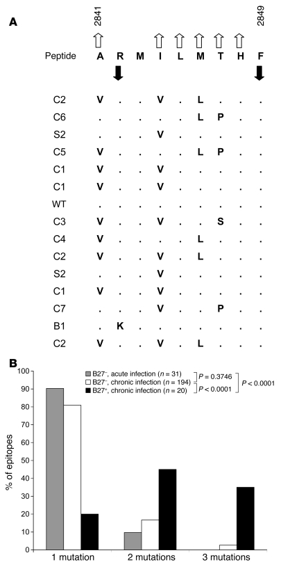
Viral escape mutations within the immunodominant HLA-B27–restricted HCV-specific CD8+ T cell epitope NS5B2841–2849 (ARMILMTHF) occur in the vast majority of patients in clusters (Figure 1A; clonal sequence data are shown in Supplemental Figure 1; supplemental material available online with this article; doi: 10.1172/JCI36587DS1); e.g., viruses analyzed in 11 of 15 patients showed 2 or 3 mutations (6 patients with 2 mutations; 5 patients with 3 mutations), while viruses in only 4 patients showed no or only 1 mutation within the otherwise highly conserved epitope region. This is a striking finding, since T cell escape mutations forced by other HLA alleles typically harbor a single (90% and 81%), less frequently 2 (10% and 17%), and rarely 3 amino acid substitutions (0% and 3%), as has been shown in comprehensive analyses of patients with acute (22–27) or chronic (24, 28–33) HCV infection (P < 0.0001; Figure 1B). In addition, escape mutations within the dominant HLA-B27 epitope were observed only in certain amino acid positions (positions 1, 4, 6, and 7). These residues are not involved in HLA-B27 binding, but rather face and interact with the TCR (16). By comparison, residues that represent major HLA-binding anchors (arginine at position 2 and the C-terminal phenylalanine) showed no variation at all, except in a single patient showing an arginine-to-lysine mutation at position 2 (Figure 1A). Of note, both phenomena, clustering of mutations within the TCR contact residues and sparing of the HLA-B27–binding anchors, have also been observed in an independent study of HLA footprints in chronic HCV infection for this epitope (32).
Figure 1. Escape mutations within the NS5B2841–2849 epitope are clustered and spare HLA-B27–binding anchors.
(A) Amino acid sequence of NS5B2841–2849. Upward-pointing open arrows indicate residues that are predicted to interact with the TCR; downward-pointing filled arrows indicate HLA-B27–binding anchors (16). Autologous viral sequences found in 15 HLA-B27+ patients with chronic genotype 1 HCV infection are shown below; points indicate homologous residues (8). (B) Numbers of amino acid substitutions per escape variant are compared in HLA-B27+ and HLA-B27– subjects. Sequences for HLA-B27– subjects were taken from the literature: for acute HCV infection, longitudinal sequences or sequences from a known donor and the patient were compared (22–27), while for chronic HCV infection, patient sequences were compared with genotype/subtype-matched consensus sequences (24, 28–33). Sequences in HLA-B27+ patients with chronic HCV infection were included from this study (Figure 1A) and from a previous, independent report (32). P values were calculated using Mann-Whitney U test.
Clustered mutations within the protective HLA-B27–restricted HCV-specific CD8+ T cell epitope are required for viral escape.
Next, we investigated the immunogenicity of the different escape mutations observed in NS5B2841–2849. We tested CD8+ T cell lines specific for the WT peptide, which were derived from 6 HLA-B27+ individuals who cleared HCV infection either spontaneously (subjects 1, 2, and 4–6) or during antiviral treatment of chronic HCV infection (subject 3). Of note, 3 of these subjects (subjects 1, 4, and 5) have been infected with a known inoculum that harbors the WT sequence within the NS5B2841–2849 epitope region (GenBank accession number AF313916; ref. 7). Interestingly, the NS5B2841–2849–specific T cell lines of the 6 subjects showed similar patterns of cross-recognition in response to the different variant peptides.
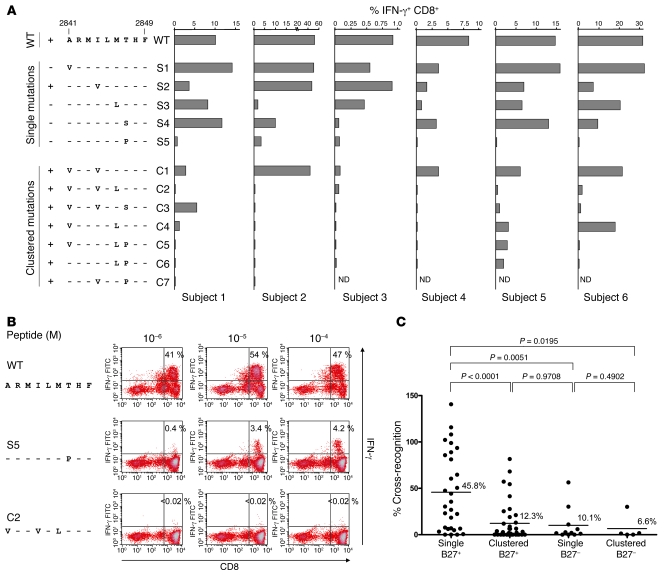
As shown in Figure 2A, variant peptides with single amino acid substitutions (S1–S5) still induced substantial levels of IFN-γ production to different degrees. The variant peptide with a proline substitution at position 7 (S5) showed only limited cross-reactivity in most subjects, although IFN-γ production in response to this variant was still easily detectable in some patients (a representative dot plot is shown in Figure 2B).
Figure 2. Clustering of mutations at TCR contact sites is required for efficient escape from the WT-specific T cell response.
(A) PBMCs from 6 HLA-B27+ individuals with HCV infection that resolved either spontaneously or through therapy were cultured for 14 days in the presence of WT peptide NS5B2841–2849. Cells were then tested for IFN-γ production after 5 hours stimulation with WT or variant peptides at a concentration of 10–5 M. Variants with single or clustered substitutions are grouped. A plus indicates that this mutation occurs in vivo, while a minus indicates that this mutation was not found in vivo. ND, not done. (B) Representative dot blots corresponding to Figure 2A (subject 2) are shown, including titration of the peptides in different concentrations as indicated. Percentages indicate IFN-γ+CD8+ T cells. (C) Levels of cross-recognition observed for each single and clustered escape variant of the HLA-B27–restricted NS5B2841–2849 epitope are compared with the levels of cross-recognition of a number of single or clustered escape variants in HLA-B27– patients described previously by our group (29). The level of cross-recognition for each variant was calculated by dividing the response to the variant peptide at a concentration of 10–5 M by the response to WT peptide at a concentration of 10–5 M. P values were calculated using Mann-Whitney U test. Bars denote mean.
Peptide variants with 2 or 3 mutations (C1–C7) showed a different pattern: While the variant peptide C1 with the combined A2841V and I2844V substitutions still showed substantial cross-recognition in most subjects, a third substitution, either M2846L (C2) or T2847S (C3), abolished T cell recognition in most subjects (Figure 2A). Only subject 1 still displayed some cross-recognition with the variant peptide containing the triple substitution A2841V, I2844V, and T2847S (C3). Variant peptides carrying the M2846L mutation in combination with A2841V (C4), T2847P (C6), or both (C5) were also not recognized in most subjects, indicating significant viral escape. The variant peptide carrying the I2844V and T2847P mutation (C7) was only tested in 2 subjects and elicited no response. Figure 2C summarizes the levels of cross-recognition observed for each single and clustered escape variant studied in the 6 patients, and these were compared with the levels of cross-recognition of a number of single or clustered escape variants in different T cell epitopes in HLA-B27– patients described previously in our laboratory (29). Of note, the level of cross-recognition was 45.8% for single escape variants of the HLA-B27–restricted epitope and thus was significantly higher than for clustered variants of the same epitope (12.3%; P < 0.0001; Mann-Whitney U test) and for single or clustered non–HLA-B27–restricted epitope variants (10.1%, P = 0.0051, and 6.6%, P = 0.0195, respectively). Strikingly, these results reveal that a clustering of mutations within the immunodominant HLA-B27–restricted epitope is required to reach a level of escape similar to that observed for single escape mutations within epitopes restricted by other HLA alleles (Figure 2C).
Taken together, these results indicate that a clustering of mutations is required for complete abrogation of T cell recognition within the dominant HLA-B27–restricted NS5B2841–2849 epitope.
Clustered mutations do not compromise or compensate viral fitness.
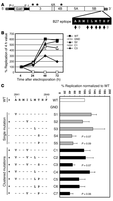
Next, we determined whether the clustered mutations observed in vivo within the highly conserved NS5B2841–2849 epitope have an impact on viral replication (fitness cost). The mutations were introduced into a subgenomic replicon (genotype 1b strain Con1) carrying a firefly luciferase reporter gene (Figure 3A). An example of the replication kinetics of 3 epitope mutants compared with WT and a replication-defective replicon (GND mutant) is shown in Figure 3B. Mutant S2 with the single mutation I2844V and mutant C1 with the double mutation A2841V/I2844V replicated to a level similar to that in WT; only mutant C3, containing a third mutation (A2841V/I2844V/T2847S), showed a marked reduction in replicative capacity. Replication data from 5 independent experiments showed that all mutants tested were still able to replicate, with most variants showing only a minor reduction in replication compared with WT (Figure 3C). A more pronounced effect on viral replication was observed for the single-amino-acid substitutions of amino acid residue 2,847 (mutants S4 and S5) that were not found as single mutations in vivo but appeared only in combination with 1 or 2 other mutations; these mutants replicated at approximately 50% of the WT level (Figure 3C). A similar impact on replication was found for 2 variants with clustered mutations containing substitutions A2841V, I2844V, and T2847S (C3) and I2844V and T2847P (C7) (Figure 3C).
Figure 3. Clustered mutations at the TCR contact sites do not substantially impair replicative capacity.
(A) Schematic overview of the subgenomic HCV reporter replicon pFKi341PiLucNS3-3ιET. Luc, luciferase gene of the firefly Photinus vulgaris; P-I, polio virus internal ribosomal entry site; E-I, encephalomyocarditis virus internal ribosomal entry site. Asterisks indicate adaptive mutations (E1202G, T1280I, K1846T). Epitope NS5B2841–2849 is highlighted. Black arrows correspond to TCR residues, gray arrows to anchor residues. (B) Representative time course of mutants S2, C1, and C3 compared with those of WT and the negative mutant GND. Replicative capacity is expressed as percentage after normalization to the 4-hour input RNA value that reflected transfection efficiency. (C) Results for all mutants at 48 hours. WT was normalized to 100%. Variants with single or clustered substitutions are grouped. A plus indicates that this mutation occurs in vivo, while a minus indicates that this mutation was not found in vivo. Data are presented as mean ± SD.
Replication levels of variants containing a single versus clustered mutations did not differ significantly (Figure 3C; mean 88.0% and 82.3% of WT replication levels; P = 0.6389, Mann-Whitney U test), indicating that the clustering of mutations did not further reduce replication or have an overall compensatory effect. Of note, no clustered mutations were observed that compensated for the replication defect of the T2847S mutation (compare S4 with C3). In contrast, some variants with clustered mutations containing the T2847P mutation replicated to a higher degree than the variant containing the T2847P mutation alone (compare S5 with C5 and C6). However, this effect was modest (~50% versus ~80%), and another variant containing clustered mutations showed no compensatory effect (C7; Figure 3C).
To exclude the possibility that the less replication-competent variants might have reverted to WT, viral RNA extracted from cells 72 hours after electroporation was amplified by RT-PCR and the nucleotide sequences of multiple molecular clones obtained from WT (5 clones) or mutants (S4, 5 clones; S5, 5 clones; C3, 3 clones) were determined. No reversion or pseudoreversion was found (data not shown).
These experiments indicate that although some mutations within the NS5B2841–2849 epitope impair RNA replication to as much as 50% of the WT level, a clustering of these mutations neither further compromises viral fitness nor has a substantial compensatory effect.
Absence of viral escape mutations within the major HLA-B27–binding anchors is due to high functional constraints.
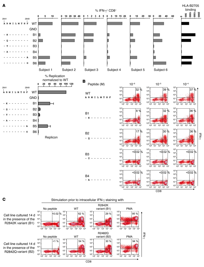
Binding of epitope peptides to HLA-B27 is mediated by 2 major HLA-B27–binding anchors: an arginine residue at position 2 and, to a lesser extent and with more flexibility, the C-terminal residue. To address the reason for the nearly complete lack of escape at these binding anchors, we tested variants with substitutions at these residues for cross-recognition by WT-specific T cell lines and their replicative capacity (Figure 4). Only one of these mutations (arginine to lysine at position 2; variant B1) occurred within a single patient of our cohort in vivo (Figure 1A); all other mutations were introduced artificially.
Figure 4. Variants with substitutions at the HLA-B27–binding anchors are either cross-recognized or show limited replication competent.
(A) PBMCs from 6 HLA-B27+ individuals with HCV infection that resolved either spontaneously or through therapy were cultured for 14 days in the presence of WT peptide NS5B2841–2849. Cells were then tested for IFN-γ production after 5 hours stimulation with WT or variant peptides containing substitutions at HLA-B27–binding anchors P2 or P9 at a concentration of 10–5 M. The far-right panel shows HLA-B2705 binding of the different peptides, and the lower-left panl shows the replicon data for anchor residue mutants 48 hours after electroporation. WT was normalized to 100%. Data are presented as mean ± SD. (B) Representative dot plots corresponding to A (subject 2) are shown, including titration of the peptides in different concentrations as indicated. Percentages indicate IFN-γ+CD8+ T cells. (C) Cell lines cultured for 14 days in the presence of the R2842K (upper panels) or R2842Q (lower panels) variant peptides showed high amounts of IFN-γ–producing CD8+ T cells after stimulation with WT or the respective variant peptide for 5 hours. Percentages indicate IFN-γ+CD8+ T cells.
First, we analyzed several variants containing mutations at the arginine residue in position 2. We substituted lysine for arginine (mutant B1), a variant that occurs within 1 patient of our cohort and that is the only other positively charged amino acid and the most frequent escape variant in the HIV-specific protective HLA-B27 epitope. Further, we replaced the arginine residue at position 2 with either glutamine or threonine (mutants B2 and B3, respectively), 2 uncharged polar residues, as well as with alanine (mutant B4), which introduces a hydrophobic residue (Figure 4A). Importantly, the variants containing lysine or glutamine (B1 or B2) exhibited substantial cross-recognition by WT-specific T cell lines (Figure 4, A and B). This finding is further supported by the observation that these variant peptides also induced specific T cell lines in vitro (Figure 4C). With respect to replication competence, the glutamine substitution (B2) severely reduced replication, while the lysine substitution (B1) reduced replication to about 40% compared with WT (Figure 4A). In contrast, the threonine or alanine substitution at position 2 (B3 and B4) abolished cross-recognition. Thus, these variants would represent excellent T cell escape mutations. However, replication competence of mutant B3 was dramatically reduced (about 20-fold lower compared with WT), while mutant B4 did not replicate at all. Taken together, these data show that these mutations result in a substantial fitness cost, which may in part explain why they are absent in vivo.
Finally, we also introduced substitutions affecting the C-terminal HLA-B27–binding anchor. For these experiments, the WT phenylalanine residue was replaced by either alanine (B5) or glycine (B6) to retain the hydrophobic character at this site. Both variants displayed strong cross-recognition by the WT-specific T cell line, strongly arguing against viral escape (Figure 4A). In addition, replication competence of mutant B5 carrying the alanine substitution was clearly reduced, to about 30% compared with the WT, whereas mutant B6 carrying the glycine substitution did not replicate to a detectable level (Figure 4A). In summary, the data indicate that mutations affecting the C-terminal anchor residue phenylalanine are likely to be limited in vivo due to the lack of T cell escape and impaired replication competence. Thus, the spectrum of possible escape variants at this position is under high functional constraints.
HLA-B27–binding assays.
Since nearly all natural HLA-B27 ligands harbor an arginine at position 2, the finding that variants with a lysine or glutamine mutation at this residue (variants B1 and B2) are still recognized by HLA-B27–restricted CD8+ T cells was surprising. We therefore directly analyzed the HLA-B27 binding affinity of the WT as well as variant peptides. Since all but 1 subject in this study were shown to carry HLA-B2705 (HLA-B27 subtyping was not successful in subject 6), HLA-B2705 heterodimers were chosen for the assay. In agreement with the results obtained from functional assays, the variants with lysine or glutamine at position 2 had an HLA-B27–binding affinity similar to that of WT, while the variants harboring threonine or alanine at this position showed only background affinity for HLA-B27 (Figure 4A). Of note, lysine and glutamine are the only residues apart from arginine that have been observed in only few natural HLA-B27 ligands at this position (34). As predicted from the functional assays, the variants containing alanine or glycine at the carboxyl terminus (B5 and B6) also showed an HLA-B27 affinity comparable to that of WT.
Structural basis for the observed functional constraints.
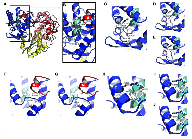
Given the stereotypic nature of the escape mutations occurring within the NS5B epitope and its conservation in the absence of immune selection, we assessed the potential impact of structural alterations on RdRp activity. We performed structure modeling of the escape mutants using NS5B of the BK strain, for which high-resolution structures are available and which has an amino acid sequence in the NS5B region identical to that of Con1 studied here. The epitope maps to NS5B helix Q (nomenclature according to ref. 35), which is the central helix in the bundle making up most of the thumb domain (Figure 5A). This helix is the central folding element of the thumb domain and functionally provides a large part of the pocket accommodating helix A of the fingertips (Figure 5, A and B). This epitope is therefore functionally important for the postulated coordination of the thumb and finger subdomains required for NS5B activity (35). Accordingly, 2 inhibitor sites have been identified in this region (36): site A is the helix A pocket itself (37), and site B corresponds to a pocket under site A that is lined in part by R2842 (Figure 5B) and M2843.
Figure 5. Structural analysis of the epitope.
(A) NS5B displayed as ribbons and colored according to individual domains (red: fingers; yellow: palm; blue: thumb). The epitope is shown in blue-green. (B) Close-up of the epitope region. Residues mutated in this study are shown in stick representation and colored according to atom type (N: blue; O: red; S: orange; C: blue-green). Four of these residues are labeled. Also labeled are helices Q (which harbors the epitope) and A (from the “fingertips”), as well as non-nucleoside inhibitor (NNI) site B. Note that helix Q is both central to the thumb’s folding domain and involved in the NNI pockets’ buildup. (C) Interactions of R2842 with neighboring residues. (D and E) Models of the same region after replacement of R2842 with lysine (variant B1) or glutamine (variant B2). (F) Interactions of I2844 with neighboring residues. (G) Model of the variant S2 containing the I2844V substitution. (H) Interactions of T2847 with neighboring residues. In C, F, and H, the neighboring residues are shown as sticks, with carbons in white; hydrogen bonds are represented as dotted yellow lines. (I and J) Models of the variants containing substitution T2847S (S4) and T2847P (S5), respectively.
Most of the epitope residues are buried in the hydrophobic core of the thumb. This is notably the case for A2841, I2844, M2846, and T2847, with the side chain hydroxyl of the latter forming hydrogen bonds with the main chain of helix Q (Figure 5H). The hydrophobic A2841, I2844, and M2846 can be replaced by hydrophobic residues of comparable size with minor adjustments in the hydrophobic core. Figure 5, F and G, illustrates this finding for residue I2844: The I2844V substitution leads to the loss of the interaction with L2909, but this is easily accommodated by small shifts of the ends of this and other neighboring hydrophobic side chains. The interaction with L2450, however, is not affected by this substitution.
The case of T2847 is somewhat different, as this residue provides both a key methyl group to the hydrophobic core and a hydrogen bond to stabilize the main chain of helix Q (Figure 5H) and therefore will be less tolerant to substitutions. The T2847S substitution (Figure 5I) involves the removal of a single methyl group in a hydrophobic environment. While the hydrogen bond to the main chain of helix Q is preserved, contacts to methyl groups close to the main chain are lost (the β carbons of R2923 and A2924) and cannot be compensated for by small shifts of these side chains. Therefore, a larger rearrangement involving either a shift in the position of helix T or an inward swinging of, for example, W2920 is necessary. Likewise, the nonconservative T2847P mutation entails a more extensive remodeling of this region. A proline substitution will normally disrupt a helix, by kinking it and disrupting its main-chain hydrogen-bonding pattern. Here the change is not so severe, as helix Q is already kinked at position 2,847. Hence, a hydrogen is donated, not by T2847’s main chain nitrogen as is normally the case, but by its side-chain hydroxyl (Figure 5H). The T2847P substitution (Figure 5J) therefore does implies not a missing main-chain hydrogen bond and the kinking of helix Q (both already there in the WT), but a pronounced modification of its immediate neighborhood. This is due to both the loss of the polar side chain hydroxyl of the threonine residue and the larger bulk of the proline residue. Thus, the T2847P mutation is accompanied by an intriguing rearrangement of this important NS5B region, which somewhat contradicts the moderate effect the substitution has on the replicative capacity of the subgenomic replicon.
In contrast to all these residues, R2842, M2843, H2848, and F2849 line pockets at the thumb surface and are only partially buried. The case of R2842 is depicted in Figure 5C. The aliphatic part of the side chain rests on the hydrophobic core, while the polar head makes 2 hydrogen bonds to conserved residues in the loop leading to helix S (L2894 and Y2897). Lysine (Figure 5D) or glutamine substitutions (Figure 5E) for the arginine at position 2,842 will disturb these hydrogen bonds with residues L2894 and Y2897, leading to a strongly modified non-nucleoside inhibitor (NNI) site B. H2848 is the main helix Q contributor to pocket A, which harbors helix A, thus explaining that histidine is highly conserved at this site. Although F2849 does not directly contribute to the formation of pocket A, it is a critical buttress for H2848, which explains why a drastic reduction of the bulk of the 2,849 side chain has such a negative effect on replication competence. These results underscore the limited flexibility of NS5B for amino acid substitutions at this site and thus the conservation of the immunodominant HLA-B27 epitope.
Discussion
In this study, we determined the mechanisms of protection against chronic HCV infection mediated by a dominant HLA-B27–restricted CD8+ T cell epitope. Indeed, we recently identified a central role of this NS5B2841–2849 epitope in spontaneous HCV clearance in the majority of HLA-B27–infected individuals (8). In patients with chronic HCV infection, analysis of the corresponding viral sequence showed a strong association between clustered sequence variations within this epitope and expression of HLA-B27, indicating allele-specific selection pressure at the population level (Figure 1A) (8, 32).
The first important finding of our study is that these clustered mutations are required for optimal T cell escape. Immunological analysis revealed that most single mutations led to only modest decreases in T cell recognition, whereas a much greater impact was observed when we used variant peptides with 2 (e.g., variants C4 and C6) or 3 (e.g., C2, C3, and C5) amino acid substitutions detected in patients. The finding that a clustering of 2 or 3 mutations is required for escape is of interest, since analyses of patients with acute or chronic HCV infection indicated that the majority of escape mutations restricted by other HLA alleles typically require only a single amino acid substitution for T cell escape (Figure 1B). Thus, we hypothesize that, as is the case in HIV infection, the clustering of mutations is directly linked to the observed protective effect of this epitope.
It has been suggested, however, that in HIV, functional constraints within the dominant HLA-B27–restricted epitope interfere with the ability of HIV to escape from the immunodominant response and that triple mutations are required as a compensation for the R264K mutation typically observed within this epitope (10, 13, 17). To address whether a similar mechanism may explain the clustering of mutations within the HCV epitope, we analyzed in the replicon model the replication efficiency of the different mutations observed in patients. Some mutants showed a replicative capacity reduced to approximately 40%–50% of the WT level, indicating fitness cost; however, replication levels did not significantly differ among single, double, and triple mutants. These results suggest that viral escape within the protective epitope does not occur easily and that the clustering of mutations does not primarily compensate for the observed fitness cost but rather is required for efficient T cell escape.
Of note, 4 of the patients described did not display clustered sequence variations, and 3 carried the combination of the A2841V and I2844V mutations (C1) that show a significant cross-reactivity with WT peptide (Figure 1A). The cause of viral persistence despite the absence of critical T cell escape mutations is not clear. Possible explanations include a primary failure to induce the dominant B27-restricted CD8+ T cell response during acute infection (4); a lack of sufficient CD4+ T cell help (38); or the subsequent loss of such an early response, possibly followed by a reversion of the mutated epitope to WT. Reversion has been documented in HIV infection (i.e., the escape mutation within a dominant HLA-B57 epitope rapidly reverted to WT in HLA-B57– newborns that had been infected by HLA-B57+ mothers; ref. 39), as well as in HCV infection (transmission of an HLA-B8–associated escape mutant to an HLA-B8– subject resulted in rapid reversion of the mutation; ref. 24). The fact that certain clustered mutations (for example, triple variant C3) have a reduced replicative capacity supports the assumption that reversion to a variant containing fewer substitutions might indeed take place once viral persistence has established and the NS5B2841–2849–specific T cell response has waned. The pattern of potential mutations in this epitope seems to be very complex and may reflect the balance between replication fitness and immune escape. Clearly, it will be important to prospectively follow HLA-B27+ patients during acute persistent HCV infection to gain further insights into the kinetics of T cell responses and viral evolution during this critical phase of infection. Since acute HCV infection is rarely symptomatic, HLA-B27 occurs in less than 10% of individuals, and viral persistence occurs only in a minority (20%) of these patients, it will be difficult to identify this subgroup of patients. The finding, however, that during acute resolving HCV infection no mutations within this dominant epitope were observed in 2 HLA-B27+ patients (data not shown) supports the concept that the absence of escape contributes to protection and that escape mutations do not occur rapidly.
Another intriguing finding of our study is the nearly complete absence of mutations at the critical binding anchors of the dominant HCV epitope. Peptides that bind to HLA-B27 almost always have an arginine at the second position that fits tightly into the B pocket of the peptide-binding groove of the HLA molecule. Escape can occur easily when this arginine is mutated. Thus, not surprisingly, the first-described and best-studied mutation within the dominant HLA-B27–restricted HIV epitope occurs at this critical position 2 (R264K) (11–13).
Our results indicate that in HCV, mutations at this critical position do not occur in vivo for at least 2 reasons. First, amino acid substitutions at position 2 that simulate the escape mutations observed in HIV infection (replacement of arginine by lysine or glutamine) are cross-recognized by the WT-specific T cell response and thus do not result in immune escape (Figure 4A). This high degree of cross-recognition is most likely due to the high degree of binding of the variant peptides to HLA-B27 heterodimers (Figure 4A).
Second, mutations at this site have a profound effect on viral fitness, as revealed by determination of the replicative capacity in the replicon system (Figure 4A). The differential impact of mutations within the NS5B epitope region could be explained by structural features of the epitope region. It is located within helix Q. This helix is functionally very important for the interaction of thumb and fingers subdomains of NS5B and flanks the 2 pockets that contain the NNI binding sites A and B. Some epitope residues (e.g., A2841, I2844, and M2846) are completely buried in the hydrophobic core of the thumb domain. These residues can be substituted with hydrophobic residues of comparable size with minor adjustments in the hydrophobic core (Figure 5, F and G), which explains the observation that these substitutions have only a minor effect on viral replication (Figure 3C). In contrast, residues R2842, M2842, H2848, and F2849 line pockets A and B at the thumb surface. Substitutions at these sites would have a much more pronounced structural effect (Figure 5, C, D, and E), explaining the stronger effect on viral replication (Figure 3C) and the high degree of conservation at these sites (Figure 1A). Epitope residue T2847 differs, since it is also buried in the hydrophobic core of the thumb domain, but has an important role in stabilization of this region (Figure 5H). Substitution of this residue by serine and, more dramatically, proline leads to rearrangement of this important NS5B region (Figure 5, I and J). Based on this observation, it is somewhat surprising that the T2847P substitution leads to only a moderate reduction in viral replication (~50% of WT; Figure 3C), suggesting that the determination of viral fitness cost in the replicon system might be limited in this case.
To date, it is not well known to what extent reduced replicative capacity in the in vitro replicon system reflects the in vivo situation. While it is reasonable to argue that variants replicating only to background levels are not viable in vivo and are thus not observed in patients, the biological significance of a reduction of replicative capacity to approximately 50% of WT is more difficult to determine. While fitness costs of this range might have important implications in the selection of variants in the acute phase of infection (40, 41), we did not observe lower viral loads in HLA-B27+ patients with chronic HCV infection in comparison with HLA-B27– patients (data not shown). Although it is intriguing to speculate that the fitness costs caused by some of the escape mutations within the NS5B2841–2849 epitope may be compensated for by mutations outside the epitope, we could not identify such compensatory mutations using the currently available resources (ref. 32 and the Los Alamos HCV Sequence Database, http://hcv.lanl.gov/content/hcv-index; ref. 42).
Based on our findings, we hypothesize that the protection conveyed by the HLA-B27–restricted epitope is at least partly due to the restrictions placed upon viral escape. Indeed, escape within the TCR contact regions requires a specific cluster of mutations and thus imposes a high genetic barrier. Immune escape by mutations affecting the binding anchors is either limited by viral fitness or does not result in reduced T cell recognition. Thus, the significant immune pressure exerted by HLA-B27 may have sufficient time to clear the virus in the majority of acutely infected patients before clustering of mutations at TCR contact residues can occur. The reasons for the failure of the HLA-B27–restricted CD8+ T cell response in the few patients who progress to chronicity remain unknown and may involve T cell dysfunction, e.g., at the site of infection (31). However, the fact that the majority of these chronically HCV-infected patients also display T cell escape mutations clearly supports the importance of viral escape in HCV persistence, as has been shown by numerous studies (23, 24, 30, 43–45), that may be partly due to limited TCR diversity and lack of CD4+ T cell help (38, 46). Taken together, our results indicate that related but distinct mechanisms contribute to the protection mediated by a single protective epitope in HIV and HCV infection. In both infections, the mutations occur predominantly in a cluster. In HIV infection, this reflects a compensatory mechanism for the reduced viral fitness induced by the R264K mutation at the HLA-B27–binding anchor. In contrast, in HCV infection, clustering is required for sufficient T cell escape, and mutations at the binding anchors do not occur due to functional constraints. Examination of immunodominant epitopes in HCV that are similarly constrained may help to identify other targets for vaccine-induced immune responses.
Methods
Study subjects.
Six HLA-B27+ subjects (subjects 1–5 expressing HLA-B2705; for subject 6, the HLA-B27 subtype could not be defined) with resolved HCV infection were included. Subjects 1, 4, and 5 were infected by a contaminated Rhesus factor D–specific Ig preparation in 1977 and cleared the HCV genotype 1b infection spontaneously (7). Subject 2 is an i.v. drug user currently participating in a polamidon substitution program. The subject is anti-HCV positive and has been determined to be HCV RNA negative by PCR at several time points. Subject 3 eliminated chronic HCV genotype 1b infection during antiviral therapy in 2005. Subject 6 was infected with genotype 1a by a contaminated tendon graft and cleared the infection spontaneously (23). Blood was drawn from the patients after receipt of written informed consent and approval by St. Vincent’s Healthcare Group Ethics and Medical Research Committee, St. James’s Hospital Research Ethics Committee (both Dublin, Ireland), and Ethik-Kommission der Albert-Ludwigs-Universität and in accordance with the Declaration of Helsinki. EDTA-anticoagulated blood was used for the isolation of PBMCs using lymphocyte separation medium density gradients (PAA).
Peptides and antibodies.
Peptides were synthesized with a free amino and carboxyl terminus by standard Fmoc chemistry by Genaxxon BioScience. The peptides were dissolved and diluted as described previously (4). Anti-CD8–PE and anti–IFN-γ–FITC antibodies as well as isotype PE and FITC (all BD Biosciences — Pharmingen) were used according to the manufacturer’s instructions.
Generation of peptide-specific T cell lines.
PBMCs (4 × 10⁶) were resuspended in 1 ml complete medium (RPMI 1640 containing 10% FCS, 1% streptomycin/penicillin, and 1.5% HEPES buffer 1 mol/l) and stimulated with peptide at a final concentration of 10 μg/ml and anti-CD28 (BD Biosciences — Pharmingen) at a final concentration of 0.5 μg/ml. On days 3 and 10, 1 ml complete medium (see above) and recombinant IL-2 (rIL-2; Hoffmann-La Roche) at a final concentration of 20 U/ml was added to each well. On day 7, the cultures were restimulated with the corresponding peptide (10 μg/ml) and 106 irradiated autologous feeder cells. On day 14, the cells were tested for peptide-specific IFN-γ production.
Intracellular IFN-γ staining.
Procedures were performed essentially as described previously (4). Briefly, peptide-specific T cell lines (0.2 × 10⁶ per well, 96-well plate) were stimulated with peptides in the concentrations indicated in the figures in the presence of 50 U/ml human rIL-2 and 1 μl/ml brefeldin A (BD Biosciences — Pharmingen). After 5 hours of incubation (37°C, 5% CO2), cells from each well were blocked with IgG1 antibodies and stained with antibodies against CD8. After permeabilization with Cytofix/Cytoperm (BD Biosciences — Pharmingen), cells were stained with antibodies against IFN-γ and fixed in 100 μl CellFIX (BD Biosciences — Pharmingen) per well before FACS analysis.
Cell culture.
Monolayers of the human hepatoma cell line Huh7-Lunet (47), which is highly permissive for HCV RNA replication, were grown in DMEM supplemented with 2 mM l-glutamine, nonessential amino acids, 100 U penicillin/ml, 100 μg streptomycin/ml, and 10% FCS (complete DMEM). Cells were routinely passaged twice a week.
Plasmid construction.
All sequence numbers refer to HCV Con1 (EMBL nucleotide sequence database accession number AJ238799) (20). The basic construct for transient-replication assays — pFKi341PiLucNS3-3′ET containing the complete HCV 5′ NTR, a 63-nucleotide spacer element, the poliovirus (PV) internal ribosomal entry site (IRES), the firefly (Photinus vulgaris) luciferase gene, the IRES of the encephalomyocarditis virus, the HCV proteins NS3 to NS5B, and the HCV 3′ NTR — has been described previously (48). This replicon carries 3 cell culture–adaptive mutations (E1202G, T1280I, K1846T) that enhance viral RNA replication. Standard recombinant DNA technologies were used for all plasmid constructions. Mutations were introduced by PCR-based site-directed mutagenesis using oligonucleotide primers carrying the necessary nucleotide alterations, and nucleotide sequences were confirmed by automated sequence analysis.
Electroporation, transient-replication assays, and nucleotide sequence analysis of recloned replicon RNAs.
In vitro transcription and electroporation were performed as described previously (49). Briefly, for transient-replication assays, 4 × 10⁶ Huh7-Lunet cells were electroporated with 5 μg purified in vitro transcripts. Transfected cells were resuspended in 12 ml complete DMEM. Subsequently, cells were seeded as follows: as either 2 ml cell suspension per well of a 6-well plate or 1 ml cell suspension per well of a 6-well plate already containing 1 ml complete DMEM. At 4, 24, 48, and 72 hours after transfection, cells were harvested and analyzed by luciferase assay as described previously (49). In each assay, we included, in addition to the mutants, the WT as positive control and a replication-deficient mutant of the NS5B RdRp active site as negative control (mutant GND). All values were normalized to the 4-hour input RNA value (set to 100%) and, if necessary, further normalized to WT.
To exclude the possibility that the less replication-competent variants might have reverted to WT, total RNA was isolated 72 hours after electroporation, and DNA was amplified by RT-PCR as described recently (50), but with minor changes. One microgram of total RNA was mixed with 50 pmol of primer A9412 (5′-CAGGATGGCCTATTGGCCTGGAG-3′) in a total volume of 10 μl. Samples were denatured for 10 minutes at 65°C and subjected to reverse transcription with the Expand RT system (Roche) in a total volume of 20 μl. After 1 hour at 42°C, 10% of the reaction mixture was used for PCR with the Expand Long Template PCR kit (Roche) according to the manufacturer’s instructions. Cycle conditions were as follows: an initial denaturation step for 3 minutes at 94°C, 33 cycles each, with one cycle consisting of 20 seconds at 94°C, 90 seconds at 48°C, and 8 minutes at 68°C. After 10 cycles, the annealing temperature was increased to 50°C for each additional cycle. Finally, the reaction mixture was incubated for 12 minutes at 68°C. PCR was performed with primers S4542-HindIII (5′-CCAAGCTTGATGAGCTCGCCGCGAAGCTGTCC-3′) and A9386-SpeI (5′-CGTACTAGTTAGCTCCCCGTTCATCGGTTGG-3′), and after restriction with HindIII and SpeI, the 4.8 kb fragment was inserted into vector pFKi389LucNS3-3′JFHdg.
Bulk and clonal sequence analyses.
The sequence analyses with sera from HLA-B27+ patients chronically infected with HCV genotype 1 were performed as described previously (51) using the following primers: genotype 1a sequences: 5′-GGATTCCAATACTCACCAGG-3′ (position 8,198), 5′-GCTATGACCAGGTACTCCG-3′ (position 8,646), 5′-TTGCCACATATGGCAGCC-3′ (position 9,155), and 5′-CCTGCAGCAAGCAGGAGTAGGC-3′ (position 9,331); genotype 1b sequences: 5′-CGCTGYTTTGACTCAACGG-3′ (position 8,297), 5′-CACGGAGGCTATGACTAGG-3′ (position 8,650), 5′-CTTCTGGCCCGATGTCTCC-3′ (position 9,132), and 5′-GCTGTGATATATGTCTCC-3′ (position 9,288).
HLA-B2705–binding assays.
Binding of peptides to HLA-B2705 heterodimers was performed by refolding assays as described in ref. 16, and the areas under the binding curves of heterodimers (confirmed by SDS gel electrophoresis) were quantified.
Statistics.
Statistical analysis was performed using GraphPad Prism (GraphPad Software) version 4 using Mann-Whitney U test (Figures 1B and 2C). For Figure 3, 2-tailed Student’s t test was used. A P value less than 0.05 was considered significant.
Structure analysis.
Since NS5B structures of the Con1 isolate that we used for the replication studies are not available, we used NS5B structures of the closely related genotype 1b isolate BK. In fact, most NS5B structures in the protein data bank are derived from the BK strain that displays 95% sequence identity to Con1 NS5B and 100% identity in the relevant region. Structure 1GX5 was used for the analysis as the highest-resolution structure available with no inhibitor bound next to the epitope. The epitope environment was analyzed qualitatively using PyMOL (http://www.pymol.org) and quantitatively using the Protein Interfaces, Surfaces, and Assemblies (PISA) service at European Bioinformatics Institute (http://www.ebi.ac.uk/msd-srv/prot_int/pistart.html) (52, 53). Mutants were generated in PyMOL using the rotamer closest to that of the initial residue. No further optimization was performed. Structural figures were generated with PyMOL.
Supplementary Material
Acknowledgments
We thank Ulrike Herian, Natalie Wischniowski, and Nadine Kersting for excellent technical assistance. This study was supported by ViroQuant Cell Networks, Bioquant, Heidelberg (E. Dazert and R. Bartenschlager), the Federal Ministry of Education and Research (01 KI 0786, project A), the Deutsche Forschungsgemeinschaft (R. Thimme, Heisenberg Professorship, TH 719/4), VIRGIL Network of Excellence (S. Bressanelli and R. Bartenschlager), the Wellcome Trust, and the National Institute for Health Research (NIHR) Biomedical Research Centre Programme (P. Klenerman).
Footnotes
Authorship note: Eva Dazert and Christoph Neumann-Haefelin contributed equally to this work.
Conflict of interest: The authors have declared that no conflict of interest exists.
Nonstandard abbreviations used: RdRp, RNA-dependent RNA polymerase.
Citation for this article: J. Clin. Invest. 119:376–386 (2009). doi:10.1172/JCI36587
References
- 1.Lauer G.M., Walker B.D. Hepatitis C virus infection. N. Engl. J. Med. 2001;345:41–52. doi: 10.1056/NEJM200107053450107. [DOI] [PubMed] [Google Scholar]
- 2.Dustin L.B., Rice C.M. Flying under the radar: the immunobiology of hepatitis C. Annu. Rev. Immunol. 2007;25:71–99. doi: 10.1146/annurev.immunol.25.022106.141602. [DOI] [PubMed] [Google Scholar]
- 3.Lechner F., et al. Analysis of successful immune responses in persons infected with hepatitis C virus. J. Exp. Med. 2000;191:1499–1512. doi: 10.1084/jem.191.9.1499. [DOI] [PMC free article] [PubMed] [Google Scholar]
- 4.Thimme R., et al. Determinants of viral clearance and persistence during acute hepatitis C virus infection. J. Exp. Med. 2001;194:1395–1406. doi: 10.1084/jem.194.10.1395. [DOI] [PMC free article] [PubMed] [Google Scholar]
- 5.Shoukry N.H., et al. Memory CD8+ T cells are required for protection from persistent hepatitis C virus infection. J. Exp. Med. 2003;197:1645–1655. doi: 10.1084/jem.20030239. [DOI] [PMC free article] [PubMed] [Google Scholar]
- 6.Cooper S., et al. Analysis of a successful immune response against hepatitis C virus. Immunity. 1999;10:439–449. doi: 10.1016/S1074-7613(00)80044-8. [DOI] [PubMed] [Google Scholar]
- 7.McKiernan S.M., et al. Distinct MHC class I and II alleles are associated with hepatitis C viral clearance, originating from a single source. Hepatology. 2004;40:108–114. doi: 10.1002/hep.20261. [DOI] [PubMed] [Google Scholar]
- 8.Neumann-Haefelin C., et al. Dominant influence of an HLA-B27 restricted CD8+ T cell response in mediating HCV clearance and evolution. Hepatology. 2006;43:563–572. doi: 10.1002/hep.21049. [DOI] [PubMed] [Google Scholar]
- 9.Hraber P., Kuiken C., Yusim K. Evidence for human leukocyte antigen heterozygote advantage against hepatitis C virus infection. Hepatology. 2007;46:1713–1721. doi: 10.1002/hep.21889. [DOI] [PubMed] [Google Scholar]
- 10.McMichael A.J. Triple bypass: complicated paths to HIV escape. J. Exp. Med. 2007;204:2785–2788. doi: 10.1084/jem.20072371. [DOI] [PMC free article] [PubMed] [Google Scholar]
- 11.Goulder P.J., et al. Evolution and transmission of stable CTL escape mutations in HIV infection. Nature. 2001;412:334–338. doi: 10.1038/35085576. [DOI] [PubMed] [Google Scholar]
- 12.Goulder P.J., et al. Late escape from an immunodominant cytotoxic T-lymphocyte response associated with progression to AIDS. Nat. Med. 1997;3:212–217. doi: 10.1038/nm0297-212. [DOI] [PubMed] [Google Scholar]
- 13.Kelleher A.D., et al. Clustered mutations in HIV-1 gag are consistently required for escape from HLA-B27-restricted cytotoxic T lymphocyte responses. J. Exp. Med. 2001;193:375–386. doi: 10.1084/jem.193.3.375. [DOI] [PMC free article] [PubMed] [Google Scholar]
- 14.Feeney M.E., et al. Immune escape precedes breakthrough human immunodeficiency virus type 1 viremia and broadening of the cytotoxic T-lymphocyte response in an HLA-B27-positive long-term-nonprogressing child. J. Virol. 2004;78:8927–8930. doi: 10.1128/JVI.78.16.8927-8930.2004. [DOI] [PMC free article] [PubMed] [Google Scholar]
- 15.Almeida J.R., et al. Superior control of HIV-1 replication by CD8+ T cells is reflected by their avidity, polyfunctionality, and clonal turnover. . J. Exp. Med. 2007;204:2473–2485. doi: 10.1084/jem.20070784. [DOI] [PMC free article] [PubMed] [Google Scholar]
- 16.Stewart-Jones G.B., et al. Crystal structures and KIR3DL1 recognition of three immunodominant viral peptides complexed to HLA-B*2705. Eur. J. Immunol. 2005;35:341–351. doi: 10.1002/eji.200425724. [DOI] [PubMed] [Google Scholar]
- 17.Schneidewind A., et al. Structural and functional constraints limit options for CTL escape in the Immunodominant HLA-B27 restricted epitope in HIV-1 capsid. J. Virol. 2008;82:5594–5605. doi: 10.1128/JVI.02356-07. [DOI] [PMC free article] [PubMed] [Google Scholar]
- 18.Schneidewind A., et al. Escape from the dominant HLA-B27-restricted cytotoxic T-lymphocyte response in Gag is associated with a dramatic reduction in human immunodeficiency virus type 1 replication. J. Virol. 2007;81:12382–12393. doi: 10.1128/JVI.01543-07. [DOI] [PMC free article] [PubMed] [Google Scholar]
- 19.Lichterfeld M., et al. A viral CTL escape mutation leading to immunoglobulin-like transcript 4-mediated functional inhibition of myelomonocytic cells. J. Exp. Med. 2007;204:2813–2824. doi: 10.1084/jem.20061865. [DOI] [PMC free article] [PubMed] [Google Scholar]
- 20.Lohmann V., et al. Replication of subgenomic hepatitis C virus RNAs in a hepatoma cell line. Science. 1999;285:110–113. doi: 10.1126/science.285.5424.110. [DOI] [PubMed] [Google Scholar]
- 21.Blight K.J., Kolykhalov A.A., Rice C.M. Efficient initiation of HCV RNA replication in cell culture. Science. 2000;290:1972–1975. doi: 10.1126/science.290.5498.1972. [DOI] [PubMed] [Google Scholar]
- 22.Cox A.L., et al. Cellular immune selection with hepatitis C virus persistence in humans. J. Exp. Med. 2005;201:1741–1752. doi: 10.1084/jem.20050121. [DOI] [PMC free article] [PubMed] [Google Scholar]
- 23.Tester I., et al. Immune evasion versus recovery after acute hepatitis C virus infection from a shared source. J. Exp. Med. 2005;201:1725–1731. doi: 10.1084/jem.20042284. [DOI] [PMC free article] [PubMed] [Google Scholar]
- 24.Timm J., et al. CD8 epitope escape and reversion in acute HCV infection. J. Exp. Med. 2004;200:1593–1604. doi: 10.1084/jem.20041006. [DOI] [PMC free article] [PubMed] [Google Scholar]
- 25.Kuntzen T., et al. Viral sequence evolution in acute HCV infection. J. Virol. 2007;81:11658–11668. doi: 10.1128/JVI.00995-07. [DOI] [PMC free article] [PubMed] [Google Scholar]
- 26.Urbani S., et al. The impairment of CD8 responses limits the selection of escape mutations in acute hepatitis C virus infection. J. Immunol. 2005;175:7519–7529. doi: 10.4049/jimmunol.175.11.7519. [DOI] [PubMed] [Google Scholar]
- 27.Guglietta S., et al. Positive selection of cytotoxic T lymphocyte escape variants during acute hepatitis C virus infection. Eur. J. Immunol. 2005;35:2627–2637. doi: 10.1002/eji.200526067. [DOI] [PubMed] [Google Scholar]
- 28.Gaudieri S., et al. Evidence of viral adaptation to HLA Class I-restricted immune pressure in chronic hepatitis C virus infection. J. Virol. 2006;80:11094–11104. doi: 10.1128/JVI.00912-06. [DOI] [PMC free article] [PubMed] [Google Scholar]
- 29.Neumann-Haefelin C., et al. Virological and immunological determinants of intrahepatic virus-specific CD8+ T-cell failure in chronic hepatitis C virus infection. Hepatology. 2008;47:1824–1836. doi: 10.1002/hep.22242. [DOI] [PubMed] [Google Scholar]
- 30.Ray S.C., et al. Divergent and convergent evolution after a common-source outbreak of hepatitis C virus. J. Exp. Med. 2005;201:1753–1759. doi: 10.1084/jem.20050122. [DOI] [PMC free article] [PubMed] [Google Scholar]
- 31.Spangenberg H.C., et al. Intrahepatic CD8(+) T-cell failure during chronic hepatitis C virus infection. Hepatology. 2005;42:828–837. doi: 10.1002/hep.20856. [DOI] [PubMed] [Google Scholar]
- 32.Timm J., et al. Human leukocyte antigen-associated sequence polymorphisms in hepatitis C virus reveal reproducible immune responses and constraints on viral evolution. Hepatology. 2007;46:339–349. doi: 10.1002/hep.21702. [DOI] [PubMed] [Google Scholar]
- 33.Komatsu H., et al. Do antiviral CD8+ T cells select hepatitis C virus escape mutants? Analysis in diverse epitopes targeted by human intrahepatic CD8+ T lymphocytes. J. Viral Hepat. 2006;13:121–130. doi: 10.1111/j.1365-2893.2005.00676.x. [DOI] [PubMed] [Google Scholar]
- 34.Lopez de Castro J.A., et al. HLA-B27: a registry of constitutive peptide ligands. Tissue Antigens. 2004;63:424–445. doi: 10.1111/j.0001-2815.2004.00220.x. [DOI] [PubMed] [Google Scholar]
- 35.Bressanelli S., et al. Crystal structure of the RNA-dependent RNA polymerase of hepatitis C virus. Proc. Natl. Acad. Sci. U. S. A. 1999;96:13034–13039. doi: 10.1073/pnas.96.23.13034. [DOI] [PMC free article] [PubMed] [Google Scholar]
- 36.De Francesco R., Carfi A. Advances in the development of new therapeutic agents targeting the NS3-4A serine protease or the NS5B RNA-dependent RNA polymerase of the hepatitis C virus. Adv. Drug Deliv. Rev. 2007;59:1242–1262. doi: 10.1016/j.addr.2007.04.016. [DOI] [PubMed] [Google Scholar]
- 37.Di Marco S., et al. Interdomain communication in hepatitis C virus polymerase abolished by small molecule inhibitors bound to a novel allosteric site. J. Biol. Chem. 2005;280:29765–29770. doi: 10.1074/jbc.M505423200. [DOI] [PubMed] [Google Scholar]
- 38.Grakoui A., et al. HCV persistence and immune evasion in the absence of memory T cell help. Science. 2003;302:659–662. doi: 10.1126/science.1088774. [DOI] [PubMed] [Google Scholar]
- 39.Leslie A.J., et al. HIV evolution: CTL escape mutation and reversion after transmission. Nat. Med. 2004;10:282–289. doi: 10.1038/nm992. [DOI] [PubMed] [Google Scholar]
- 40.Salloum S., et al. Escape from HLA-B*08 restricted CD8 T cells by hepatitis C virus is associated with fitness costs. J. Virol. 2008;82:11803–11812. doi: 10.1128/JVI.00997-08. [DOI] [PMC free article] [PubMed] [Google Scholar]
- 41.Uebelhoer L., et al. Stable cytotoxic T cell escape mutation in hepatitis C virus is linked to maintenance of viral fitness. PLoS Pathog. 2008;4:e1000143. doi: 10.1371/journal.ppat.1000143. [DOI] [PMC free article] [PubMed] [Google Scholar]
- 42.Kuiken C., Yusim K., Boykin L., Richardson R. The Los Alamos HCV Sequence Database. Bioinformatics. 2005;21:379–384. doi: 10.1093/bioinformatics/bth485. [DOI] [PubMed] [Google Scholar]
- 43.Bowen D.G., Walker C.M. Mutational escape from CD8+ T cell immunity: HCV evolution, from chimpanzees to man. J. Exp. Med. 2005;201:1709–1714. doi: 10.1084/jem.20050808. [DOI] [PMC free article] [PubMed] [Google Scholar]
- 44.Chang K.M., et al. Immunological significance of cytotoxic T lymphocyte epitope variants in patients chronically infected by the hepatitis C virus. J. Clin. Invest. 1997;100:2376–2385. doi: 10.1172/JCI119778. [DOI] [PMC free article] [PubMed] [Google Scholar]
- 45.Erickson A.L., et al. The outcome of hepatitis C virus infection is predicted by escape mutations in epitopes targeted by cytotoxic T lymphocytes. Immunity. 2001;15:883–895. doi: 10.1016/S1074-7613(01)00245-X. [DOI] [PubMed] [Google Scholar]
- 46.Meyer-Olson D., et al. Limited T cell receptor diversity of HCV-specific T cell responses is associated with CTL escape. J. Exp. Med. 2004;200:307–319. doi: 10.1084/jem.20040638. [DOI] [PMC free article] [PubMed] [Google Scholar]
- 47.Friebe P., Boudet J., Simorre J.P., Bartenschlager R. Kissing-loop interaction in the 3ι end of the hepatitis C virus genome essential for RNA replication. J. Virol. 2005;79:380–392. doi: 10.1128/JVI.79.1.380-392.2005. [DOI] [PMC free article] [PubMed] [Google Scholar]
- 48.Lohmann V., Hoffmann S., Herian U., Penin F., Bartenschlager R. Viral and cellular determinants of hepatitis C virus RNA replication in cell culture. J. Virol. 2003;77:3007–3019. doi: 10.1128/JVI.77.5.3007-3019.2003. [DOI] [PMC free article] [PubMed] [Google Scholar]
- 49.Friebe P., Bartenschlager R. Genetic analysis of sequences in the 3ι nontranslated region of hepatitis C virus that are important for RNA replication. J. Virol. 2002;76:5326–5338. doi: 10.1128/JVI.76.11.5326-5338.2002. [DOI] [PMC free article] [PubMed] [Google Scholar]
- 50.Pietschmann T., et al. Persistent and transient replication of full-length hepatitis C virus genomes in cell culture. J. Virol. 2002;76:4008–4021. doi: 10.1128/JVI.76.8.4008-4021.2002. [DOI] [PMC free article] [PubMed] [Google Scholar]
- 51.Neumann-Haefelin C., et al. Analysis of the evolutionary forces in an immunodominant CD8 epitope in hepatitis C virus at a population level. . J. Virol. 2008;82:3438–3451. doi: 10.1128/JVI.01700-07. [DOI] [PMC free article] [PubMed] [Google Scholar]
- 52.Krissinel E. On the relationship between sequence and structure similarities in proteomics. Bioinformatics. 2007;23:717–723. doi: 10.1093/bioinformatics/btm006. [DOI] [PubMed] [Google Scholar]
- 53.Krissinel E., Henrick K. Inference of macromolecular assemblies from crystalline state. J. Mol. Biol. 2007;372:774–797. doi: 10.1016/j.jmb.2007.05.022. [DOI] [PubMed] [Google Scholar]
Associated Data
This section collects any data citations, data availability statements, or supplementary materials included in this article.


