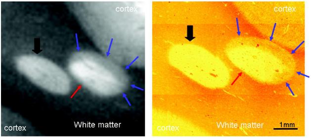
In multiple sclerosis (MS), remyelination may restore conduction and prevent axonal degeneration1. Ability to monitor remyelination in MS in vivo would benefit natural history studies and clinical trials of novel drugs2. High-field MRI (≥3T) is a promising tool to detect remyelination. We scanned a block of post mortem MS brain at 9.4T. Histology revealed two areas of demyelination, and one showing remyelination. These findings corresponded to distinct changes visible on the T2 weighted MRI (figure). As human high-field MRI systems become increasingly widespread, remyelination in patients with MS may become detectable on T2 weighted scans.
Demyelinated (block arrow) and partially remyelinated (red arrow= demyelinated; blue arrows= remyelinated) lesions in post mortem MS brain. Spin-echo MRI (relaxation time= 3000ms, echo time= 60ms, field of view= 30×30mm2, matrix size 256×256 [∼117μm2 in-plane resolution], 16 averages). The corresponding histological section was immuno-stained for myelin basic protein.
Footnotes
Disclosure: The authors report no conflicts of interest.
References
- 1.Rodriguez M. Effectors of Demyelination and Remyelination in the CNS: Implications for Multiple Sclerosis. Brain Pathol. 2007;17:219–29. doi: 10.1111/j.1750-3639.2007.00065.x. [DOI] [PMC free article] [PubMed] [Google Scholar]
- 2.Zhao C, Zawadzka M, Roulois AJA, Bruce CC, Franklin RJM. Promoting remyelination in multiple sclerosis by endogenous adult neural stem/precursor cells: Defining cellular targets. J Neurol Sci. 2008;265:12–16. doi: 10.1016/j.jns.2007.05.008. [DOI] [PubMed] [Google Scholar]


