Abstract
Deletion of the lipooligosaccharide biosynthesis region (Cj1132c to Cj1152c) from the genome of Campylobacter jejuni NCTC11168 shows that the core is not required for viability. The mutant was attenuated for growth and has increased sensitivity to antibiotics and detergents. Natural transformation and invasion of cultured host cells was abolished.
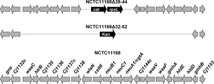
Campylobacter jejuni is the leading cause of bacterial food-borne gastroenteritis worldwide and consequently is a significant public health, social, and economic burden (1, 11). Lipooligosaccharide (LOS) is an important constituent of the bacterial outer membrane, and by acting as a barrier, it maintains cellular integrity (19). The Campylobacter LOS molecule consists of a lipid A moiety and a nonrepeating unit of inner and outer core oligosaccharides (OS) (3, 10). The outer core OS of NCTC11168 LOS mimics GM1 ganglioside (16, 22, 23). Such molecular mimicry may form a strategy for the avoidance of host immune defenses by C. jejuni and is the likely explanation for its association with the neuropathies Guillain-Barré syndrome and Miller Fisher syndrome (2, 15). The core biosynthesis genes are present in a large cluster in the genome (10, 17) (Fig. 1), and modulation of LOS structure arises due to gene content diversity, allelic variation, and high-frequency sequence variation of homopolymeric tracts (10). The function of some genes in the cluster has been elucidated, and individual mutations in waaC and waaF have yielded deep rough mutants (7, 8, 16). Here, we describe the construction and phenotype of a mutant in which the core LOS biosynthesis gene cluster has been removed.
FIG. 1.
Genetic organization of the NCTC11168, NCTC11168Δ38-44 and NCTC11168Δ32-52 LOS biosynthesis regions. The extent of the genetic deletion in the NCTC11168Δ38-44 and NCTC11168Δ32-52 strains is indicated by the broken line with the inserted antibiotic resistance and rpsL genes shown in black. NCTC11168 genes are shown in gray and labeled according to the reannotated genomes (AL111168).
Mutant construction.
An insertion cassette conferring resistance to chloramphenicol was inserted into plasmid pGLM9 that contained the deletion-flanking sequences Cj1138c and Cj1144c (Table 1; Fig. 1). An intermediate mutant strain was first constructed by electroporation (25) of pGLM9 into NCTC11168 (Table 1), and transformants were selected for chloramphenicol resistance. The resultant mutant (NCTC11168Δ38-44) (Table 1; Fig. 1) containing the deletion between Cj1138c and Cj1144c was verified by PCR, Southern blot, and DNA sequencing analysis (data not shown). An insertion cassette conferring resistance to kanamycin was inserted into plasmid pGLM14 that contained the deletion-flanking sequences Cj1132c and Cj1152c (Table 1). The NCTC11168Δ38-44 mutant was transformed by electroporation with pGLM14 and selected on medium containing kanamycin. The Cj1132c-to-Cj1152c deletion mutant (NCTC11168Δ32-52) (Table 1; Fig. 1) was confirmed (data not shown) using primers corresponding to genes external to the deletion, Cj1130F and Cj1153R (Table 2), which confirmed the presence of a 16.9-kb deletion (Fig. 1). Southern hybridization data using internal (htrB- and gmhA-specific probes) and flanking (Cj1132c and gmhB from pGLM14) sequences supported the presence of a large deletion (Fig. 1) in the core LOS biosynthesis gene cluster (data not shown). DNA sequence analysis confirmed the integrity of the genome flanking the deletion and the boundary with the kanamycin resistance cassette (data not shown). Direct transformation of NCTC11168 to create the NCTC11168Δ32-52 mutant and the use of rpsL-based counterselection (6) to remove the insertion cassette between Cj1132c and Cj1152c were not successful. An attempt was also made to adapt the rpsL cassette-based strategy (6) to include the recombinational loss of the inserted cassette by incorporating the imperfect inverted repeats flanking wlaJ (14,26). Unfortunately, this recombination event was not found to take place at a detectable frequency.
TABLE 1.
Bacterial strains and plasmidsa
| Plasmid or strain | Characteristics | Source |
|---|---|---|
| Strains | ||
| NCTC11168 | HS:2 | NCTC; 17 |
| NCTC11168s | Streptomycin-resistant derivative | This study |
| NCTC11168Δ38-44 | Streptomycin sensitive, Catr; genes between Cj1138c and Cj1144c deleted | This study |
| NCTC11168Δ32-52 | Streptomycin resistant, Kanr; genes between Cj1132c and Cj1152c (gmhB) deleted | This study |
| Plasmids | ||
| pUC19 | Ampicillin resistant | New England BioLabs |
| pGLM8 | pUC19 with a fragment containing BamHI-digested Catr and XbaI-digested rpsLWT | This study |
| pGLM9 | pUC19 with a fragment of XbaI/XhoI-digested Cj1138c, XhoI/KpnI-digested Cj1144c, and XhoI-digested CatrrpsLWT cassette from pGLM8 | This study |
| pGLM13 | pUC19 with a fragment of XbaI/XhoI-digested Cj1132c, XhoI/KpnI-digested Cj1152c, and XhoI-digested CatrrpsLWT cassette from pGLM8 | This study |
| pGLM14 | pGLM13 digested with XhoI and then blunted and ligated with EcoRV-digested Kanr | This study |
NCTC, National Collection of Type Cultures; WT, wild type.
TABLE 2.
Primers
| Primer name | Nucleotide sequence (5′ to 3′)a | Product or annealing siteb |
|---|---|---|
| Strep(XbaI)F | GCTCTAGAGCTCAAAGAAAGGAATTATTGTGCCTACC | rpsL |
| Strep(XbaI)R | GCTCTAGAGCTCTAACGGATTTGTCTGTATGAATG | rpsL |
| wlaPF(XbaI) | GCTCTAGAGCTTAGATTCTGATGATTATTGGGAATTAAAC | Cj1138c |
| wlaPR(XhoI) | CCGCTCGAGCGGTACAAATCGCACTAATTCTAAACTG | Cj1138c |
| wlaSCF(XhoI) | CCGCTCGAGCGGGTTGTCCATAAACTCCCATTTG | Cj1144c |
| wlaSCR(KpnI) | GGGGTACCCCGGTATGGGTAGATCTTGATATG | Cj1144c |
| CATrpsLF(XhoI) | CCGCTCGAGCGGTTTTCATACCAATTTTTAAGCTCTGCTCGGCGGTGTTCCTTTC | CatrrpsLWT cassette |
| CATrpsLR(XhoI) | CCGCTCGAGCGGTTGTTGTTCTAACAATAATTTTAGCTCTAACGGATTTGTCTGTATGAATG | CatrrpsLWT cassette |
| CATrpsLF1(XhoI) | CCGCTCGAGCGGTTTTAATAACAATCTTGTTGTTCTGCTCGGCGGTGTTCCTTTCCAAG | CatrrpsLWT cassette |
| CATrpsLR1(XhoI) | CCGCTCGAGCGGAAAAGTATGGTTAAAAATTGGAAGCTCTAACGGATTTGTCTGTATGAATG | CatrrpsLWT cassette |
| CATrpsLR2(XhoI) | CCGCTCGAGCGGTTTTAATAACAATCTTGTTGTTAGCTCTAACGGATTTGTCTGTATGAATG | CatrrpsLWT cassette |
| CATrpsLF2(XhoI) | CCGCTCGAGCGGAAAAGTATGGTTAAAAATTGGAAGCTCTAACGGATTTGTCTGTATGAATG | CatrrpsLWT cassette |
| wlaAF(XbaI) | GCTCTAGAGCCATTACCTCATCGCCATGCACATC | Cj1132c |
| wlaAR(XhoI) | CCGCTCGAGCGGGTGCCAGATGTTGAGCTTATCC | Cj1132c |
| Cj1152F(XhoI) | CCGCTCGAGCGGAAGTGCCAATATCAGCATTTAATCC | Cj1152c |
| wlaTR(KpnI) | GGGGTACCCCGGTTTAGATAGAGACGGTGTGATTAATATAG | Cj1152c |
The additional 5′ oligonucleotide sequence is underlined.
WT, wild type.
Mutant LOS phenotype.
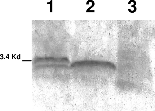
LOS was extracted from both mutants and visualized as previously described (16). A truncated core was detected by silver staining of the sodium dodecyl sulfate (SDS)-polyacrylamide gel electrophoresis-separated LOS extract of the NCTC11168Δ38-44 mutant, but no LOS core was detected for the NCTC11168Δ32-52 mutant (Fig. 2). Western transfer onto polyvinylidene difluoride and the use of the cholera toxin B subunit as a probe for the GM1 epitope (data not shown) verified that this epitope, present in the outer LOS core in NCTC11168 (16), was not present in either mutant strain or the non-GM1 phase variant band in the wild type (Fig. 2).
FIG. 2.
Deletions of the LOS biosynthesis region truncates or removes the core polysaccharide. SDS-polyacrylamide gel electrophoresis (18% [vol/vol]) gel with LOS extracts from NCTC11168s (lane 1), NCTC11168Δ38-44 (lane 2), and NCTC11168Δ32-52 (lane 3) mutants stained with silver nitrate. The wild-type LOS extract shows phase variation switching.
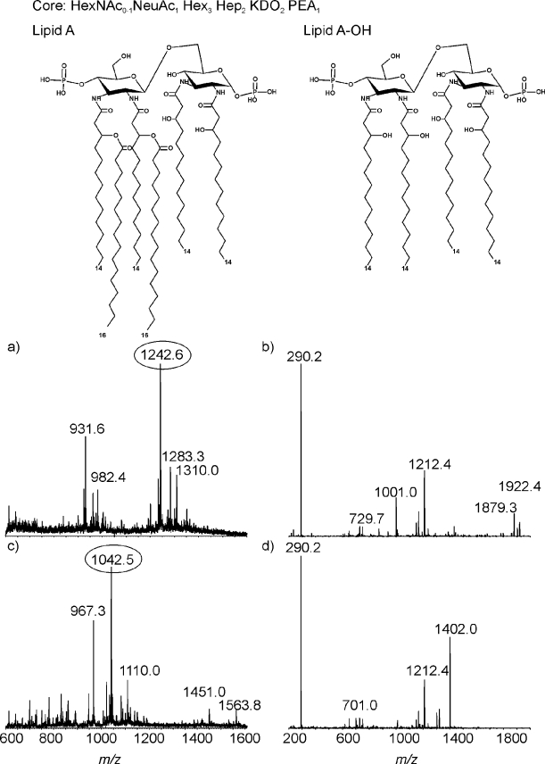
Capillary electrophoresis mass spectroscopy (CE-MS) analysis of both mutants was carried out to investigate LOS structure (Fig. 3 and 4). LOS was analyzed directly or after O deacylation by CE-MS in negative-ion mode. The extracted MS spectrum of NCTC11168Δ38-44 LOS (Fig. 3a) showed triple-charged ions at m/z 1,242.6, 1,283.3, and 1,310.1 together with their corresponding quadruple-charge ions at m/z 931.6, 962.3, and 982.4, respectively. The compositions for each glycoform were derived from the mass spectrum as Neu5Ac1·Hex3·Hep2·Kdo2·lipid A, Neu5Ac1·Hex3·Hep2·PEtn1·Kdo2·lipid A, and HexNAc1·Neu5Ac1·Hex3·Hep2·PEtn1·Kdo2·lipid A, respectively. The tandem MS (MS-MS) spectrum of the ion at m/z 1,242.6 is presented in Fig. 3b. The fragment ion at m/z 290.2 revealed the existence of Neu5Ac. Other major fragment ions are derived from the lipid A portion, such as a doubly charged ion at m/z 1001.0 and singly charged ions at m/z 1,922.4 and 1,879.3, corresponding to the consecutive loss of PPEtn and two PEtn residues, respectively. The ion at m/z 1,212.4 corresponds to the composition Hex3·Hep2·PEtn1·Kdo1. The MS analysis of O-deacylated LOS obtained from the NCTC11168Δ38-44 mutant indicated a major triply charged ion at m/z 1,042.5 corresponding to the composition Neu5Ac1·Hex3·Hep2·Kdo2·lipid A-OH. In addition, a minor ion at m/z 1,110.0 corresponds to a glycoform containing additional HexNAc. The MS-MS of an ion at m/z 1,042.5 (Fig. 3d) showed a singly charged fragment ion at m/z 1,402.0, which revealed that the lipid A-OH consisted of disaccharide [β-d-GlcN3N-(1→6)-β-d-GlcN3N], to which two P- and four N-linked 3-OH-14:0 fatty acids were attached. The fragment ion at m/z 1,212.4 indicates a core region with the composition Hex3·Hep2·PEtn1·Kdo1. This perhaps shows that although the GM1 epitope is missing, there has been partial complementation by genes of similar function from other polysaccharide biosynthesis pathways (10). The absence of wlaN (Cj1139) would account for the loss of the terminal galactose, but no other hexoses should be absent, as the mutant has the heptose inner core OS and the transferases Cj1135-Cj1138 (10) were not deleted; mutant polarity or the involvement of other pathways may be responsible.
FIG. 3.
CE-MS analysis of the LOS from the NCTC11168Δ38-44 mutant. (a) MS spectrum of intact LOS; (b) MS-MS spectrum of ion at m/z 1,242.6; (c) MS spectrum of O-deacylated LOS; (d) MS-MS spectrum of ion at m/z 1,042.5.
FIG. 4.
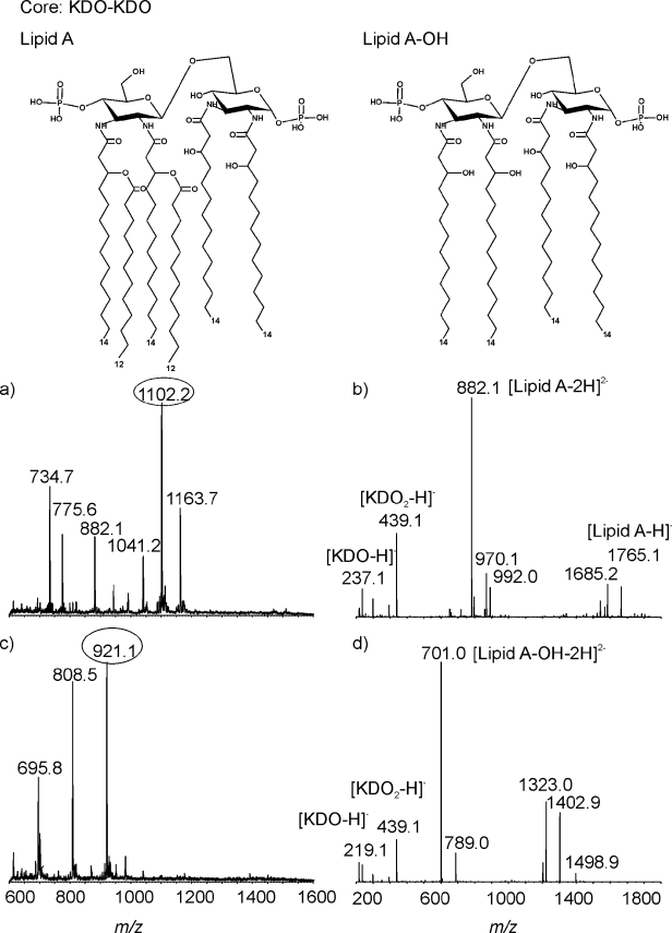
CE-MS analysis of the LOS from the NCTC11168Δ32-52 mutant. (a) MS spectrum of intact LOS; (b) MS-MS spectrum of ion at m/z 1,102.2; (c) MS spectrum of O-deacylated LOS; (d) MS-MS spectrum of ion at m/z 921.1.
Figure 4a and b show the mass spectrum obtained from the NCTC11168Δ32-52 mutant, which is dominated by a molecular ion corresponding to triply and doubly charged ions. The ion at m/z 1,102.2 represents a glycoform with the composition of Kdo2·lipid A. The ion at m/z 1,163.7 corresponds to the same glycoform but with an additional PEtn moiety. The MS-MS spectrum of the doubly charged ion at m/z 1,102.2 shows major fragment ions at m/z 882.1 (doubly charged) and 1,765.1 (singly charged), providing information on lipid A composition. For this strain, the lipid A portion is composed of a disaccharide β-d-GlcN3N-(1→6)-β-d-GlcN3N carrying two phosphate groups and with six fatty acid chains (four N-linked 3-OH-14:0 linked in 2′, 2, 3, and 3′ positions and two O-linked 3-OH-12:0 linked to the 3-OH-14:0 acyl chain in 2′ and 3′ positions) attached. The fragment ions at m/z 237.1 and 439.1 give evidence for the existence of Kdo and Kdo-Kdo. The MS spectrum of O-deacylated LOS (Fig. 4c) showed doubly charged ions at m/z 921.1, 808.5, and 695.8. The mass difference of 226 Da indicated that an incomplete lipid A-OH resulted from a missing 3-OH-14:0 fatty acid. The observation of fragment ions at m/z 219.12 and 439.1 in the MS-MS spectrum of ion m/z 921.1 suggests the presence of Kdo and Kdo-Kdo (Fig. 4d). The doubly charged fragment ion at m/z 701.0 revealed that lipid A-OH consisted of a disaccharide of N-acylated 3-OH-14:0 glucosamine residues, each residue being replaced with a phosphate group.
The core structure of the NCTC11168Δ32-52 large deletion mutant was expected to contain one Kdo. In contrast to the predicted results, the outer core of the NCTC11168Δ32-52 mutant contained two Kdo moieties. The alterations in lipid A involving the presence of O-linked 12:0 instead of the 16:0 fatty acids found in wild-type NCTC11168 and the NCTC11168Δ38-44 mutant are expected, as the late acetyltransferase gene, htrB (lpxL), has been deleted. Phongsisay et al. (18) were unable to mutate htrB, but the NCTC11168Δ32-52 mutant described here and an htrB insertion deletion mutant constructed previously (L. A. Millar and J. M. Ketley, unpublished data) confirm that the loss of htrB is not lethal, at least in NCTC11168. CE-MS analysis of the LOS from the htrB mutant also showed that the lipid A molecule was altered, but the extent of the alteration was not determined. Deletion of Cj1152 (gmhB) may not be possible, as the gene encodes a phosphatase required by both the capsule and LOS biosynthetic pathways (9). The predicted structure of the LOS of this mutant was expected to be the same as that seen by Kanipes et al. (8), where only the Kdo was present with no proximal sugars. The core structure of the NCTC11168Δ32-52 mutant was shown to contain two Kdo moieties and no proximal sugars. The different structures may reflect the use of different strains of C. jejuni. In the 81-176 waaC mutant (8), the presence of one Kdo and the loss of the 3-O-methyl group from the capsule imply that this capsular change allows the strain to remain viable. The capsule biosynthesis region of 81-176 is smaller than that in NCTC11168 but may express four different structures, whereas NCTC11168 has only one potential capsule structure (9). The effect of the LOS core deletion in the NCTC11168Δ32-52 deletion strain on the capsule remains to be explored and may provide information about the interaction between these polysaccharides.
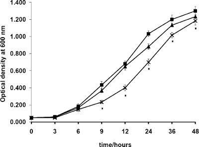
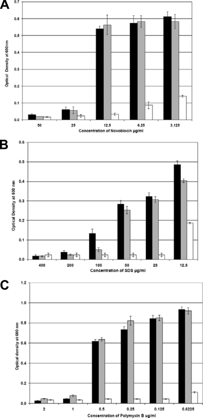
LOS mutants in Neisseria meningitidis have been shown to have reduced growth rates compared to that of the wild type (21). No differences in growth rate have been seen for C. jejuni waaC, htrB, or waaF mutants (8, 16) (Millar and Ketley, unpublished), yet recovery of the NCTC11168Δ32-52 mutant after electroporation was noted to take much longer than is normal. Figure 5 shows the growth of the NCTC11168Δ32-52 mutant compared to the parent strain over 48 h; the mutant initially has a reduced growth rate. Deep rough mutants are also hypersensitive to detergents and hydrophobic antibiotics (21, 24) due to perturbation of the outer membrane (19), and likewise, the NCTC11168Δ32-52 mutant was found to be hypersensitive to SDS, novobiocin, and polymyxin B (Fig. 6). Assessment of the ability of the NCTC11168Δ32-52 mutant to utilize iron provided further evidence of the general perturbation of the outer membrane (data not shown). The loss of the core OS affected ferrous iron uptake and hemin uptake, which enter the periplasm via nonspecific porins and the specific OM receptor ChuA (20), respectively. The invasive potential of the NCTC11168Δ32-52 mutant was examined in a Caco-2 cell invasion assay (12) and shown to be completely abolished (below the limit of detection) compared to that of the wild-type strain (1.78 × 10² CFU/ml). Interestingly, the NCTC11168Δ38-44 mutant was found to be more invasive (1.22 × 10³ CFU/ml, eightfold that of the wild-type level), perhaps indicating that partial loss of the outer core and notably the absence of the ganglioside-like epitope (Fig. 1) are advantageous for invasion. Even in the context of the low level of invasion of strain NCTC11168, our data support the findings of Guerry et al. (5), in which the loss of a GM3-like epitope in 81-176 due to phase variation of the cgtA gene increased invasion of INT407 cells without affecting adherence.
FIG. 5.
Deletion of the LOS biosynthesis region affects growth. Growth assays were conducted with the NCTC11168s (▪), NCTC11168Δ38-44 (▴), and NCTC11168Δ32-52 (×) mutants in Mueller-Hinton broth incubated microaerobically with agitation at 42°C for 48 h. Each data point is the mean of results of six replicate experiments, and the standard error is indicated. Measurements where the difference between results for the wild type and the NCTC11168Δ32-52 mutant is significant are indicated by asterisks.
FIG. 6.
Removal of the core polysaccharide increases sensitivity to detergent and hydrophobic antibiotics. Sensitivity assays were conducted with the NCTC11168s (black), NCTC11168Δ38-44 (gray), and NCTC11168Δ32-52 (white) mutants grown in Mueller-Hinton broth, supplemented with a range of concentrations of novobiocin (A), SDS (B), and polymyxin B (C) at 42°C microaerobically for 16 h. The assays were performed in triplicate, and the error bars indicate the standard errors, where n = 3.
The deletion of the LOS genes in the NCTC11168Δ32-52 mutant provides an opportunity to examine the different LOS biosynthesis gene content, (4) in the same genetic background and provides the ability to investigate gene function by the reintroduction of genes in a stepwise manner. Unfortunately, multiple attempts to naturally transform the NCTC11168Δ32-52 mutant with NCTC11168, RM1221, and NCTC11168Δ38-44 LOS clusters, as well as a positive control (cheV::cat; average transformation efficiency of 1.6 × 10⁴ CFU/μg [O. Bridle, personal communication]) were unsuccessful. Therefore, deletion of the LOS cluster in the NCTC11168Δ32-52 mutant results in the loss of natural competence; this may be due to the disruption of the OM. It is possible that second-site mutations contributed to the recovery of the NCTC11168Δ32-52 mutant and the resulting phenotype; unfortunately, the loss of genetic competence renders complementation unachievable.
In conclusion, the deletion of the LOS cluster in the NCTC11168Δ32-52 mutant has confirmed predictions of the minimal LOS gene content required for viability in campylobacters. Absence of the LOS outer core is detrimental to C. jejuni growth and undermines outer membrane integrity. The inability of the NCTC11168Δ32-52 large deletion mutant to invade Caco-2 further supports a role for LOS in adherence and invasion (7, 8, 13).
Acknowledgments
G.L.M. was funded by an MRC CASE studentship to J.M.K. and A.J.L.
We thank Claire Miller for technical assistance in performing the iron uptake and growth assay and also Monika Dzieciatkowska for preparing LOS samples and performing CE-MS analysis.
Footnotes
Published ahead of print on 30 January 2009.
REFERENCES
- 1.Adak, G. K., S. M. Long, and S. J. O'Brien. 2002. Trends in indigenous foodborne disease and deaths, England and Wales: 1992 to 2000. Gut 51832. [DOI] [PMC free article] [PubMed] [Google Scholar]
- 2.Allos, B. M. 1997. Association between Campylobacter infection and Guillain-Barré syndrome. J. Infect. Dis. 176S125-S128. [DOI] [PubMed] [Google Scholar]
- 3.Aspinall, G. O., C. M. Lynch, H. Pang, R. T. Shaver, and A. P. Moran. 1995. Chemical structures of the core region of Campylobacter jejuni O:3 lipopolysaccharide and an associated polysaccharide. Eur. J. Biochem. 231570-578. [PubMed] [Google Scholar]
- 4.Gilbert, M., M.-F. Karwaski, S. Bernatchez, N. M. Young, E. Taboada, J. Michniewicz, A.-M. Cunningham, and W. W. Wakarchuk. 2002. The genetic bases for the variation in the lipo-oligosaccharide of the mucosal pathogen, Campylobacter jejuni: biosynthesis of sialylated ganglioside mimics in the core oligosaccharide. J. Biol. Chem. 277327-337. [DOI] [PubMed] [Google Scholar]
- 5.Guerry, P., C. M. Szymanski, M. M. Prendergast, T. E. Hickey, C. P. Ewing, D. L. Pattarini, and A. P. Moran. 2002. Phase variation of Campylobacter jejuni 81-176 lipooligosaccharide affects ganglioside mimicry and invasiveness in vitro. Infect. Immun. 70787-793. [DOI] [PMC free article] [PubMed] [Google Scholar]
- 6.Hendrixson, D. R., B. J. Akerley, and V. J. DiRita. 2001. Transposon mutagenesis of Campylobacter jejuni identifies a bipartite energy taxis system required for motility. Mol. Microbiol. 40214-224. [DOI] [PubMed] [Google Scholar]
- 7.Kanipes, M. I., L. C. Holder, A. T. Corcoran, A. P. Moran, and P. Guerry. 2004. A deep-rough mutant of Campylobacter jejuni 81-176 is noninvasive for intestinal epithelial cells. Infect. Immun. 722452-2455. [DOI] [PMC free article] [PubMed] [Google Scholar]
- 8.Kanipes, M. I., E. Papp-Szabo, P. Guerry, and M. A. Monteiro. 2006. Mutation of waaC, encoding heptosyltransferase I in Campylobacter jejuni 81-176, affects the structure of both lipooligosaccharide and capsular carbohydrate. J. Bacteriol. 1883273-3279. [DOI] [PMC free article] [PubMed] [Google Scholar]
- 9.Karlyshev, A. V., O. L. Champion, C. Churcher, J.-R. Brisson, H. C. Jarrell, M. Gilbert, D. Brochu, F. St. Michael, J. Li, W. W. Wakarchuk, I. Goodhead, M. Sanders, K. Stevens, B. White, J. Parkhill, B. Wren, and C. Szymanski. 2005. Analysis of Campylobacter jejuni capsular loci reveals multiple mechanisms for the generation of structural diversity and the ability to form complex heptoses. Mol. Microbiol. 5590-103. [DOI] [PubMed] [Google Scholar]
- 10.Karlyshev, A. V., J. M. Ketley, and B. W. Wren. 2005. The Campylobacter jejuni glycome. FEMS Microbiol. Rev. 29377-390. [DOI] [PubMed] [Google Scholar]
- 11.Ketley, J. 1997. Pathogenesis of enteric infection by Campylobacter. Microbiology 1435-21. [DOI] [PubMed] [Google Scholar]
- 12.MacCallum, A. J., D. Harris, G. Haddock, and P. H. Everest. 2006. Campylobacter jejuni-infected human epithelial cell lines vary in their ability to secrete interleukin-8 compared to in vitro-infected primary human intestinal tissue. Microbiology 1523661-3665. [DOI] [PubMed] [Google Scholar]
- 13.McSweegan, E., and R. I. Walker. 1986. Identification and characterization of two Campylobacter jejuni adhesins for cellular and mucous substrates. Infect. Immun. 53141-148. [DOI] [PMC free article] [PubMed] [Google Scholar]
- 14.Misawa, N., K. Kawashima, F. Kondo, B. M. Allos, and M. J. Blaser. 2001. DNA diversity of the wla gene cluster among serotype HS:19 and non-HS:19 Campylobacter jejuni strains. J. Endotoxin Res. 7349-358. [PubMed] [Google Scholar]
- 15.Moran, A., B. Appelmelk, and G. Aspinall. 1996. Molecular mimicry of host structures by lipopolysaccharides of Campylobacter and Helicobacter spp.: implications in pathogenesis. J. Endotoxin Res. 3521-531. [Google Scholar]
- 16.Oldfield, N. J., A. P. Moran, L. A. Millar, M. M. Prendergast, and J. M. Ketley. 2002. Characterization of the Campylobacter jejuni heptosyltransferase II gene, waaF, provides genetic evidence that extracellular polysaccharide is lipid A core independent. J. Bacteriol. 1842100-2107. [DOI] [PMC free article] [PubMed] [Google Scholar]
- 17.Parkhill, J., B. W. Wren, K. Mungall, J. M. Ketley, C. Churcher, D. Basham, T. Chillingworth, R. M. Davies, T. Feltwell, S. Holroyd, K. Jagels, A. V. Karlyshev, S. Moule, M. J. Pallen, C. W. Penn, M. A. Quail, M. A. Rajandream, K. M. Rutherford, A. H. van Vliet, S. Whitehead, and B. G. Barrell. 2000. The genome sequence of the food-borne pathogen Campylobacter jejuni reveals hypervariable sequences. Nature 403665-668. [DOI] [PubMed] [Google Scholar]
- 18.Phongsisay, V., V. N. Perera, and B. N. Fry. 2006. Exchange of lipooligosaccharide synthesis genes creates potential Guillain-Barré syndrome-inducible strains of Campylobacter jejuni. Infect. Immun. 741368-1372. [DOI] [PMC free article] [PubMed] [Google Scholar]
- 19.Raetz, C. R. H., and C. Whitfield. 2002. Lipopolysaccharide endotoxins. Annu. Rev. Biochem. 71635-700. [DOI] [PMC free article] [PubMed] [Google Scholar]
- 20.Ridley, K. A., J. D. Rock, Y. Li, and J. M. Ketley. 2006. Heme utilization in Campylobacter jejuni. J. Bacteriol. 1887862-7875. [DOI] [PMC free article] [PubMed] [Google Scholar]
- 21.Steeghs, L., H. de Cock, E. Evers, B. Zomer, J. Tommassen, and P. van der Ley. 2001. Outer membrane composition of a lipopolysaccharide-deficient Neisseria meningitidis mutant. EMBO J. 206937-6945. [DOI] [PMC free article] [PubMed] [Google Scholar]
- 22.St. Michael, F., C. Szymanski, J. Li, K. H. Chan, N. H. Khieu, A. Larocque, W. W. Wakarchuk, J.-R. Brisson, and M. A. Monteiro. 2002. The structures of the lipooligosaccharide and capsule polysaccharide of Campylobacter jejuni genome sequenced strain NCTC 11168. Eur. J. Biochem. 2695119-5136. [DOI] [PubMed] [Google Scholar]
- 23.Szymanski, C. M., F. S. Michael, H. C. Jarrell, J. Li, M. Gilbert, S. Larocque, E. Vinogradov, and J. R. Brisson. 2003. Detection of conserved N-linked glycans and phase-variable lipooligosaccharides and capsules from campylobacter cells by mass spectrometry and high resolution magic angle spinning NMR spectroscopy. J. Biol. Chem. 27824509-24520. [DOI] [PubMed] [Google Scholar]
- 24.Tzeng, Y.-L., A. Datta, V. K. Kolli, R. W. Carlson, and D. S. Stephens. 2002. Endotoxin of Neisseria meningitidis composed only of intact lipid A: inactivation of the meningococcal 3-deoxy-d-manno-octulosonic acid transferase. J. Bacteriol. 1842379-2388. [DOI] [PMC free article] [PubMed] [Google Scholar]
- 25.van Vliet, A. H. M., A. C. Wood, J. Henderson, K. G. Wooldridge, and J. M. Ketley. 1998. Genetic manipulation of enteric Campylobacter species, p. 407-420. In P. Williams, J. Ketley, and G. Salmond (ed.), Bacterial pathogenesis, vol. 27. Academic Press, London, United Kingdom. [Google Scholar]
- 26.Wood, A. C., N. J. Oldfield, C. A. O'Dwyer, and J. M. Ketley. 1999. Cloning, mutation and distribution of a putative lipopolysaccharide biosynthesis locus in Campylobacter jejuni. Microbiology 145379-388. [DOI] [PubMed] [Google Scholar]


