Abstract
Sustained intracellular Ca2+ elevation is a well-established contributor to neuronal injury following excessive activation of NMDA-type glutamate receptors. Zn2+ can also be involved in excitotoxic degeneration, but the relative contributions of these two cations to the initiation and progression of excitotoxic injury is not yet known. We previously concluded that extended NMDA exposure led to sustained Ca2+ increases that originated in apical dendrites of CA1 neurons and then propagated slowly throughout neurons and caused rapid necrotic injury. However the fluorescent indicator used in those studies (Fura-6F) may also respond to Zn2+, and in the present work we examine possible contributions of Zn2+ to indicator signals and to the progression of degenerative signaling along CA1 dendrites. Selective chelation of Zn2+ with N,N,N′,N′-tetrakis(2-pyridylmethyl)ethylenediamine (TPEN) significantly delayed, but did not prevent the development and progression of sustained high-level Fura-6F signals from dendrites to somata. Rapid indicator loss during the Ca2+ overload response, which corresponds to rapid neuronal injury, was also not prevented by TPEN. The relationship between cytosolic Zn2+ and Ca2+ levels was assessed in single CA1 neurons co-loaded with Fura-6F and the Zn2+-selective indicator FluoZin-3. NMDA exposure resulted in significant initial increases in FluoZin-3 increases that were prevented by TPEN, but not by extracellular Zn2+ chelation with Ca-EDTA. Consistent with this result, Ca-EDTA did not delay the progression of Fura-6F signals during NMDA. Removal of extracellular Ca2+ reduced, but did not prevent FluoZin-3 increases. These results suggest that sustained Ca2+ increases indeed underlie Fura-6F signals that slowly propagate throughout neurons, and that Ca2+ (rather than Zn2+) increases are ultimately responsible for neuronal injury during NMDA. However, mobilization of Zn2+ from endogenous sources leads to significant neuronal Zn2+ increases, that in turn contribute to mechanisms of initiation and progression of progressive Ca2+ deregulation.
Keywords: Hippocampal slice, FluoZin-3, Fura, TPEN, NMDA
Introduction
There has been longstanding interest in neuronal Ca2+ loading following both normal and excessive glutamate receptor activation, with much evidence demonstrating that sustained Ca2+ accumulation can trigger neuronal injury in a variety of experimental models (Choi, 1988; Nicholls and Budd, 2000; Connor and Shuttleworth, 2001; Arundine and Tymianski, 2003). Zn2+ is also present at significant concentrations in the brain, and can contribute to neuronal death following its mobilization from synaptic vesicles and/or intracellular binding sites (Choi and Koh, 1998; Weiss et al., 2000; Frederickson et al., 2005). At present there is controversy over the relative importance of each ion in the complex reactions that initiate neuronal injury. We therefore considered it an important goal to determine the extent to which Ca2+- and Zn2+-dependent mechanisms may interact to initiate neuronal injury.
In an effort to test the roles of Ca2+ in initiation of excitotoxic injury, we recently used a low-affinity Ca2+-sensitive indicator (Fura-6F) to examine responses of CA1 neuron dendrites during extended exposures to NMDA. We concluded that very large Ca2+ increases originated in distal CA1 apical dendrites and propagated slowly throughout the neuron and arrival of sustained Ca2+ elevations at somata triggered relatively rapid cell injury (Dietz et al., 2007; Vander Jagt et al., 2008). Indicators of the Fura family are also highly sensitive to Zn2+ with affinities, in vitro, higher than for Ca2+ (e.g. Grynkiewicz et al., 1985; Hechtenberg and Beyersmann, 1993; Simons, 1993; Atar et al., 1995; Sensi et al., 1997; Cheng and Reynolds, 1998; Martin et al., 2006). When measured against physiological Ca2+ concentrations in intact neurons, the apparent sensitivity of Fura indicators for Zn2+ is greatly decreased (Marchi et al., 2000; Devinney et al., 2005) and Ca2+ and Zn2+ signals can be effectively discriminated (Devinney et al., 2005; Dietz et al., 2008). However, it is not yet known whether Zn2+ binding to the indicator could contribute to the very large Fura-6F signals observed to propagate along CA1 dendrites and furthermore whether Zn2+ increases could contribute to mechanisms triggering Ca2+ deregulation.
Previous studies of cell culture neurons have established that glutamate receptor activation in the presence of exogenous Zn2+ can lead to intracellular Zn2+ increases and toxicity, as a result of influx through channels and transporters that are better known for their flux of Ca2+ (Weiss et al., 1993; Sensi et al., 1997; Choi and Koh, 1998; Weiss et al., 2000). Zn2+ can also bind to NMDA receptors and inhibit function (Peters et al., 1987; Westbrook and Mayer, 1987) but also directly permeates the channel, although the process is relatively slow and can appear as a flicker-type block of channel activity (Christine and Choi, 1990). Exposure to NMDA could also lead to Zn2+ influx via other channels as a secondary consequence of depolarization during NMDA exposure, for example voltage-dependent Ca2+ channels, or possibly reverse-operation of sodium calcium exchangers (Sensi et al., 1997; Choi and Koh, 1998; Weiss et al., 2000). Zn2+ can be present in the extracellular space as a result of contaminant from solution preparation (Kay, 2004), but there is also evidence that significant amounts of endogenous Zn2+ are released in the CA1 region following stimulation of Schaffer collateral fibers in murine hippocampal slices (Qian and Noebels, 2006). In addition to influx from the extracellular space, Zn2+ can also be mobilized from intracellular binding proteins following excitotoxic or oxidative stress (Aizenman et al., 2000; Lee et al., 2003; Bossy-Wetzel et al., 2004) and it is possible that one or both sources of Zn2+ could be mobilized following extended NMDA exposures in slice.
The present study measured Zn2+ dynamics in single CA1 neurons in murine hippocampal slices during extended NMDA exposures, using combinations of fluorescent indicators and Zn2+-selective chelators in order to probe for possible contributions of Zn2+ accumulation to subsequent Ca2+ deregulation and neuronal damage. Some of these results have been presented in Abstract form (Vander Jagt et al., 2007).
Experimental Procedures
Animals
Male FVB/N mice were obtained from Harlan (Bar Harbor, Maine) at 4 weeks of age and housed under standard conditions (12hr/12hr light/dark cycle) before sacrifice at 4-6 weeks of age. Mice were deeply anesthetized with a mixture of ketamine and xylazine (85mg/ml and 15mg/ml, respectively; 150μl s.c.) and decapitated. Brains were quickly removed and placed in ice cold cutting solution (see below for composition). Experiments were carried out in accordance with the National Institute of Health guidelines for the humane treatment of laboratory animals, and the protocol for these procedures was reviewed annually by the Institutional Animal Care and Use Committee at the University of New Mexico School of Medicine.
Slice Preparation and single-cell indicator loading
The procedures for slice preparation and loading of fluorescent indicators were identical to those recently described (Vander Jagt et al., 2008) and are summarized here. A Vibratome (St. Louis, MO) was used to prepare coronal slices (350μm), which were then warmed to 35°C for 1 hour and then held at room temperature until used for recording. Recordings were made on a fixed stage microscope (Olympus BX51WI) with a Warner Instruments recording chamber (RC-27L) and slices were superfused with warmed (32°C), oxygenated ACSF at 2-2.5 ml/min. Single CA1 neurons were visualized by using DIC optics and briefly loaded with fluorescent indicators via patch pipettes in the “whole-cell” recording mode. As previously described, neurons were used for analysis only if the current required to maintain a holding voltage of -70 mV exceeded was <100pA and membrane resistance was between 100 and 250MΩ. In studies of cells loaded with Fura-6F alone, 1mM indicator was included in the pipette solution (Vander Jagt et al., 2008) and for experiments co-loading FluoZin-3 and Fura-6F, initial pipette indicator concentrations were 0.6mM and 0.5mM, respectively. After obtaining whole-cell access, indicator loading was strictly limited to 3 minutes to minimize loss of diffusible cellular components and final cellular indicator concentrations in neurons was estimated at ≤ 200μM (see Vander Jagt et al., 2008). Loading pipettes were then carefully removed, and cells were discarded if soma Ca2+ elevations were detected during the pipette removal process. Cells were then allowed to recover for 25 minutes after pipette removal before NMDA challenge.
Imaging
Fluorescence data were captured using a CCD-based system (Till Imago SensiCam, Polychrome IV monochrometer, TillVision v4.1 software, Till Photonics Pleasanton, CA). In studies of Fura-6F alone, indicator was excited at 350/380nm (25-50ms) and emission detected with a 400nm / 510±40nm dichromatic mirror / emission filter combination. Co-loaded neurons were excited sequentially at 350/380/490nm, (100-150ms) and emission of both indicators detected with a single dichromatic mirror / emission filter set (505nm / 535±25nm), similar to previous reports (Devinney et al., 2005; Dietz et al., 2008). Acquisition rate was 0.2Hz for each indicator (single image or image pair). Fura-6F ratios were converted to estimated Ca2+ concentrations, using procedures previously described (Vander Jagt et al., 2008) and data for FluoZin-3 was presented as Δ F/F0 after background subtraction, except where noted.
Reagents and Solutions
ACSF contained (in mM): 126 NaCl, 2 KCl, 1.25 NaH2PO4, 1 MgSO4, 26 NaHCO3, 2 CaCl2, and 10 glucose, equilibrated with 95%O2 / 5%CO2. Slice cutting solution contained (in mM): 2 KCl, 1.25 NaH2PO4, 6 MgSO4, 26 NaHCO3, 0.2 CaCl2, 10 glucose, 220 sucrose and 0.43 ketamine. Cutting and recording solutions were all 315-320 mOsmol. Whole-cell ppipette solutions contained (in mM): 135 K-gluconate, 8 NaCl, 1 MgCl2, 10 HEPES, 2 Mg2+-ATP and was pH (7.25) adjusted with KOH. NMDA exposures were made in modified ACSF lacking added Mg2+ and supplemented with nimodipine (10μM), as described in (Vander Jagt et al., 2008). Fluorescent indicators were from Invitrogen-Molecular Probes (Eugene, OR) and other reagents were from Sigma Chemical Co. (St. Louis, MO). Nimodipine was prepared in ethanol (10mM stock) and NMDA stocks were prepared in de-ionized water (5mM) and stored at -20°C until use. Two Zn2+-selective chelators were used: N,N,N′,N′-Tetrakis (2-pyridylmethyl) ethylenediamine (TPEN) which has KD(Zn2+) = 2.95×10⁻¹⁶M and KD(Ca2+) of 4.0×10⁻⁵ (Arslan et al., 1985) and Ca-EDTA, which exchanges its bound Ca2+ for Zn2+ with a KD of 8×10-14M (Lee et al., 2002). TPEN was prepared fresh prior to each use in DMSO (50mM) and Ca-EDTA was prepared in ACSF. Tests of possible effects of matched vehicle DMSO concentrations (max 0.1%) showed no effects on NMDA-stimulated responses in these experiments.
Statistics
Group data are presented as mean±SEM, and “n” values refer to the number of neurons examined. Only one neuron was studied in each slice. For each experimental condition, no more than two slices were examined from a single animal. Differences between group data were analyzed with unpaired Student's t-tests, with p<0.05 being considered significant. Each set of experiments presented was internally controlled, using similar numbers of control slices from interleaved experiments to compare with each experimental condition. This was important to control for possible differences in animals, tissue handling or other unknown variables that may contribute to small changes in average response times.
Results
1. Effects of Zn2+ chelation with TPEN on overall Fura-6F responses
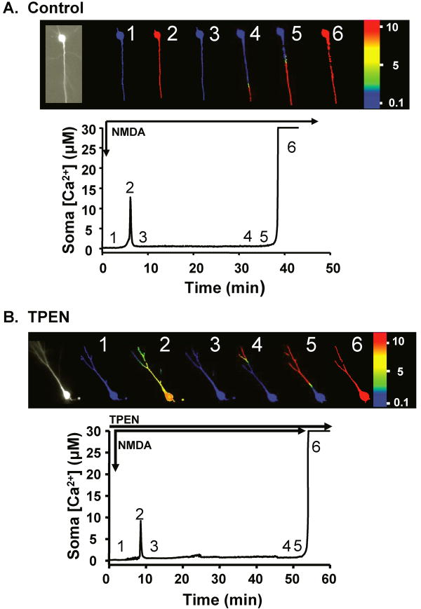
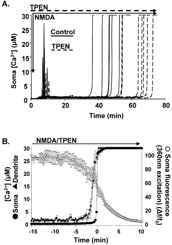
Figure 1A shows the characteristic responses of a single CA1 neuron to extended NMDA exposure, monitored using the low-affinity indicator Fura-6F. As previously described (Dietz et al., 2007; Vander Jagt et al., 2008), the Fura-6F signal showed an initial spike followed by recovery to near-baseline for about 25 min and then a larger and sustained increase originating in the distal dendrite that propagated slowly and invaded the soma. Arrival of the sustained response at the soma was accompanied by rapid loss of indicator, suggesting acute neuronal injury (see below). Figure 1B shows a neuron from the same experimental group, but in this case the membrane-permeable, Zn2+-selective chelator, TPEN was present throughout (50μM; with 20 min pre-exposure). Although TPEN did have significant quantitative effects (see below), it did not change the basic characteristics of the compound Fura-6F response. There was still an initial Fura-6F spike ∼6 min after NMDA onset, followed by recovery to near-baseline levels. In TPEN, the propagating sustained Fura-6F increase was still sufficiently large to saturate the indicator, and progressed apparently normally along the apical dendrite to the soma. This result, when taken together with previous observations that Fura-6F signals were abolished by extracellular Ca2+ removal, implies that Ca2+ (rather than Zn2+) increases are primarily responsible for the large propagating Fura-6F signals associated with neuronal degeneration. Figure 2A shows recordings from a group of experiments with and without TPEN, and reveals that TPEN produced a significant decrease in amplitude of the initial spikes, and a consistent delay in the arrival of dendritic Ca2+ overload responses at somata.
Figure 1. TPEN did not prevent progressive Ca2+ overload.
A: Fura-6F responses in a CA1 neuron during NMDA exposure (5μM). Left panel; 380nm excitation image of filled neuron. Subsequent panels are pseudo-color images, masked and calibrated to estimated Ca2+ levels before (1) and during NMDA exposure (2-6). An initial Ca2+ spike (2) was followed by recovery to near basal levels (3) throughout the neuron. After a significant delay, sustained Ca2+ increases appeared in distal dendrite locations (4-5) and slowly propagated into the soma (6). Plot shows data extracted from a region of interest in the soma. B: As in “A,” except that the slice was pre-exposed to the Zn2+ chelator TPEN (50μM). The general characteristics of the Fura-6F response appeared very similar, with an initial transient increase, recovery and long delay before progression of sustained Ca2+ increase from apical dendrite into the soma.
Figure 2. TPEN delayed the arrival of sustained dendritic Ca2+ increases at the soma.
A: Each trace represents the Fura-6F response recorded from the soma of a single neuron, calibrated to estimated Ca2+ levels. Studies in a set of control slices (solid lines) were interleaved with slices slices pre-exposed to TPEN (50μM; dashed lines). TPEN significantly delayed the arrival of sustained Ca2+ increases at somata (p<0.01 n=7 each). B: TPEN did not prevent rapid neuronal injury after establishment of Ca2+ overload in somata. The rising phases of sustained Ca2+ increases in somata were aligned (time 0; filled circles, n=5) and the preceding responses from a region of apical proximal dendrite (40-60 μm from somata; triangles) are also included for 3 neurons where dendrites were sufficiently within the plane of focus. Fluorescence intensity at the isobestic point for Fura-6F (360nm; open circles) was monitored to determine indicator loss, and despite the presence of TPEN, showed dramatic loss as sustained Ca2+ levels became established in somata.
2. Effects of TPEN on initial Fura-6F spikes
Initial Fura-6F spikes were significantly decreased (15.3±2.8 vs. 5.5±1.8μM, control and TPEN respectively; p<0.01, n=6,7), but there was no difference in the latency of these initial events (7.9±0.3 vs. 8.2±0.5min; control and TPEN respectively; p=0.6, n=6,7).
While TPEN has a very high affinity for Zn2+ (KD 2.9×10⁻¹⁶M), it still binds Ca2+ (KD 4×10⁻⁵M (Arslan et al., 1985)), and it is possible that 50μM TPEN could buffer intracellular Ca2+ levels and thereby decrease initial Fura-6F spike responses. This seems unlikely since when we reduced the TPEN concentration to 10μM, there was still a very similar reduction of initial spike amplitude (15.1±2.6 vs. 6.7±1.8μM, control and 10μM TPEN respectively, p=0.03, n=6 each) which would not be expected if Ca2+ buffering were the main effect. Given evidence that intracellular Zn2+ increases occurs at the same time (see below), it seems more likely that Zn2+ binding to Fura-6F or downstream consequences of Zn2+ mobilization modulates the initial Fura-6F spikes.
3. Effects of TPEN on propagating Ca2+ overload responses
The average time taken for Ca2+ overload to reach the soma after NMDA application (see Fig 2A) was significantly increased by TPEN (45.5±2.2 vs. 63.9±2.7min, control and TPEN respectively, p<0.01, n=6,7). This increase was not related to the differences in the initial spike, since it has been shown that abolishing the spike altogether with transient removal of extracellular Ca2+ did not influence the timing of dendritic Ca2+ deregulation (Vander Jagt et al., 2008b). This was confirmed in a set of experiments where TPEN was applied immediately after the initial spike responses, instead of pre-exposing slices to the chelator. Under these conditions, there was still a significantly increased delay in arrival of Ca2+ overload responses at somata (38.6±3.4 vs. 54.1±4.3min, control and TPEN respectively; p<0.02, n=6,7). The effect here was smaller than in slices pre-exposed to the chelator, and this is presumably due to fact that it takes some time for the chelator to accumulate to effective concentrations in neurons in slice, while activation of deleterious processes by NMDA is already ongoing.
When measured at the isobestic point for Ca2+ (360nm), loss of Fura-6F fluorescence can provide an effective measure of acute membrane failure, as the charged indicator of 836 MW is rapidly lost from the loaded cell after Ca2+ overload occurs ((Vander Jagt et al., 2008) and see also (Randall and Thayer, 1992)). TPEN did not modify this injury response, following invasion of somata by Ca2+ overload responses. Figure 2C shows Fura-6F fluorescence loss associated with sustained Ca2+ increases in the presence of TPEN. Indicator loss from somata slightly preceded increases in Fura-6F ratio in this compartment, and this is likely due to leakage of indicator from distal dendrites that experienced the sustained Ca2+ response earlier. The degree of indicator loss in TPEN was very similar to that seen in interleaved controls. Fura-6F (360nm) fluorescence measured 5 min after somatic Ca2+ deregulation was 14±2% of levels just prior to Ca2+ overload in controls vs. 15±2% in TPEN (p=0.64, n=5 each). Under both conditions levels declined to <10% by 10 min after somatic deregulation.
4. Zn2+ elevations evaluated with FluoZin-3
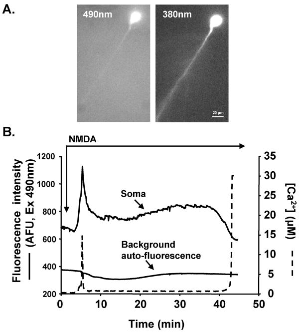
The relationship between Zn2+ and Ca2+ increases was evaluated in neurons co-loaded with FluoZin-3 and Fura-6F. FluoZin-3 fluorescence signals were difficult to detect in dendritic processes (Fig 3A) and therefore the analysis below is restricted to neuronal somata, where reliable measurements could be made. Figure 3B shows a representative example of responses to NMDA. Coincident with the initial Fura-6F spike there was a rapidly increasing FluoZin-3 signal. Unlike the Fura-6F spike, the FluoZin-3 signal did not reset to baseline but remained at an elevated level for the next 25 min. When Fura-6F ratio increases then reported the arrival of sustained Ca2+ elevations in the soma, FluoZin-3 levels did not increase, but decreased rapidly. The rate of FluoZin-3 fluorescence loss around this time point was similar to the loss of single wavelength Fura-6F fluorescence (measured at 360nm, Fig 2C), suggesting that the late FluoZin-3 fluorescence decrease was due to loss of indicator from the cell, rather than precipitous decreases in cellular Zn2+ levels.
Figure 3. Intracellular Zn2+ increases during NMDA exposure.
A: Fluorescence images of a single CA1 neuron co-loaded with FluoZin-3 (490nm) and Fura-6F (360nm). B: Data extracted from the soma of the neuron shown in A, during NMDA exposure. An initial FluoZin-3 increase (solid line, top trace) was observed soon after NMDA addition, coincident with the early spike in the Fura-6F signal (dashed line). After a partial recovery, Fluozin-3 fluorescence remained elevated during the latent period, and then decreased to below baseline levels when the sustained Ca2+ response arrived at the soma. Autofluorescence (490nm) from a region of stratum pyramidale outside the neuron (solid line, bottom trace) showed no initial transient, but a slow fluctuation during the latent period.
Slice autofluorescence at 490nm is due in part to tissue flavoproteins, and since flavoprotein oxidation state can change in an activity-dependent manner under some conditions (Shibuki et al., 2003; Shuttleworth et al., 2003) autofluorescence changes could potentially complicate analysis of the FluoZin-3 signals. As illustrated in Fig 3, there was no demonstrable autofluorescence change coincident with the initial FluoZin-3 transient, but a slower change was observed that overlapped with the time course of the sustained fluorescence increase after the transient. For this reason, all subsequent FluoZin-3 signals were corrected by subtraction of autofluorescence signals at the same time points, from an adjacent region of stratum pyramidale.
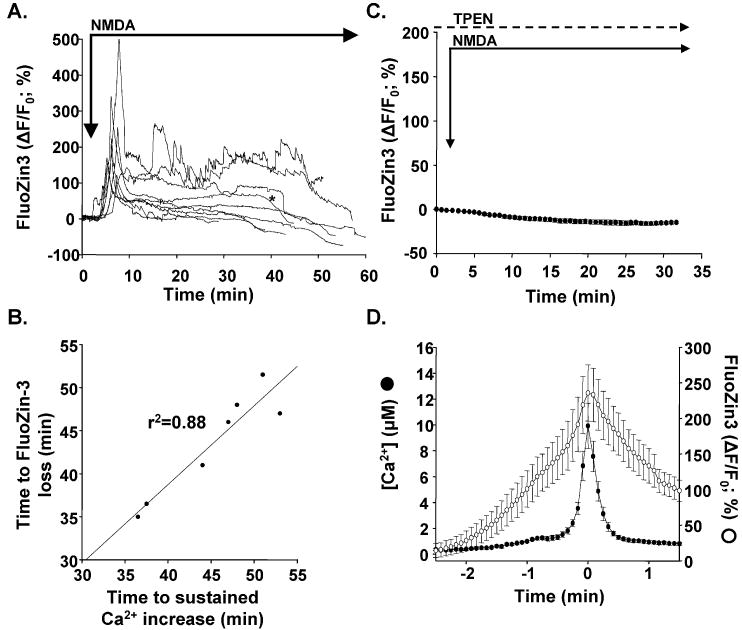
The FluoZin-3 signals from 9 preparations (including the cell from Fig 3) are shown in Fig 4A. Although there was considerable cell-cell variability in the absolute amplitude of responses, all cells showed the same overall pattern; an early FluoZin-3 fluorescence increase followed by variable recovery during a latent period before rapid loss of fluorescence 30-40 min later. The plateau level of FluoZin-3 increase during the latent period (between initial spike and ultimate Ca2+ overload) was quite variable (range -8 to 177%, mean 61±20%; n=9). Figure 4B shows the relationship between FluoZin-3 fluorescence loss and arrival of the sustained Ca2+ increase and supports the conclusion that FluoZin-3 indicator loss occurs around the time that the Ca2+ overload responses arrive.
Figure 4. Characteristics of Zn2+ increases.
A: FluoZin-3 responses during NMDA exposure from 9 preparations, corrected for slow autofluorescence changes. All show initial transient FluoZin-3 increases, which then recover to variable degrees for ∼30 min before fluorescence levels decline sharply. B: Late FluoZin-3 signal loss correlated strongly with sustained Ca2+ increases in somata. Clear increases in the rate of FluoZin-3 fluorescence loss was detectable in 7/9 neurons. For these neurons, the time at which 50% FluoZin-3 fluorescence intensity (compared with plateau levels prior to arrival of Ca2+ overload) is lost is plotted against the time of half-maximal Ca2+ increase as the sustained Ca2+ response invaded somata. C: TPEN prevented FluoZin-3 fluorescence increases. TPEN (50μM) was added 20 min prior to NMDA and remained throughout the experiment. No initial or delayed fluorescence increases were observed during NMDA (mean±SEM, n=4). D: Initial transient FluoZin-3 increases from the responses illustrated in A are shown at an expanded time base (open circles), together with initial Fura-6F transients from the same neurons (filled circles). Records were aligned with the timing of the peak Fura-6F response in each cell (n=9) and show that both signals peaked at the same time, although the duration of the Fura-6F transients appears significantly shorter.
In a set of slices exposed to TPEN (50μM) FluoZin-3 fluorescence did not increase during NMDA exposure (Fig 4C). When taken together with the insensitivity of FluoZin-3 to Ca2+ reported by many groups (see Discussion) this strongly supports the interpretation that FluoZin-3 signals are due to Zn2+, and will be referred to as Zn2+ signals in the sections below.
5. Initial transient Zn2+ and Fura-6F increases
Figure 4D compares the population averages of the initial Zn2+ transient with the initial Fura-6F spike increases for the 9 co-loaded neurons shown in Figure 4A. The records for the different neurons have been aligned with respect to the peak of Fura-6F spike, and show that peak of the Zn2+ and Fura-6F increases are coincident, but the Zn2+ transient is considerably broader. It is possible that the rise times of Ca2+ and Zn2+ increases are actually very similar, since the low affinity Fura-6F makes Ca2+ increases below about 500nM undetectable. These initial smaller changes have been measured using Fura-2 and occupy a period of 2-3 min (Fig 2A Vander Jagt et al., 2008). However it is also possible that Zn2+ increases precede and outlast Ca2+ increases, and the full relationship between the two signals may require development of high affinity Ca2+ indicators that retain apparent insensitivity to Zn2+ when tested in neurons. The same considerations apply to the apparently slower and less complete recovery of FluoZin-3 responses after the initial peak responses.
6. Extracellular Zn2+ chelation using Ca-EDTA
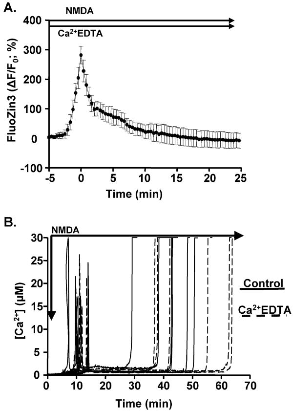
To assess the source(s) of Zn2+ responsible for FluoZin-3 signals, we examined the effects of selective chelation of Zn2+ in the extracellular space, by using Ca-EDTA. Ca-EDTA has high affinity for Zn2+ (KD=8×10-14M) (Lee et al., 2002), but unlike TPEN is not membrane permeable. Extended exposures to Ca-EDTA alone (up to 30 min, n=3) did not modify initial FluoZin-3 fluorescence levels (data not shown). This suggests that there is not substantial depletion of cytosolic Zn2+ as a result of extracellular chelation, in contrast to the described loss of fluorescence attributed to Zn2+ in synaptic vesicles by similar procedures (Frederickson et al., 2002). Figure 5A shows that (unlike TPEN) Ca-EDTA (1 mM) did not block initial intracellular Zn2+ increases during NMDA. Peak Zn2+ increases were of very similar amplitude (281±30%) and latency (6.4±1.1min, n=5) to those observed in control cells (Figure 4). Taken together with TPEN data, these results suggest that the source(s) of Zn2+ responsible for initial transients may be due to liberation from intracellular binding proteins. An alternative possibility is that Zn2+ may enter from the extracellular space from release sites very close to Zn2+ entry routes, since the relatively slow binding kinetics of Ca-EDTA (Kay, 2003) may not be sufficient rapid to chelate Zn2+ before neuronal entry (as considered in Dietz et al., 2008)).
Figure 5. Ca-EDTA did not reduce initial Zn2+ increases, nor significantly delay the dendritic Ca2+ overload response.
A: Slices were pre-exposed to Ca-EDTA for 20 min prior to NMDA, and the chelator was then maintained in the superfusate throughout (n=5). Initial Zn2+ increases were similar to those described above in control conditions (Fig 4), but on average recovered to baseline within ∼15min of the peak response, unlike the plateau in normal ACSF (Fig 4). B: In separate experiments, compound Fura-6F responses were not modified by Ca-EDTA. Control (solid lines) and Ca-EDTA-treated preparations (dashed lines) were interleaved, and did not reveal a significant delay in arrival of Ca2+ overload (p=0.2; n=6 each).
The recovery of FluoZin-3 fluorescence after initial transients was more complete in Ca-EDTA than under control conditions. Thus there was recovery to 7±29% by 15 min after the spike, compared to the larger (but variable) plateau levels observed in control conditions. This raises the possibility that sustained influx of Zn2+ from the extracellular space may contribute to the plateau responses in many cells. Ca-EDTA did not significantly reduce initial Fura-6F spikes (19.4±3.6 vs. 15.8±3.4μM, control and Ca-EDTA respectively; p=0.5, n=6 each). Likewise, the arrival of Ca2+ overload responses at somata was not significantly delayed (40.6±3.1 vs. 48.4±5.0 min, control and Ca-EDTA respectively, p=0.2, n=6 each).
8. Effects of Ca2+ removal on intracellular Zn2+ increases
We addressed the question of whether Zn2+ increases might be dependent on cytosolic Ca2+ accumulation. Extracellular Ca2+ was washed out (without addition of any chelators) 10 min prior to onset of NMDA exposure, and 2mM Ca2+ was not re-introduced until after initial spike responses should have been completed (i.e. 15 min after addition of NMDA). From measurements of Ca2+ in the recording chamber, full restoration of extracellular [Ca2+] required an additional ∼2 min.
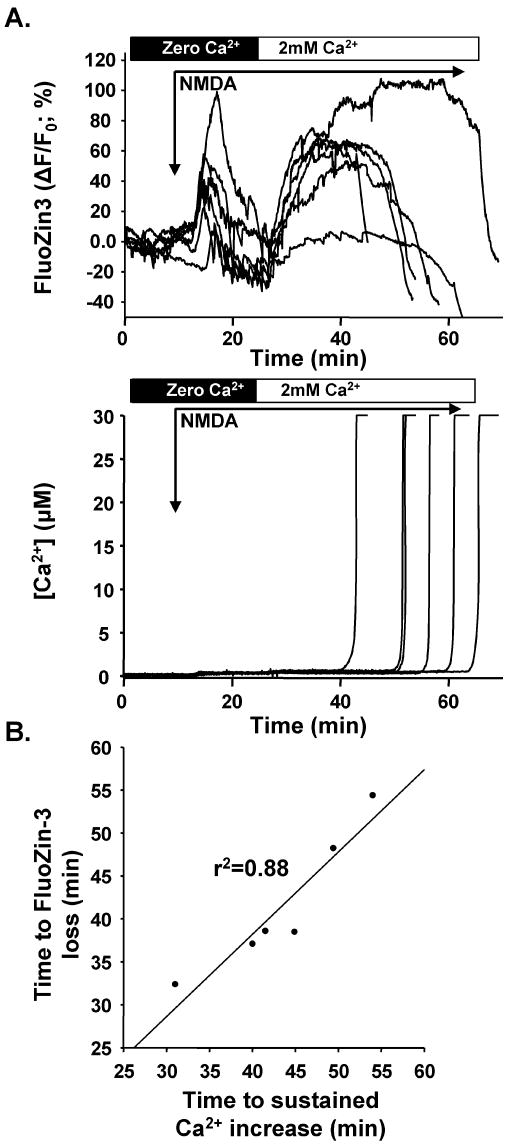
Figure 6 shows that reducing Ca2+ influx during the initial period reduced, but did not prevent intracellular Zn2+ increases. Initial transient FluoZin-3 increases reached 47.3±11.8 % (n=6), a substantially lower level than in control conditions (compare with Figs. 4A&5A). FluoZin-3 fluorescence then returned to near-baseline levels until reestablishment of 2mM extracellular Ca2+, when Zn2+ levels promptly increased to a new plateau level (mean 55.0±12.2% n=6). The plots in Figure 6A are aligned (upper panel: FluoZin-3, lower panel: Fura-6F) so that it can be seen that FluoZin-3 signals dropped sharply as sustained Ca2+ increases arrived at the soma. Figure 6A (lower panel) also shows a small residual increase in Fura-6F signal prior to re-introduction of Ca2+ (at t∼15 min), possibly the result of enhanced interaction of Zn2+ with Fura-6F in Ca2+-free conditions (see (Devinney et al., 2005)).
Figure 6. Effects of extracellular Ca2+ removal.
A: Near-simultaneous FluoZin-3 (top) and Fura-6F (bottom) records from co-loaded neurons. Extracellular Ca2+ was washed out for 10 min before exposure to NMDA in the nominally Ca2+-free solution. Subsequently 2mM Ca2+ was introduced 15 min into the NMDA exposure. In the nominal absence of extracellular Ca2+, initial FluoZin-3 increases were reduced but not abolished (compare with Figure 4). Reintroduction of Ca2+ produced prompt and sustained FluoZin-3 fluorescence increases. The lower panel of A shows that Ca2+ removal virtually abolished Fura-6F responses, until sustained increases were observed after reintroduction of Ca2+. B: As in control conditions, late FluoZin-3 loss was coincident with arrival of sustained Ca2+ increases in somata. The relationship between the time at which 50% FluoZin-3 fluorescence intensity (compared with plateau levels prior to arrival of Ca2+ overload) is lost is plotted against the time of half-maximal Ca2+ increase as the sustained Ca2+ response invaded somata for the 6 neurons shown in A.
Discussion
1. General
We draw three main conclusions from these results. Firstly, Zn2+ binding to Fura-6F does not appear responsible for previously-reported sustained Ca2+ increases, that originate in distal locations in apical dendrites and then propagate to somata. These dendritic signals appear to be due to excessive Ca2+ influx and accumulation, ultimately leading to Ca2+ overload in somata and causing acute injury to these neurons (Vander Jagt et al., 2008). Secondly, sustained NMDA exposure leads to significant increases in neuronal Zn2+ accumulation that are detectable using single neurons loaded with FluoZin-3. Zn2+ increases derive from endogenous sources within the slice, and are chelatable by TPEN but are largely unaffected by the slow membrane-impermeant chelator Ca-EDTA. Thirdly, Zn2+ increases appear to contribute to the progression of degenerative Ca2+ signals along apical dendrites, and we speculate that detrimental effects of Zn2+ accumulation leads to progressive metabolic dysfunction, ultimately contributing to regional loss of Ca2+ homeostasis.
2. Potential interference of Zn2+ with Fura-6F signals
A high-affinity interaction of Zn2+ with the widely-utilized Ca2+-indicator Fura-2 has been noted for more than 2 decades (Grynkiewicz et al., 1985; Cheng and Reynolds, 1998) and the high sensitivity of related Ca2+ indicators to Zn2+ has been reported in subsequent studies in free solution (Simons, 1993; Atar et al., 1995; Sensi et al., 1997; e.g. Hyrc et al., 2000; Devinney et al., 2005). However when these indicators are loaded into neurons at concentrations suitable for live cell imaging, the sensitivity to Zn2+ appears to decrease substantially. An important contributor to this effect is likely to be the relatively high indicator concentrations that are usually required for cellular studies, since as indicator concentration exceeds KD, the former makes progressively larger contributions to binding, and large differences in affinity become less meaningful (Dineley et al., 2002). When recordings are then made with physiological Ca2+ concentrations, the contribution of Zn2+ binding to the total indicator signal is small (Devinney et al., 2005). It is also possible that a range of intracellular constituents could also contribute to diminishing apparent Zn2+ sensitivity within cells (Marchi et al., 2000), although the impact of such interactions in neuronal recordings has been questioned (Devinney et al., 2005). These observations are important, since they emphasize that the Zn2+ and Ca2+ signals can be distinguished within neurons with existing fluorescent indicators, despite concerns raised from affinity studies in free solution (Martin et al., 2006). In a recent study of CA1 neurons co-loaded with Fura-6F and FluoZin-3, we detected Zn2+ increases during ouabain exposures without demonstrable increases in Fura-2 or Fura-6F, clearly showing that Zn2+ did not meaningfully interact with Fura-type indicators under those conditions (Dietz et al., 2008). However the present study is a little different, because NMDA produced an initial transient increase in both Fura-6F and FluoZin-3 signals, and selective intracellular chelation of Zn2+ significantly reduced the amplitude of Fura-6F spikes (see Fig 2A). Thus the potential contributions of Zn2+ to initial Fura-6F spikes needs to be considered (see below). In contrast to the initial Fura-6F spikes, it is important to emphasize that the sustained Fura-6F increases that propagate along dendritic processes can not be explained by Zn2+ accumulation, since they were only slowed but otherwise unaffected by TPEN and prevented by Ca2+ removal. The Ca2+ levels achieved during the overload response readily saturated the low affinity indicator used, and because of this it is not yet known whether TPEN may have some effect on the final Ca2+ concentrations that are achieved. However, it is likely rapid membrane compromise results in very high intracellular Ca2+ levels during this phase of the response, and that Ca2+ overload remains the logical explanation for these propagating Fura-6F signals associated with degeneration.
3. The early Fura-6F spike
We previously showed that initial Fura-6F spike responses were almost abolished by nominally Ca2+-free media (Vander Jagt et al., 2008) and see also Figure 6), however from this result alone it can not be concluded that Ca2+ is solely responsible for these transients. For example, it has been suggested that un-avoidable contamination of Ca2+ stock solutions with Zn2+ could make significant contributions to indicator signals (Kay, 2004) and it is thus possible that removal of Ca2+ from the media also removes an amount of Zn2+ (Dineley, 2007). This does not appear to be the case here, since the chelator Ca-EDTA (1mM) should remove contaminating Zn2+ from the ACSF, and did not reduce any component of Fura-6F signals.
Although Zn2+ binding to Fura-6F will be limited by the factors described above, Zn2+ may still bind to Fura-6F during initial spikes, but we note that this would not necessarily be expected to contribute to increases in Fura-6F ratio. The spectra produced by Ca2+ and Zn2+ binding to Fura indicators are slightly shifted relative to each other, so that the 380nm fluorescence changes are significantly less with Zn2+ than with Ca2+. This has the consequence of actually decreasing cumulative Fura ratio changes, when measurements are made in the presence of Ca2+ (Devinney et al., 2005). Under the conditions of our experiments, we would therefore predict that TPEN should produce an increase in Fura-6F ratio during the initial spike response, if Zn2+ binding to Fura-6F were significant during this event. In fact the converse was observed, suggesting that a stronger effect is that Zn2+ elevations somehow contribute to mechanisms underlying Ca2+ increases.
4. FluoZin-3 signals
In contrast to potential concerns about Zn2+ binding to Fura-indicators, the converse situation appears more simple, since a number of groups have reported that Ca2+ binding to FluoZin-3 is negligible, even with very high Ca2+ concentrations (Gee et al., 2002; Kay, 2004; Devinney et al., 2005). A recent report has questioned this conclusion (Martin et al., 2006) and possible reasons for the discrepancy in this report have been debated (Dineley, 2007). The present work supports a lack of Ca2+-sensitivity of FluoZin-3. Selective chelation of Zn2+ with TPEN completely abolished initial FluoZin-3 signals, despite the fact that large Ca2+ elevations (∼5μM) were still occurring and were readily detected with Fura-6F. Thus it appears that significant amounts of Zn2+ are mobilized together with Ca2+ during early transient responses.
Figure 4 demonstrates that Zn2+ levels remain above baseline in many neurons during the latent period before arrival of Ca2+ overload responses at the soma. These sustained elevations are probably not related to subsequent Ca2+ overload responses, since Ca-EDTA prevented the sustained Zn2+ elevations but did not have a significant effect on the timing of degenerative Ca2+ signals. This suggests that the early transient responses may be more important in degeneration.
After irrecoverable Ca2+ overload responses are established, neurons leak charged indicators and fluorescence is lost from the cell. This has been described previously for Ca2+ indicators, where sustained ratio increases (indicating persistently high Ca2+ levels) are observed in the face of loss of indicator fluorescence levels (Randall and Thayer, 1992; Shuttleworth and Connor, 2001; Avignone et al., 2005; Vander Jagt et al., 2008). A similar loss of FluoZin-3 fluorescence was observed in the present study, where FluoZin-3 signals in somata promptly decreased as the Ca2+ overload response approached the region (Figures 3 & 6). This loss of indicator fluorescence makes it difficult to determine whether Zn2+ levels actually increase first during the period of severe Ca2+ overload, or whether Zn2+ elevations may occur after this time point as a consequence of Ca2+ toxicity. The development of ratiometric indicators for Zn2+ with appropriate sensitivity and selectivity to address this question would be helpful for future studies.
5. Contribution of Zn2+ to the mechanism(s) of degenerative signaling along dendrites
Prevention of intracellular Zn2+ accumulation with TPEN produced a relatively small, but significant delay in the arrival of dendritic Ca2+ increases at somata (Fig. 2), suggesting that intracellular Zn2+ increases do influence the initiation and/or propagation of mechanisms responsible for Ca2+ deregulation along the dendrite.
Zn2+ is known to inhibit NMDA receptors (Peters et al., 1987; Westbrook and Mayer, 1987). However, this can not explain the longer delay in Ca2+ overload response with TPEN, since an increase in NMDA activation and accelerated injury should result if this were the dominant effect of Zn2+ in these experiments. Zn2+-dependent inhibition of metabolism is a likely mechanism participating in the origin of the injurious Ca2+ overload seen here. We recently showed that the progression of Ca2+ overload along dendrites was not due to a “feed-forward” Ca2+-dependent mechanism, whereby excessive Ca2+ loading was initially responsible for causing deregulation of adjacent segments of dendrite. Instead, the metabolic consequences of excessive Na+ loading during extended NMDA exposures appeared to be a more likely candidate, with Ca2+ overload following, presumably after ATP supply in a region of dendrite became irrecoverably lost (Vander Jagt et al., 2008b). Zn2+ has been shown to inhibit mitochondrial function (Skulachev et al., 1967; Sensi et al., 2003; Gazaryan et al., 2007) and also impair glycolysis by inhibition of glyceraldyhyde-3 phosphate dehydrogenase (Sheline et al., 2000). Zn2+ could also contribute to Ca2+ overload by blocking Na+/K+ ATPase activity (Donaldson et al., 1971; Manzerra et al., 2001) and thereby contributing to excessive accumulation of intracellular Na+ and reversal of sodium-calcium exchangers. However, we have found that sodium-calcium exchange reversal does not appear to be a major Ca2+ influx pathway in this model of NMDA exposure (Dietz et al., 2007). As noted above, even with chelation of Zn2+, the responses are merely delayed and were not abolished. Thus any deleterious effects of Zn2+ impairing ATP re-supply are likely to be additive with Zn2+-independent mechanisms of ATP depletion, including persistent Na+-dependent activation of Na+/K+/ATPase activity.
6. Sources of Zn2+
As noted in the Introduction, mobilization of Zn2+ from presynaptic sources, and/or liberation from intracellular binding proteins are two major sources of endogenous Zn2+ that can be considered for the Zn2+ increases described here. Zn2+ accumulates in synaptic vesicles following accumulation through ZnT3 transporters (Palmiter et al., 1996), and can be liberated following synaptic stimulation in the hippocampus (Assaf and Chung, 1984; Howell et al., 1984; Qian and Noebels, 2006). It is possible that NMDA releases Zn2+ from synaptic vesicles in the CA1 region, and Zn2+ then is taken up by CA1 neurons. The fact that the extracellular Zn2+ chelator (Ca-EDTA) did not prevent Zn2+ increases tends to argue against this possibility, however it is possible that the kinetics of Ca-EDTA are too slow to prevent effective sequestration, if the synaptic release sites were very close to postsynaptic influx routes. Such a scenario was proposed recently to explain the lack of effect of Ca-EDTA on Zn2+ increases that were deduced to be due to influx from the extracellular space (Dietz et al., 2008). If synaptic release and influx were involved and followed by intracellular transport to the soma, then a reduction in Ca2+-dependent vesicle fusion during NMDA exposure could explain the inhibitory effect of Ca2+ removal on the amplitude of measured Zn2+ increases. We also note that a number of factors influence the availability of synaptically-released Zn2+ and it is quite possible that synaptic contributions to initial Zn2+ increases may be larger in preparations from older animals, or when recording temperatures are increased (Frederickson et al., 2006).
An alternative possibility is that the source of free intracellular Zn2+ in these studies may be predominantly from intracellular binding proteins. Neurons have relatively large stores of Zn2+ that are normally associated with binding proteins such as Metallothionein III. This Zn2+ can be readily mobilized by excitotoxic or oxidative insults (Aizenman et al., 2000; Lee et al., 2003; Sensi et al., 2003; Bossy-Wetzel et al., 2004). Micromolar Ca2+ increases could cause liberation of Zn2+ from intracellular binding sites and (together with synaptic effects discussed above) could contribute to the partial Ca2+-dependence of Zn2+ increases observed in the present work. Zn2+ release from metallothioneins could be triggered by reactive oxygen species generated in a Ca2+-dependent manner by mitochondria, nitric oxide synthase or phospholipase C (Frederickson et al., 2004; Dineley et al., 2008). In addition, Ca2+-dependent acidification caused by NMDA receptor activation could play a significant role, since intracellular pH decreases are well-established to mobilize Zn2+ from intracellular stores (Jiang et al., 2000; Frazzini et al., 2007).
7. Conclusion
Simultaneous imaging of changes in intracellular Ca2+ and Zn2+ in single CA1 neurons, using Fura-6F and FlouZin-3, revealed significant accumulation of both Zn2+ and Ca2+ during extended NMDA exposures. Excessive somatic Ca2+ accumulation (following apical dendrite Ca2+ deregulation) appeared to be ultimately responsible for neuronal injury, but Zn2+ increases may contribute to initiation and progression of degenerative Ca2+ signaling.
Acknowledgments
Supported by NIH grants NS051288 (C.W.S.) and NS36548 (J.H.W.).
Abbreviations
- ACSF
artificial cerebrospinal fluid
- DMSO
Dimethyl sulfoxide
- EDTA
Ethylenediaminetetraacetate
- NMDA
N-Methyl-D-aspartic acid
- TPEN
N,N,N′,N′-Tetrakis(2-pyridylmethyl) ethylenediamine
Footnotes
Publisher's Disclaimer: This is a PDF file of an unedited manuscript that has been accepted for publication. As a service to our customers we are providing this early version of the manuscript. The manuscript will undergo copyediting, typesetting, and review of the resulting proof before it is published in its final citable form. Please note that during the production process errors may be discovered which could affect the content, and all legal disclaimers that apply to the journal pertain.
References
- Aizenman E, Stout AK, Hartnett KA, Dineley KE, McLaughlin B, Reynolds IJ. Induction of neuronal apoptosis by thiol oxidation: putative role of intracellular zinc release. J Neurochem. 2000;75:1878–1888. doi: 10.1046/j.1471-4159.2000.0751878.x. [DOI] [PubMed] [Google Scholar]
- Arslan P, Di Virgilio F, Beltrame M, Tsien RY, Pozzan T. Cytosolic Ca2+ homeostasis in Ehrlich and Yoshida carcinomas. A new, membrane-permeant chelator of heavy metals reveals that these ascites tumor cell lines have normal cytosolic free Ca2+ J Biol Chem. 1985;260:2719–2727. [PubMed] [Google Scholar]
- Arundine M, Tymianski M. Molecular mechanisms of calcium-dependent neurodegeneration in excitotoxicity. Cell Calcium. 2003;34:325–337. doi: 10.1016/s0143-4160(03)00141-6. [DOI] [PubMed] [Google Scholar]
- Assaf SY, Chung SH. Release of endogenous Zn2+ from brain tissue during activity. Nature. 1984;308:734–736. doi: 10.1038/308734a0. [DOI] [PubMed] [Google Scholar]
- Atar D, Backx PH, Appel MM, Gao WD, Marban E. Excitation-transcription coupling mediated by zinc influx through voltage-dependent calcium channels. J Biol Chem. 1995;270:2473–2477. doi: 10.1074/jbc.270.6.2473. [DOI] [PubMed] [Google Scholar]
- Avignone E, Frenguelli BG, Irving AJ. Differential responses to NMDA receptor activation in rat hippocampal interneurons and pyramidal cells may underlie enhanced pyramidal cell vulnerability. Eur J Neurosci. 2005;22:3077–3090. doi: 10.1111/j.1460-9568.2005.04497.x. [DOI] [PubMed] [Google Scholar]
- Bossy-Wetzel E, Talantova MV, Lee WD, Scholzke MN, Harrop A, Mathews E, Gotz T, Han J, Ellisman MH, Perkins GA, Lipton SA. Crosstalk between nitric oxide and zinc pathways to neuronal cell death involving mitochondrial dysfunction and p38-activated K+ channels. Neuron. 2004;41:351–365. doi: 10.1016/s0896-6273(04)00015-7. [DOI] [PubMed] [Google Scholar]
- Cheng C, Reynolds IJ. Calcium-sensitive fluorescent dyes can report increases in intracellular free zinc concentration in cultured forebrain neurons. J Neurochem. 1998;71:2401–2410. doi: 10.1046/j.1471-4159.1998.71062401.x. [DOI] [PubMed] [Google Scholar]
- Choi DW. Calcium-mediated neurotoxicity: relationship to specific channel types and role in ischemic damage. Trends Neurosci. 1988;11:465–469. doi: 10.1016/0166-2236(88)90200-7. [DOI] [PubMed] [Google Scholar]
- Choi DW, Koh JY. Zinc and brain injury. Annu Rev Neurosci. 1998;21:347–375. doi: 10.1146/annurev.neuro.21.1.347. [DOI] [PubMed] [Google Scholar]
- Christine CW, Choi DW. Effect of zinc on NMDA receptor-mediated channel currents in cortical neurons. J Neurosci. 1990;10:108–116. doi: 10.1523/JNEUROSCI.10-01-00108.1990. [DOI] [PMC free article] [PubMed] [Google Scholar]
- Connor JA, Shuttleworth CW. Intracellular Ca2+ signals underlying rapid and delayed neuronal death in mature CNS neurons. In: Lin RCS, editor. New concepts in cerebral ischemia. CRC Press; 2001. pp. 113–134. [Google Scholar]
- Devinney MJ, 2nd, Reynolds IJ, Dineley KE. Simultaneous detection of intracellular free calcium and zinc using fura-2FF and FluoZin-3. Cell Calcium. 2005;37:225–232. doi: 10.1016/j.ceca.2004.10.003. [DOI] [PubMed] [Google Scholar]
- Dietz RM, Kiedrowski L, Shuttleworth CW. Contribution of Na(+)/Ca(2+) exchange to excessive Ca(2+) loading in dendrites and somata of CA1 neurons in acute slice. Hippocampus. 2007;17:1049–1059. doi: 10.1002/hipo.20336. [DOI] [PubMed] [Google Scholar]
- Dietz RM, Weiss JH, Shuttleworth CW. Zn2+ influx is critical for some forms of spreading depression in brain slices. J Neurosci. 2008;28:8014–8024. doi: 10.1523/JNEUROSCI.0765-08.2008. [DOI] [PMC free article] [PubMed] [Google Scholar]
- Dineley KE. On the use of fluorescent probes to distinguish Ca2+ from Zn2+ in models of excitotoxicity. Cell Calcium. 2007;42:341–342. doi: 10.1016/j.ceca.2007.01.004. author reply 343-344. [DOI] [PubMed] [Google Scholar]
- Dineley KE, Malaiyandi LM, Reynolds IJ. A reevaluation of neuronal zinc measurements: artifacts associated with high intracellular dye concentration. Mol Pharmacol. 2002;62:618–627. doi: 10.1124/mol.62.3.618. [DOI] [PubMed] [Google Scholar]
- Dineley KE, Devinney MJ, 2nd, Zeak JA, Rintoul GL, Reynolds IJ. Glutamate mobilizes [Zn2+] through Ca2+ -dependent reactive oxygen species accumulation. J Neurochem. 2008;106:2184–2193. doi: 10.1111/j.1471-4159.2008.05536.x. [DOI] [PMC free article] [PubMed] [Google Scholar]
- Donaldson J, St Pierre T, Minnich J, Barbeau A. Seizures in rats associated with divalent cation inhibition of NA + -K + -ATP'ase. Can J Biochem. 1971;49:1217–1224. doi: 10.1139/o71-175. [DOI] [PubMed] [Google Scholar]
- Frazzini V, Rapposelli IG, Corona C, Rockabrand E, Canzoniero LM, Sensi SL. Mild Acidosis Enhances Ampa Receptor-Mediated Intracellular Zinc Mobilization in Cortical Neurons. Mol Med. 2007 doi: 10.2119/2007-00047.Frazzini. [DOI] [PMC free article] [PubMed] [Google Scholar]
- Frederickson CJ, Maret W, Cuajungco MP. Zinc and excitotoxic brain injury: a new model. Neuroscientist. 2004;10:18–25. doi: 10.1177/1073858403255840. [DOI] [PubMed] [Google Scholar]
- Frederickson CJ, Koh JY, Bush AI. The neurobiology of zinc in health and disease. Nat Rev Neurosci. 2005;6:449–462. doi: 10.1038/nrn1671. [DOI] [PubMed] [Google Scholar]
- Frederickson CJ, Suh SW, Koh JY, Cha YK, Thompson RB, LaBuda CJ, Balaji RV, Cuajungco MP. Depletion of intracellular zinc from neurons by use of an extracellular chelator in vivo and in vitro. J Histochem Cytochem. 2002;50:1659–1662. doi: 10.1177/002215540205001210. [DOI] [PubMed] [Google Scholar]
- Frederickson CJ, Giblin LJ, 3rd, Balaji RV, Masalha R, Frederickson CJ, Zeng Y, Lopez EV, Koh JY, Chorin U, Besser L, Hershfinkel M, Li Y, Thompson RB, Krezel A. Synaptic release of zinc from brain slices: factors governing release, imaging, and accurate calculation of concentration. J Neurosci Methods. 2006;154:19–29. doi: 10.1016/j.jneumeth.2005.11.014. [DOI] [PubMed] [Google Scholar]
- Gazaryan IG, Krasinskaya IP, Kristal BS, Brown AM. Zinc irreversibly damages major enzymes of energy production and antioxidant defense prior to mitochondrial permeability transition. J Biol Chem. 2007;282:24373–24380. doi: 10.1074/jbc.M611376200. [DOI] [PubMed] [Google Scholar]
- Gee KR, Zhou ZL, Ton-That D, Sensi SL, Weiss JH. Measuring zinc in living cells. A new generation of sensitive and selective fluorescent probes. Cell Calcium. 2002;31:245–251. doi: 10.1016/S0143-4160(02)00053-2. [DOI] [PubMed] [Google Scholar]
- Grynkiewicz G, Poenie M, Tsien RY. A new generation of Ca2+ indicators with greatly improved fluorescence properties. J Biol Chem. 1985;260:3440–3450. [PubMed] [Google Scholar]
- Hechtenberg S, Beyersmann D. Differential control of free calcium and free zinc levels in isolated bovine liver nuclei. Biochem J. 1993;289(Pt 3):757–760. doi: 10.1042/bj2890757. [DOI] [PMC free article] [PubMed] [Google Scholar]
- Howell GA, Welch MG, Frederickson CJ. Stimulation-induced uptake and release of zinc in hippocampal slices. Nature. 1984;308:736–738. doi: 10.1038/308736a0. [DOI] [PubMed] [Google Scholar]
- Hyrc KL, Bownik JM, Goldberg MP. Ionic selectivity of low-affinity ratiometric calcium indicators: mag-Fura-2, Fura-2FF and BTC. Cell Calcium. 2000;27:75–86. doi: 10.1054/ceca.1999.0092. [DOI] [PubMed] [Google Scholar]
- Jiang LJ, Vasak M, Vallee BL, Maret W. Zinc transfer potentials of the alpha - and beta-clusters of metallothionein are affected by domain interactions in the whole molecule. Proc Natl Acad Sci U S A. 2000;97:2503–2508. doi: 10.1073/pnas.97.6.2503. [DOI] [PMC free article] [PubMed] [Google Scholar]
- Kay AR. Evidence for chelatable zinc in the extracellular space of the hippocampus, but little evidence for synaptic release of Zn. J Neurosci. 2003;23:6847–6855. doi: 10.1523/JNEUROSCI.23-17-06847.2003. [DOI] [PMC free article] [PubMed] [Google Scholar]
- Kay AR. Detecting and minimizing zinc contamination in physiological solutions. BMC Physiol. 2004;4:4. doi: 10.1186/1472-6793-4-4. [DOI] [PMC free article] [PubMed] [Google Scholar]
- Lee JM, Zipfel GJ, Park KH, He YY, Hsu CY, Choi DW. Zinc translocation accelerates infarction after mild transient focal ischemia. Neuroscience. 2002;115:871–878. doi: 10.1016/s0306-4522(02)00513-4. [DOI] [PubMed] [Google Scholar]
- Lee JY, Kim JH, Palmiter RD, Koh JY. Zinc released from metallothionein-iii may contribute to hippocampal CA1 and thalamic neuronal death following acute brain injury. Exp Neurol. 2003;184:337–347. doi: 10.1016/s0014-4886(03)00382-0. [DOI] [PubMed] [Google Scholar]
- Manzerra P, Behrens MM, Canzoniero LM, Wang XQ, Heidinger V, Ichinose T, Yu SP, Choi DW. Zinc induces a Src family kinase-mediated up-regulation of NMDA receptor activity and excitotoxicity. Proc Natl Acad Sci U S A. 2001;98:11055–11061. doi: 10.1073/pnas.191353598. [DOI] [PMC free article] [PubMed] [Google Scholar]
- Marchi B, Burlando B, Panfoli I, Viarengo A. Interference of heavy metal cations with fluorescent Ca2+ probes does not affect Ca2+ measurements in living cells. Cell Calcium. 2000;28:225–231. doi: 10.1054/ceca.2000.0155. [DOI] [PubMed] [Google Scholar]
- Martin JL, Stork CJ, Li YV. Determining zinc with commonly used calcium and zinc fluorescent indicators, a question on calcium signals. Cell Calcium. 2006;40:393–402. doi: 10.1016/j.ceca.2006.04.008. [DOI] [PubMed] [Google Scholar]
- Nicholls DG, Budd SL. Mitochondria and neuronal survival. Physiol Rev. 2000;80:315–360. doi: 10.1152/physrev.2000.80.1.315. [DOI] [PubMed] [Google Scholar]
- Palmiter RD, Cole TB, Quaife CJ, Findley SD. ZnT-3, a putative transporter of zinc into synaptic vesicles. Proc Natl Acad Sci U S A. 1996;93:14934–14939. doi: 10.1073/pnas.93.25.14934. [DOI] [PMC free article] [PubMed] [Google Scholar]
- Peters S, Koh J, Choi DW. Zinc selectively blocks the action of N-methyl-D-aspartate on cortical neurons. Science. 1987;236:589–593. doi: 10.1126/science.2883728. [DOI] [PubMed] [Google Scholar]
- Qian J, Noebels JL. Exocytosis of vesicular zinc reveals persistent depression of neurotransmitter release during metabotropic glutamate receptor long-term depression at the hippocampal CA3-CA1 synapse. J Neurosci. 2006;26:6089–6095. doi: 10.1523/JNEUROSCI.0475-06.2006. [DOI] [PMC free article] [PubMed] [Google Scholar]
- Randall RD, Thayer SA. Glutamate-induced calcium transient triggers delayed calcium overload and neurotoxicity in rat hippocampal neurons. J Neurosci. 1992;12:1882–1895. doi: 10.1523/JNEUROSCI.12-05-01882.1992. [DOI] [PMC free article] [PubMed] [Google Scholar]
- Sensi SL, Canzoniero LM, Yu SP, Ying HS, Koh JY, Kerchner GA, Choi DW. Measurement of intracellular free zinc in living cortical neurons: routes of entry. J Neurosci. 1997;17:9554–9564. doi: 10.1523/JNEUROSCI.17-24-09554.1997. [DOI] [PMC free article] [PubMed] [Google Scholar]
- Sensi SL, Ton-That D, Sullivan PG, Jonas EA, Gee KR, Kaczmarek LK, Weiss JH. Modulation of mitochondrial function by endogenous Zn2+ pools. Proc Natl Acad Sci U S A. 2003;100:6157–6162. doi: 10.1073/pnas.1031598100. [DOI] [PMC free article] [PubMed] [Google Scholar]
- Sheline CT, Behrens MM, Choi DW. Zinc-induced cortical neuronal death: contribution of energy failure attributable to loss of NAD(+) and inhibition of glycolysis. J Neurosci. 2000;20:3139–3146. doi: 10.1523/JNEUROSCI.20-09-03139.2000. [DOI] [PMC free article] [PubMed] [Google Scholar]
- Shibuki K, Hishida R, Murakami H, Kudoh M, Kawaguchi T, Watanabe M, Watanabe S, Kouuchi T, Tanaka R. Dynamic imaging of somatosensory cortical activity in the rat visualized by flavoprotein autofluorescence. J Physiol. 2003;549:919–927. doi: 10.1113/jphysiol.2003.040709. [DOI] [PMC free article] [PubMed] [Google Scholar]
- Shuttleworth CW, Connor JA. Strain-dependent differences in calcium signaling predict excitotoxicity in murine hippocampal neurons. J Neurosci. 2001;21:4225–4236. doi: 10.1523/JNEUROSCI.21-12-04225.2001. [DOI] [PMC free article] [PubMed] [Google Scholar]
- Shuttleworth CW, Brennan AM, Connor JA. NAD(P)H fluorescence imaging of postsynaptic neuronal activation in murine hippocampal slices. J Neurosci. 2003;23:3196–3208. doi: 10.1523/JNEUROSCI.23-08-03196.2003. [DOI] [PMC free article] [PubMed] [Google Scholar]
- Simons TJ. Measurement of free Zn2+ ion concentration with the fluorescent probe mag-fura-2 (furaptra) J Biochem Biophys Methods. 1993;27:25–37. doi: 10.1016/0165-022x(93)90065-v. [DOI] [PubMed] [Google Scholar]
- Skulachev VP, Chistyakov VV, Jasaitis AA, Smirnova EG. Inhibition of the respiratory chain by zinc ions. Biochem Biophys Res Commun. 1967;26:1–6. doi: 10.1016/0006-291x(67)90242-2. [DOI] [PubMed] [Google Scholar]
- Vander Jagt TA, Weiss JH, Shuttleworth CW. Contribution of intracellular Zn2+ to progressive loss of Ca+ homeostasis in CA1 neurons in hippocampal slices. 2007 Meeting Planner Society for Neuroscience; 2007; 2007. Online 55.5. [Google Scholar]
- Vander Jagt TA, Connor JA, Shuttleworth CW. Localized loss of Ca2+ homeostasis in neuronal dendrites is a downstream consequence of metabolic compromise during extended NMDA exposures. J Neurosci. 2008;28:5029–5039. doi: 10.1523/JNEUROSCI.5069-07.2008. [DOI] [PMC free article] [PubMed] [Google Scholar]
- Weiss JH, Sensi SL, Koh JY. Zn(2+): a novel ionic mediator of neural injury in brain disease. Trends Pharmacol Sci. 2000;21:395–401. doi: 10.1016/s0165-6147(00)01541-8. [DOI] [PubMed] [Google Scholar]
- Weiss JH, Hartley DM, Koh JY, Choi DW. AMPA receptor activation potentiates zinc neurotoxicity. Neuron. 1993;10:43–49. doi: 10.1016/0896-6273(93)90240-r. [DOI] [PubMed] [Google Scholar]
- Westbrook GL, Mayer ML. Micromolar concentrations of Zn2+ antagonize NMDA and GABA responses of hippocampal neurons. Nature. 1987;328:640–643. doi: 10.1038/328640a0. [DOI] [PubMed] [Google Scholar]


