Abstract
Despite the type I insulin-like growth factor receptor (IGF-IR) being highly expressed in more than 80% of human lung tumors, a transgenic model of IGF-IR overexpression in the lung has not been created. We produced two novel transgenic mouse models in which IGF-IR is overexpressed in either lung type II alveolar cells (surfactant protein C [SPC]-IGFIR) or Clara cells (CCSP-IGFIR) in a doxycycline-inducible manner. Overexpression of IGF-IR in either cell type caused multifocal adenomatous alveolar hyperplasia with papillary and solid adenomas. These tumors expressed thyroid transcription factor 1 and Kruppel-like factor 5 in most tumor cells. Similar to our previous work with lung tumors that developed in the mouse mammary tumor virus-IGF-II transgenic mice, the lung tumors that develop in the SPC-IGFIR and CCSP-IGFIR transgenic mice expressed high levels of the cyclic adenosine monophosphate response element binding protein that was localized primarily to the nucleus. Although elevated IGF-IR expression can initiate lung tumor development, tumors can become independent of IGF-IR signaling as IGF-IR down-regulation in established tumors produced tumor regression in some, but not all, of the tumors. These findings implicate IGF-IR as an important initiator of lung tumorigenesis and suggest that the SPC-IGFIR and CCSP-IGFIR transgenic mice can be used to further our understanding of human lung cancer and the role IGF-IR plays in this disease.
Introduction
Lung cancer is the leading cause of cancer-related deaths for both men and women in Canada and the United States (Canadian Cancer Statistics 2007 and National Cancer Institute Statistics 2007). The devastatingly high mortality rate (approximately 80%) of this disease is largely attributable to difficulties in early diagnosis and lack of effective therapies [1–3].
Lung cancer is associated with a number of different epithelial cell types and broadly classified into small cell lung cancer (SCLC) and non-small cell lung cancer (NSCLC). Non-small cell lung cancer cells, which comprise approximately 75% of the cases of lung cancer, are further subdivided into squamous cell carcinoma, adenocarcinoma, and large cell carcinoma [4–7]. These tumors are grouped together based on their similar response to treatment. Squamous cell carcinomas frequently arise from segmental bronchi, whereas adenocarcinomas typically arise in peripheral regions of the lung and may express differentiated features of type II alveolar cells or Clara cells [5,8]. Large cell carcinomas frequently arise in the distal tubules. Small cell carcinomas are usually found in central endobronchial locations [6]. A number of gene abnormalities have been associated with NSCLC including mutations in p53 [9–14], c-erbB2 (Her2/neu) [15–18], and K-ras [19–23]. In addition to genetic alterations, there is evidence that inappropriate activation of the insulin-like growth factor (IGF) signaling may contribute to lung tumor development.
The IGF family consists of two ligands, insulin-like growth factor I (IGF-I) and insulin-like growth factor II (IGF-II), the IGF-I receptor (IGF-IR) and IGF-II receptor (IGF-IIR), and at least six IGF binding proteins (IGFBP-1 to IGFBP-6) [24–27]. Insulin-like growth factor signaling has been implicated in promoting growth, development, and metastasis in many different types of cancer (reviewed in [28–32]). In the normal lung, IGF-I and IGF-II are expressed by a number of lung cell types in including epithelial cells, fibroblasts and macrophages [33]. Insulin-like growth factors are expressed in the lung during alveolarization and are required for increasing proliferation of mesenchymal and epithelial cells during lung morphogenesis [34].
With respect to lung cancer, high plasma IGF-I levels and low plasma IGFBP-3 levels have been associated with increased lung cancer risk [35]. In addition, IGF-IR is expressed at high levels in 95% of human SCLCs and 80% of NSCLCs [36–38], whereas adenocarcinoma patients with a higher number of IGF-II-positive cells were reported to have a worse prognosis than patients with lung tumors containing low IGF-II staining [39]. Studies on human lung tumor cell lines have also implicated the IGF family in lung tumorigenesis because administration of IGF-I or IGF-II promotes proliferation of SCLC and NSCLC cell lines [40–42], whereas targeting the IGF-IR inhibits lung tumor cell proliferation and sensitizes lung tumor cells to chemotherapy and radiotherapy [42–48].
A more complete understanding of the function of the IGF family in lung tumorigenesis has been hampered by the paucity of appropriate animal models. Our laboratory has demonstrated that transgenic overexpression of IGF-II in the lung was sufficient to induce lung tumors [49]. These tumors were associated with enhanced activation of Erk1/2, p38 mitogen-activated protein kinase, and the transcription factor cAMP response element binding protein (CREB) [49]. The usefulness of the IGF-II transgenic model is limited by indirect transgene targeting (IGF-II transgene expression is regulated by the mouse mammary tumor virus [MMTV] promoter which directs transgene expression primarily to the mammary epithelium but can also drive expression in other tissues such as the lung), prolonged tumor latency (∼18 months), and incomplete penetrance of the tumor phenotype.
To overcome the aforementioned limitations of the IGF-II transgenic model and to provide a model to directly assess the importance of IGF signaling in lung tumorigenesis, our laboratory created transgenic mice that expressed high levels of the human IGF-IR complementary DNA in type II alveolar cells or Clara cells of the lung. Characterization of these lung-specific IGF-IR transgenic mice in the current study revealed that elevated IGF-IR expression in either type II alveolar or Clara cells induced epithelial hyperplasia and focal tumors. These tumors also expressed high levels of CREB, thyroid transcription factor 1 (TTF-1), and Kruppel-like factor 5 (KLF5). Interestingly, tumors initiated by IGF-IR can become independent of IGF-IR signaling. In these IGF-IR-independent tumors, CREB protein expression remained high suggesting that this protein may be critical for continued tumor growth.
Materials and Methods
Mice
Surfactant protein C (SPC)-reverse tetracycline transactivator (rtTA) and Clara cell secretory protein (CCSP)-rtTA mice were generated previously as described [50]. Tetracycline response element (TRE)-IGFIR transgenic mice were generated previously in our laboratory [51]. The SPC-rtTA and CCSP-rtTA transgenic mice were then mated with TRE-IGFIR transgenic mice producing the desired double transgenic mice designated as SPC-IGFIR and CCSP-IGFIR. Doxycycline-treated mice were fed chow supplemented with 2 g of doxycycline per kilogram of rodent chow (Bio-Serv, Frenchtown, NJ) beginning at 21 days of age. Mice were maintained and cared for following Canadian Council for Animal Care guidelines.
Western Blot Analysis
Western blot analysis was performed as described in Jones et al. [51] and Linnerth et al. [52]. All primary antibodies, anti-IGF-IR (R&D Systems, Inc., Minneapolis, MN), anti-phospho-IGFIR, anti-phospho-Akt, anti-Akt, anti-phospho-CREB, anti-CREB (Cell Signaling Technology, Beverly, MA), and antitubulin (Santa Cruz Technologies, Santa Cruz, CA), were used at a 1:1,000 dilution. Secondary antibodies (Cell Signaling Technology) were used at a 1:2,000 dilution. Images were captured on a FluorChem 9900 gel documentation system (Alpha Innotech, San Leandro, CA).
Immunohistochemistry and Immunofluorescence
Two different procedures were used for immunohistochemistry. Immunohistochemistry for TTF-1, KLF5, pro-SPC, and CCSP were performed in Dr. Jeffery Whitsett's laboratory using previously described methods [53–56]. Immunohistochemistry for IGF-IR, pAkt, and CREB were performed in our laboratory using previously described methods [52]. The primary antibodies were used at a 1:100 dilution, and the pAkt and CREB antibodies were obtained from Cell Signaling Technology, whereas the IGF-IR antibody was obtained from R&D Systems, Inc. Immunofluorescence was performed in our laboratory using previously described methods [52]. The pro-SPC and CCSP antibodies (Abcam, Cambridge, MA) were used at a 1:500 dilution for the immunofluorescence experiments. All experiments included primary and secondary antibody omission controls.
Tissue Array
A lung cancer tissue array (US Biomax, Inc., Rockville, MD) containing lung tumors from 192 patients and 8 cases of normal tissue was used for this study. The immunohistochemical procedure was previously described in Linnerth et al. [52]. The anti-CREB antibody (Cell Signaling Technology) was used at a dilution of 1:50 in 5% BSA. CREB staining intensity was measured using ImageScope analysis software (Aperio, Vista, CA) and categorized as less than 10% of the tumor cells staining positive for CREB, 11% to 20%, 21% to 30%, or more than 30% staining positive for CREB. CREB nuclear localization was scored by the investigator and was grouped into the classifications listed above.
Results
Generation and Characterization of Doxycycline-Inducible IGF-IR Mice
To prevent complications arising from altered gene expression during lung morphogenesis, we have used a doxycycline inducible system to express the IGF-IR transgene. This system uses two activator constructs (SPC-rtTA or CCSP-rtTA) and one target construct (TRE-IGFIR). The 3.7-kb human SPC gene promoter drives expression of the rtTA primarily in type II alveolar cells, and the 2.3-kb rat CCSP gene promoter drives expression of the rtTA primarily in Clara cells [50]. TRE-IGFIR transgenic mice have been described previously [51]. In the absence of doxycycline, the rtTA protein is unable to bind to the TRE, and thus, only transgenic mice bearing both an activator construct (SPC-rtTA or CCSP-rtTA) and the target construct (TRE-IGFIR) in the presence of doxycycline express the IGF-IR transgene.
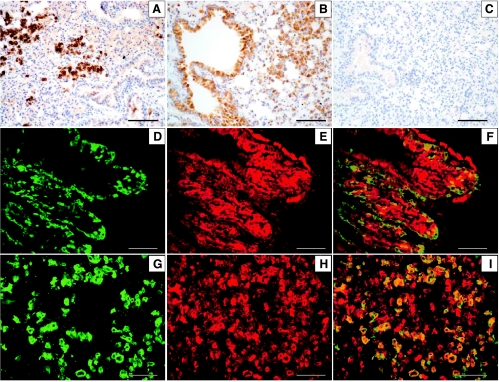
To determine the effect of IGF-IR expression in type II alveolar cells or Clara cells, SPC-IGFIR and CCSP-IGFIR transgenic mice were administered doxycycline beginning at 21 days of age. Lungs from SPC-IGFIR and CCSP-IGFIR were then collected at various intervals after the initiation of doxycycline treatment. At the earliest time point examined (1 month after the initiation of doxycycline treatment), cells expressing IGF-IR were observed in all doxycycline-treated SPC-IGFIR and CCSP-IGFIR transgenic mice. In SPC-IGFIR mice, alveolar epithelial cells were found to express high levels of IGF-IR (Figure 1A). In CCSP-IGFIR mice, cells expressing high levels of IGF-IR were found in the bronchiolar epithelium and in the alveolar epithelium (Figure 1B). Only low levels of IGF-IR expression were observed in wild-type or single transgenic mice treated with doxycycline (Figure 1C). Double transgenic mice not exposed to doxycycline displayed IGF-IR levels similar to lungs from wild-type mice (data not shown).
Figure 1.
Immunohistochemistry for IGF-IR in SPC-IGFIR (A), CCSP-IGFIR (B), or wild-type (C) mouse treated with doxycycline for 2 months. Immunofluorescence for IGF-IR (D) and CCSP (E) in a CCSP-IGFIR mouse and IGF-IR (G) and pro-SPC (H) in an SPC-IGFIR mouse treated with doxycycline. Merged images of IGF-IR/CCSP (F) and IGF-IR/SPC (I). Scale bars, 100 µm.
To further assess the cell type expressing high levels of IGF-IR, dual-labeling immunofluorescence was used. In CCSP-IGFIR transgenic mice, IGF-IR protein was found to be expressed in cells lining the bronchi that also stained positive for CCSP (Figure 1, D–F) and in alveolar epithelial cells (data not shown). In SPC-IGFIR transgenic mice, IGF-IR protein was expressed in pro-SPC-positive, type II alveolar cells (Figure 1, G–I).
IGF-IR Transgene Expression Induces Alveolar Epithelial Hyperplasia and Adenomas
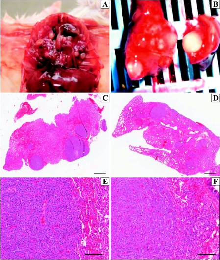
Macroscopic tumors were visible on the lung surface in both SPC-IGFIR and CCSP-IGFIR mice as early as 90 days after the initiation of IGF-IR transgene expression (data not shown). In SPC-IGFIR and CCSP-IGFIR mice treated with doxycycline for up to 9 months, macroscopically visible lung tumors (2–30 tumors) were detected in most mice. Macroscopic lesions presented as white, solid masses, varying in size and location on the lung (Figure 2, A and B). Histologically, lungs from all SPC-IGFIR and CCSP-IGFIR mice treated with doxycycline for 9 months displayed multifocal adenomatous alveolar hyperplasia (AAH) with papillary and solid adenomas. Bronchiolar hyperplasia was not observed in either genotype (Figure 2, C–F).
Figure 2.
(A, B) Macroscopic images of lung tumors from a 9-month-old SPC-IGFIR transgenic mouse. Hematoxylin and eosin-stained lung sections from a CCSP-IGFIR transgenicmouse (C, E) and an SPC-IGFIR transgenic mouse (D, F). Scale bars, 800 µm (C, D) and 100 µm (E, F).
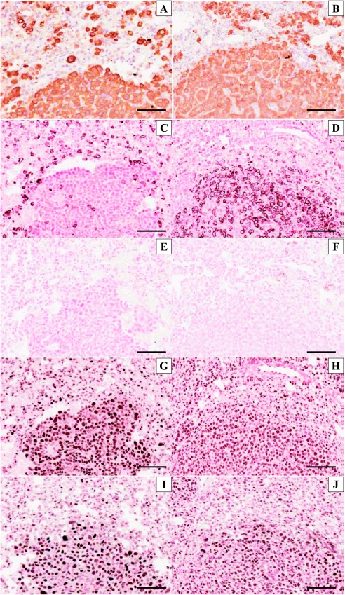
Immunohistochemistry for IGF-IR, pro-SPC, and CCSP revealed that the hyperplasia/tumors expressed high levels of IGF-IR (Figure 3, A and B), variable levels of pro-SPC (Figure 3, C and D), and no detectable CCSP protein (Figure 3, E and F). Thyroid transcription factor 1 (Figure 4, G and H) and KLF5 (Figure 3, I and J) were expressed in almost all of the tumor cells.
Figure 3.
Immunohistochemistry for IGF-IR (A, B), pro-SPC (C, D), CCSP (E, F), TTF-1 (G, H), and KLF5 (I, J) in a lung tumor froma doxycycline treated CCSP-IGFIR transgenicmouse (A, C, E, G, I) or a doxycycline-treated SPC-IGFIR transgenicmouse (B, D, F, H, J). Scale bars, 100 µm.
Figure 4.
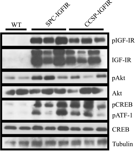
Western blot of phosphorylated IGF-IR (pIGF-IR), IGF-IR, phosphorylated Akt (pAkt), Akt, phosphorylated CREB (pCREB), CREB, and phosphorylated ATF-1 (pATF-1) in wild-type (WT), SPC-IGFIR, or CCSP-IGFIR transgenic mice administered doxycycline for 8 months. Tubulin served as a loading control.
IGF-IR-Induced Lung Tumors Contain High Levels of Nuclear CREB
Although there was some variability, most of the lung tissue collected from SPC-IGFIR or CCSP-IGFIR transgenic mice treated with doxycycline for 8 months contained higher levels of IGF-IR, phospho-IGFIR, phospho-Akt, and phospho-CREB compared with doxycycline-treated wild-type mice (Figure 4). The phospho-CREB antibody also detects phospho-ATF-1 (a CREB binding partner), and this phosphorylated protein was also elevated in the transgenic lung tissue compared with wild-type controls.
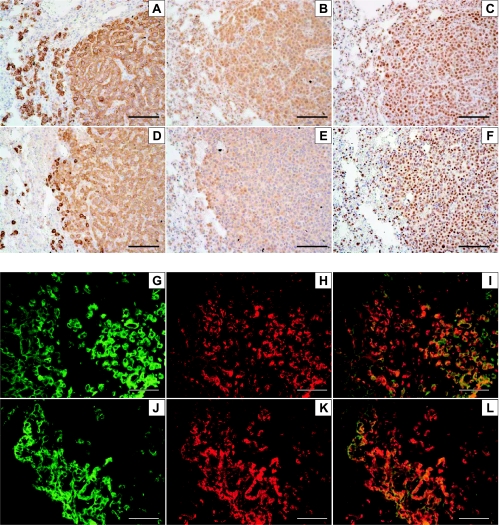
Immunohistochemistry and immunofluorescence revealed that IGF-IR (Figure 5, A and D), phospho-Akt (Figure 5, B and E), and phospho-CREB were highly expressed in the lung tumor cells compared with the normal surrounding cells (Figure 5, C and F). Phospho-p38 mitogen-activated protein kinase, but not pErk1/2, was also highly expressed in the lung tumor cells (data not shown). Tumor cells expressing high levels of phospho-Akt (Figure 5H) or phospho-CREB (Figure 5K) coexpressed high levels of IGF-IR (Figure 5, G, I, J, and L).
Figure 5.
Immunohistochemistry for IGF-IR (A, D), phosphorylated Akt (B, E), and phosphorylated CREB (C, F) in lung tumors from SPC-IGFIR (A–C) and CCSP-IGFIR (D–F) mice treated with doxycycline for at least 8 months. Immunofluorescence for IGF-IR (G, J), phosphorylated Akt (H), and phosphorylated CREB (K) in a lung tumor from an SPC-IGFIR transgenic mouse treated with doxycycline for at least 8 months. Merged images of IGF-IR/phosphorylated Akt (I) and IGF-IR/phosphorylated CREB (L). Scale bars, 100 µm.
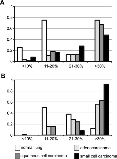
As high levels of CREB were observed in lung tumors arising in MMTV-IGF-II [49], SPC-IGFIR, and CCSP-IGFIR transgenic mice, CREB expression was examined in human lung tissue. Most of the human lung adenocarcinomas, squamous cell carcinomas, and small cell carcinomas expressed moderate or high levels of CREB (Figure 6A) with most of this CREB being localized to the nucleus (Figure 6B).
Figure 6.
Quantification of CREB protein levels (A) and nuclear localization (B) in normal lung or lung tumors cores.
Incomplete Tumor Regression after IGF-IR Down-regulation
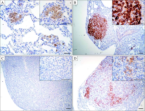
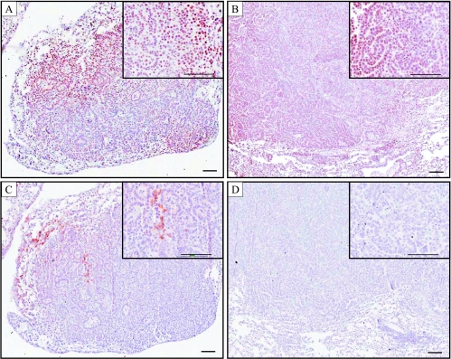
To determine whether IGF-IR-induced tumors remained dependent on IGF-IR signaling, doxycycline was withdrawn from SPC-IGFIR and CCSP-IGFIR transgenic mice after an 8-month treatment period. Lung tissue was then collected 96 hours, 2 weeks or 1 month after the removal of doxycycline. Of the three mice examined (two CCSP-IGFIR and one SPC-IGFIR) after a 96-hour doxycycline withdrawal, the lungs of all of the mice displayed either IGF-IR-positive cells or IGF-IR-positive tumors (data not shown). Of the three mice examined after a 2-week doxycycline withdrawal period, one CCSP-IGFIR transgenic mouse lung contained IGF-IR-positive cells and several IGF-IR-positive tumors, one SPC-IGFIR transgenic mouse lung contained IGF-IR-positive cells and at least one tumor with both IGF-IR-positive and -negative cells, whereas one CCSP-IGFIR transgenic mouse lung contained IGF-IR-positive tumors and a few areas that resembled completely regressed tumor lesions (data not shown). Eight mice (six SPC-IGFIR and two CCSP-IGFIR) were examined after a 1-month doxycycline withdrawal period. Lung tissue from one of the CCSP-IGFIR transgenic mice did not contain any IGF-IR-positive cells or apparent tumors but did contain several regions that resembled regressed tumor lesions (Figure 7A). These areas of tumor regression are similar to the mammary tumors that regressed after doxycycline withdrawal in MMTV-IGFIR transgenic mice (unpublished observations). Three of the mice (all SPC-IGFIR) contained only tumors that were primarily negative for IGF-IR staining (Figure 7C), whereas two of the mice (both SPC-IGFIR) contained either IGF-IR-positive cells or IGF-IR-positive tumors (Figure 7B). The remaining two mice (one SPC-IGFIR and one CCSP-IGFIR) contained both IGF-IR-positive and -negative tumors. Some of the tumors in the lungs of these mice also demonstrated a mixture of IGF-IR-positive and IGF-IR-negative cells (Figure 7D). Immunohistochemistry revealed that CREB (Figure 8, A and B) and KLF5 (data not shown) were expressed at high levels in tumors where IGF-IR levels were mixed (Figure 8C) or absent (Figure 8D).
Figure 7.
Immunohistochemistry for IGF-IR in the lungs from mice treated with doxycycline for 8 months followed by a 1 month withdrawal of doxycycline. (A) Mouse with apparent regressed tumor lesions, (B) a tumor containing primarily IGF-IR-positive cells, (C) a tumor containing exclusively IGF-IR-negative cells, and (D) a number containing both IGF-IR-positive and IGF-IR-negative cells. The inset is a higher magnification of the stained slide. Scale bars, 50 µm; insets, 100 µm.
Figure 8.
Immunohistochemistry for CREB (A, B) in lung tumors after doxycycline withdrawal that display mixed IGF-IR expression (C) or no IGF-IR expression (D).
Discussion
The IGF-IR has been associated with human SCLC and NSCLC in that the IGF-IR is highly expressed in approximately 95% of SCLC and 80% of NSCLC cases [36–38]. In addition, studies on human lung cancer cell lines demonstrated that IGF signaling promotes lung tumor proliferation and inhibits apoptosis, whereas down-regulation of IGF-IR inhibits lung tumor cell proliferation, promotes apoptosis, and sensitizes lung cancer cells to chemotherapy and radiotherapy [42–48]. Although these studies demonstrate that IGF signaling is an important regulator of lung tumorigenesis, they do not address whether IGF signaling mediates transformation of normal lung cells and is thus involved in tumor initiation or whether IGF-IR primarily mediates lung tumor progression. Our characterization of SPC-IGFIR and CCSP-IGFIR transgenic mice demonstrated that elevated levels of IGF-IR and hyperactive IGF-IR signaling are sufficient to promote lung tumor initiation. This finding supports our previous transgenic model where elevated levels of IGF-II induced pulmonary tumorigenesis [49].
Interestingly, IGF-IR overexpression in Clara cells produced solid adenomas that were largely negative for the Clara cell marker CCSP. Loss of CCSP expression has been observed in at least two other transgenic models of lung tumorigenesis, the CC10TAg transgenic mice and the CCSP-rtTA-FGF10 transgenic mice [57,58]. There are at least three possible explanations for the loss of Clara cell protein markers in lung tumors arising from CCSP-targeted transgenes. First, it is possible that upon transformation, Clara cells undergo changes in gene expression leading to very low levels of the Clara cell marker [59]. An alternative explanation is that the CCSP promoter is expressed in both Clara cells and type II alveolar cells, and only type II alveolar cells are transformed and thus give rise to tumors [57]. As we observed elevated IGF-IR levels in Clara cells as well as cells in the lung periphery in the CCSP-IGFIR transgenic mice, and there is no apparent bronchiolar hyperplasia, it is possible that type II alveolar cells are the cells of origin for the tumors in both the SPC-IGFIR and CCSP-IGFIR transgenic mice. However, the solid adenomas in the CCSP-IGFIR transgenic mice are largely negative for SPC staining, whereas the adenomas in the SPC-IGFIR transgenic mice are primarily SPC-positive. A final explanation involves putative bronchioalveolar stem cells (BASCs). Bronchioalveolar stem cells are cells located between the conducting and respiratory epithelium in a region termed the bronchioalveolar duct junction [60]. Bronchioalveolar stem cells have been identified as cells expressing both SPC and CCSP proteins that increase in number in response to lung injury. Therefore, it is possible that the IGF-IR transgene may be expressed in the proposed BASC “stem” cells as well as in Clara cells and type II alveolar cells. Proliferation of BASC cells expressing high levels of IGF-IR may differentiate toward lung cells expressing SPC or neither SPC nor CCSP.
Most of the tumor cells in both SPC-IGFIR and CCSP-IGFIR transgenic mice expressed high levels of CREB regardless of their expression of SPC or CCSP proteins. High levels of CREB were also a consistent finding in our MMTV-IGF-II induced lung tumors [49]. The CREB in all three transgenicmodels primarily localized to the nuclei of the tumor cells suggesting that the CREB was phosphorylated. Moreover, our study found a high level of CREB and nuclear localized CREB in human lung cancer samples suggesting that CREB is an important mediator of lung tumorigenesis.
cAMP response element binding protein was first implicated in lung tumorigenesis by Wardlaw et al. [61] when this group demonstrated that lung tumors induced by NNK [4-(methylnitrosamino)-1-(3-pyridal)-1-butanone] had mutations in the CREB/ATF binding site which resulted in the inhibition of COX-2 promoter activity. A number of additional studies have also demonstrated that CREB is an important mediator of lung tumorigenesis in human cell lines and human lung tumor samples. Aggarwal et al. [62] reported that targeting CREB using a R0-31-8220 inhibitor resulted in suppression of cell growth and tumorigenicity in NSCLC cells. We demonstrated that dominant-negative CREB vectors inhibited soft agar growth of NSCLC cell lines [52]. More recently, a study has shown that CREB is elevated in human NSCLC and is associated with a poor prognosis [63]. Our studies on CREB in human lung tumor tissue agree with this study [63] in that CREB staining was higher in the lung tumors compared with normal tissue. Thus, CREB is emerging as an important regulator of human lung tumorigenesis and may serve as a therapeutic target or biomarker for lung cancer; two areas being actively investigated in our SPC-IGFIR and CCSP-IGFIR transgenic mice.
In addition to CREB, TTF-1 and KLF5 were highly expressed in a majority of tumor cells in the SPC-IGFIR and CCSP-IGFIR mice. Thyroid transcription factor 1, a transcription factor critical for airway formation and epithelial differentiation, is often expressed at high levels in NSCLC [64–70]. In contrast, KLF5 has not previously been described in lung tumors. Kruppel-like factor 5 is a member of the zinc finger transcription factor family Sp1 and was originally identified in the intestinal epithelium [53]. Since its discovery, KLF5 has also been found in the tongue, epidermis, trachea, and bronchial epithelial cells. The importance of KLF5 in normal lung development was demonstrated in mice where the KLF5 gene was conditionally deleted in lung epithelial cells [53]. Loss of lung epithelial KLF5 disrupted lung development during the saccular stage, and mice died of respiratory distress immediately after birth [53]. The saccules of the KLF5 null mice were hypercellular and were not as dilated as the saccules of lungs from wild-type mice. Interestingly, a similar phenotype was observed in the lungs of mice lacking IGF-II [71]. Although KLF5 has not been associated with lung tumorigenesis, it has been observed in colon, gastric, and intestinal tumors [72–74]. The function of KLF5 in IGF-IR-induced lung tumorigenesis and whether IGF-IR signaling directly regulates KLF5 expression is currently under investigation in our laboratory.
Lung tumor development in transgenic mice that express high levels of IGF-IR demonstrates the importance of IGF-IR in tumor initiation. However, the IGF-IR may only represent a viable therapeutic target if the lung tumors remain dependent on IGF-IR expression for continued growth and survival. Down-regulation of IGF-IR transgene expression (through doxycycline withdrawal) in mice treated with doxycycline for 8 months revealed that some tumor cells remained despite little or no IGF-IR expression. Because our IGF-IR antibody detects endogenous and transgenic IGF-IR, these tumors remained in the absence of elevated IGF-IR expression. Intriguingly, CREB and KLF5 are still expressed in these IGF-IR-negative tumors suggesting that they may be important regulators of lung tumorigenesis. These findings suggest that some tumors that are initiated by IGF-IR overexpression acquire additional genetic alterations that render the tumor cells independent of IGF-IR signaling for growth and survival. In the case of the lung tumors, this IGF-IR independence may be associated with CREB and/or KLF5 activation through an alternative signaling pathway. In our mammary tumor model, tumors initiated by IGF-IR overexpression also display IGF-IR-independent tumor growth [75] indicating that this escape from IGF-IR dependence is not restricted to lung tumors. Development of IGF-IR independence has important clinical ramifications because it suggests that some lung tumors could either acquire or be intrinsically resistant to IGF-IR targeting agents.
In summary, using IGF-IR transgenic mice, we have shown that IGF-IR overexpression in type II alveolar cells or Clara cells is sufficient to initiate lung tumor development; however, continued growth of these lung tumors is not explicitly dependent on continued IGF-IR expression. Two proteins that may mediate IGF-IR induced lung tumorigenesis and possibly tumor escape from IGF-IR dependence are CREB and KLF5. Therefore, CREB and KLF5 may represent more viable therapeutic targets for the treatment of lung cancer than IGF-IR.
Acknowledgments
The authors thank the technical assistance of Michelle Ross, Helen Coates, and the Pulmonary Biology Morphology Core at Cincinnati Children's Hospital Medical Center directed by Susan Wert. The pathology reports on some of the lung tumors were provided by Kathryn Wikenheiser-Brokamp (Cincinnati Children's Hospital Medical Center).
Abbreviations
- AAH
adenomatous alveolar hyperplasia
- BASC
bronchioalveolar stem cell
- CREB
cyclic adenosine monophosphate response element binding protein
- CCSP
Clara cell secretory protein
- IGF
insulin-like growth factor
- IGFBP
insulin-like growth factor binding protein
- IGF-IR
type I insulin-like growth factor receptor
- KLF5
Kruppel-like factor 5
- MMTV
mouse mammary tumor virus
- NSCLC
non-small cell lung cancer
- SCLC
small cell lung cancer
- SPC
surfactant protein C
- TTF-1
thyroid transcription factor 1
- TRE
tetracycline response element
- rtTA
reverse tetracycline transactivator
Footnotes
This work was supported by a Canadian Institutes of Health Research operating grant awarded to R.A.M. and a Natural Sciences and Engineering Research Council scholarship awarded to N.M.L. and by the National Institutes of Health (HL090156) to J.A.W.
References
- 1.Butnor KJ. Avoiding underdiagnosis, overdiagnosis, and misdiagnosis of lung carcinoma. Arch Pathol Lab Med. 2008;132:1118–1132. doi: 10.5858/2008-132-1118-AUOAMO. [DOI] [PubMed] [Google Scholar]
- 2.Molina JR, Yang P, Cassivi SD, Schild SE, Adjei AA. Non-small cell lung cancer: epidemiology, risk factors, treatment, and survivorship. Mayo Clin Proc. 2008;83:584–594. doi: 10.4065/83.5.584. [DOI] [PMC free article] [PubMed] [Google Scholar]
- 3.Dubey S, Powell CA. Update in lung cancer 2007. Am J Respir Crit Care Med. 2008;177:941–946. doi: 10.1164/rccm.200801-107UP. [DOI] [PMC free article] [PubMed] [Google Scholar]
- 4.Beasley MB, Brambilla E, Travis WD. The 2004 World Health Organization classification of lung tumors. Semin Roentgenol. 2005;40:90–97. doi: 10.1053/j.ro.2005.01.001. [DOI] [PubMed] [Google Scholar]
- 5.Maggiore C, Mule A, Fadda G, Rossi ED, Lauriola L, Vecchio FM, Capelli A. Histological classification of lung cancer. Rays. 2004;29:353–355. [PubMed] [Google Scholar]
- 6.Giaccone G, Smit E. Lung cancer. Cancer Chemother Biol Response Modif. 2003;21:445–483. doi: 10.1016/s0921-4410(03)21022-x. [DOI] [PubMed] [Google Scholar]
- 7.Watanabe Y. TNM classification for lung cancer. Ann Thorac Cardiovasc Surg. 2003;9:343–350. [PubMed] [Google Scholar]
- 8.Gazdar AF, Linnoila RI. The pathology of lung cancer—changing concepts and newer diagnostic techniques. Semin Oncol. 1988;15:215–225. [PubMed] [Google Scholar]
- 9.Rom WN, Hay JG, Lee TC, Jiang Y, Tchou-Wong KM. Molecular and genetic aspects of lung cancer. Am J Respir Crit Care Med. 2000;161:1355–1367. doi: 10.1164/ajrccm.161.4.9908012. [DOI] [PubMed] [Google Scholar]
- 10.Harris CC, Hollstein M. Clinical implications of the p53 tumor-suppressor gene. N Engl J Med. 1993;329:1318–1327. doi: 10.1056/NEJM199310283291807. [DOI] [PubMed] [Google Scholar]
- 11.Levine AJ. p53, the cellular gatekeeper for growth and division. Cell. 1997;88:323–331. doi: 10.1016/s0092-8674(00)81871-1. [DOI] [PubMed] [Google Scholar]
- 12.Quinlan DC, Davidson AG, Summers CL, Warden HE, Doshi HM. Accumulation of p53 protein correlates with a poor prognosis in human lung cancer. Cancer Res. 1992;52:4828–4831. [PubMed] [Google Scholar]
- 13.Carbone DP, Minna JD. In vivo gene therapy of human lung cancer using wild-type p53 delivered by retrovirus. J Natl Cancer Inst. 1994;86:1437–1438. doi: 10.1093/jnci/86.19.1437. [DOI] [PubMed] [Google Scholar]
- 14.Fukuyama Y, Mitsudomi T, Sugio K, Ishida T, Akazawa K, Sugimachi K. K-ras and p53 mutations are an independent unfavourable prognostic indicator in patients with non-small-cell lung cancer. Br J Cancer. 1997;75:1125–1130. doi: 10.1038/bjc.1997.194. [DOI] [PMC free article] [PubMed] [Google Scholar]
- 15.Pastorino U, Andreola S, Tagliabue E, Pezzella F, Incarbone M, Sozzi G, Buyse M, Menard S, Pierotti M, Rilke F. Immunocytochemical markers in stage I lung cancer: relevance to prognosis. J Clin Oncol. 1997;15:2858–2865. doi: 10.1200/JCO.1997.15.8.2858. [DOI] [PubMed] [Google Scholar]
- 16.Veale D, Kerr N, Gibson GJ, Kelly PJ, Harris AL. The relationship of quantitative epidermal growth factor receptor expression in non-small cell lung cancer to long term survival. Br J Cancer. 1993;68:162–165. doi: 10.1038/bjc.1993.306. [DOI] [PMC free article] [PubMed] [Google Scholar]
- 17.Fontanini G, Boldrini L, Vignati S, Chine S, Basolo F, Silvestri V, Lucchi M, Mussi A, Angeletti CA, Bevilacqua G. Bcl2 and p53 regulate vascular endothelial growth factor (VEGF)-mediated angiogenesis in non-small cell lung carcinoma. Eur J Cancer. 1998;34:718–723. doi: 10.1016/s0959-8049(97)10145-9. [DOI] [PubMed] [Google Scholar]
- 18.Giatromanolaki A, Gorgoulis V, Chetty R, Koukourakis MI, Whitehouse R, Kittas C, Veslemes M, Gatter KC, Iordanoglou I. C-erbB-2 oncoprotein expression in operable non-small cell lung cancer. Anticancer Res. 1996;16:987–993. [PubMed] [Google Scholar]
- 19.Slebos RJ, Kibbelaar RE, Dalesio O, Kooistra A, Stam J, Meijer CJ, Wagenaar SS, Vanderschueren RG, van Zandwijk N, Mooi WJ. K-ras oncogene activation as a prognostic marker in adenocarcinoma of the lung. N Engl J Med. 1990;323:561–565. doi: 10.1056/NEJM199008303230902. [DOI] [PubMed] [Google Scholar]
- 20.Rosell R, Li S, Skacel Z, Mate JL, Maestre J, Canela M, Tolosa E, Armengol P, Barnadas A, Ariza A. Prognostic impact of mutated K-ras gene in surgically resected non-small cell lung cancer patients. Oncogene. 1993;8:2407–2412. [PubMed] [Google Scholar]
- 21.Westra WH, Slebos RJ, Offerhaus GJ, Goodman SN, Evers SG, Kensler TW, Askin FB, Rodenhuis S, Hruban RH. K-ras oncogene activation in lung adenocarcinomas from former smokers. Evidence that K-ras mutations are an early and irreversible event in the development of adenocarcinoma of the lung. Cancer. 1993;72:432–438. doi: 10.1002/1097-0142(19930715)72:2<432::aid-cncr2820720219>3.0.co;2-#. [DOI] [PubMed] [Google Scholar]
- 22.Noda N, Matsuzoe D, Konno T, Kawahara K, Yamashita Y, Shirakusa T. Risk for K-ras gene mutations in smoking-induced lung cancer is associated with cytochrome P4501A1 and glutathione S-transferase micro1 polymorphisms. Oncol Rep. 2004;12:773–779. [PubMed] [Google Scholar]
- 23.Rodenhuis S, Slebos RJ. The ras oncogenes in human lung cancer. Am Rev Respir Dis. 1990;142:S27–S30. doi: 10.1164/ajrccm/142.6_Pt_2.S27. [DOI] [PubMed] [Google Scholar]
- 24.Werner H, Le Roith D. New concepts in regulation and function of the insulin-like growth factors: implications for understanding normal growth and neoplasia. Cell Mol Life Sci. 2000;57:932–942. doi: 10.1007/PL00000735. [DOI] [PMC free article] [PubMed] [Google Scholar]
- 25.Werner H, Le Roith D. The role of the insulin-like growth factor system in human cancer. Adv Cancer Res. 1996;68:183–223. doi: 10.1016/s0065-230x(08)60354-1. [DOI] [PubMed] [Google Scholar]
- 26.Wetterau LA, Moore MG, Lee KW, Shim ML, Cohen P. Novel aspects of the insulin-like growth factor binding proteins. Mol Genet Metab. 1999;68:161–181. doi: 10.1006/mgme.1999.2920. [DOI] [PubMed] [Google Scholar]
- 27.Wood TL, Yee D. IGFs and IGFBPs in the normal mammary gland and in breast cancer. J Mam Gland Biol Neoplasia. 2000;5:1–5. doi: 10.1023/a:1009580913795. [DOI] [PubMed] [Google Scholar]
- 28.Frasca F, Pandini G, Scalia P, Sciacca L, Mineo R, Costantino A, Goldfine ID, Belfiore A, Vigneri R. Insulin receptor isoform A, a newly recognized, high-affinity insulin-like growth factor II receptor in fetal and cancer cells. Mol Cell Biol. 1999;19:3278–3288. doi: 10.1128/mcb.19.5.3278. [DOI] [PMC free article] [PubMed] [Google Scholar]
- 29.Yuen JS, Macaulay VM. Targeting the type 1 insulin-like growth factor receptor as a treatment for cancer. Expert Opin Ther Targets. 2008;12:589–603. doi: 10.1517/14728222.12.5.589. [DOI] [PubMed] [Google Scholar]
- 30.Ryan PD, Goss PE. The emerging role of the insulin-like growth factor pathway as a therapeutic target in cancer. Oncologist. 2008;13:16–24. doi: 10.1634/theoncologist.2007-0199. [DOI] [PubMed] [Google Scholar]
- 31.Tao Y, Pinzi V, Bourhis J, Deutsch E. Mechanisms of disease: signaling of the insulin-like growth factor 1 receptor pathway—therapeutic perspectives in cancer. Nat Clin Pract Oncol. 2007;4:591–602. doi: 10.1038/ncponc0934. [DOI] [PubMed] [Google Scholar]
- 32.Hartog H, Wesseling J, Boezen HM, van der Graaf WT. The insulin-like growth factor 1 receptor in cancer: old focus, new future. Eur J Cancer. 2007;43:1895–1904. doi: 10.1016/j.ejca.2007.05.021. [DOI] [PubMed] [Google Scholar]
- 33.Narasaraju TA, Chen H, Weng T, Bhaskaran M, Jin N, Chen J, Chen Z, Chinoy MR, Liu L. Expression profile of IGF system during lung injury and recovery in rats exposed to hyperoxia: a possible role of IGF-1 in alveolar epithelial cell proliferation and differentiation. J Cell Biochem. 2005;33:561–565. doi: 10.1002/jcb.20653. [DOI] [PubMed] [Google Scholar]
- 34.Moats-Staats BM, Price WA, Xu L, Jarvis HW, Stiles AD. Regulation of the insulin-like growth factor system during normal rat lung development. Am J Respir Cell Mol Biol. 1995;12:56–64. doi: 10.1165/ajrcmb.12.1.7529031. [DOI] [PubMed] [Google Scholar]
- 35.Yu H, Spitz MR, Mistry J, Gu J, Hong WK, Wu X. Plasma levels of insulin-like growth factor-I and lung cancer risk: a case-control analysis. J Natl Cancer Inst. 1999;91:151–156. doi: 10.1093/jnci/91.2.151. [DOI] [PubMed] [Google Scholar]
- 36.Reeve JG, Morgan J, Schwander J, Bleehen NM. Role for membrane and secreted insulin-like growth factor-binding protein-2 in the regulation of insulin-like growth factor action in lung tumors. Cancer Res. 1993;53:4680–4685. [PubMed] [Google Scholar]
- 37.Quinn KA, Treston AM, Unsworth EJ, Miller MJ, Vos M, Grimley C, Battey J, Mulshine JL, Cuttitta F. Insulin-like growth factor expression in human cancer cell lines. J Biol Chem. 1996;271:11477–11483. doi: 10.1074/jbc.271.19.11477. [DOI] [PubMed] [Google Scholar]
- 38.Viktorsson K, De Petris L, Lewensohn R. The role of p53 in treatment responses of lung cancer. Biochem Biophys Res Commun. 2005;331:868–880. doi: 10.1016/j.bbrc.2005.03.192. [DOI] [PubMed] [Google Scholar]
- 39.Takanami I, Imamuma T, Hashizume T, Kikuchi K, Yamamoto Y, Yamamoto T, Kodaira S. Insulin-like growth factor-II as a prognostic factor in pulmonary adenocarcinoma. J Surg Oncol. 1996;61:205–208. doi: 10.1002/(SICI)1096-9098(199603)61:3<205::AID-JSO8>3.0.CO;2-F. [DOI] [PubMed] [Google Scholar]
- 40.Nakanishi Y, Mulshine JL, Kasprzyk PG, Natale RB, Maneckjee R, Avis I, Treston AM, Gazdar AF, Minna JD, Cuttitta F. Insulin-like growth factor-I can mediate autocrine proliferation of human small cell lung cancer cell lines in vitro. J Clin Invest. 1988;82:354–359. doi: 10.1172/JCI113594. [DOI] [PMC free article] [PubMed] [Google Scholar]
- 41.Rotsch M, Maasberg M, Erbil C, Jaques G, Worsch U, Havemann K. Characterization of insulin-like growth factor I receptors and growth effects in human lung cancer cell lines. J Cancer Res Clin Oncol. 1992;118:502–508. doi: 10.1007/BF01225264. [DOI] [PubMed] [Google Scholar]
- 42.Zia F, Jacobs S, Kull F, Jr, Cuttitta F, Mulshine JL, Moody TW. Monoclonal antibody alpha IR-3 inhibits non-small cell lung cancer growth in vitro and in vivo. J Cell Biochem Suppl. 1996;24:269–275. doi: 10.1002/jcb.240630522. [DOI] [PubMed] [Google Scholar]
- 43.Warshamana-Greene GS, Litz J, Buchdunger E, Garcia-Echeverria C, Hofmann F, Krystal GW. The insulin-like growth factor-I receptor kinase inhibitor, NVP-ADW742, sensitizes small cell lung cancer cell lines to the effects of chemotherapy. Clin Cancer Res. 2005;11:1563–1571. doi: 10.1158/1078-0432.CCR-04-1544. [DOI] [PubMed] [Google Scholar]
- 44.Goetsch L, Gonzalez A, Leger O, Beck A, Pauwels PJ, Haeuw JF, Corvaia N. A recombinant humanized anti-insulin-like growth factor receptor type I antibody (h7C10) enhances the antitumor activity of vinorelbine and anti-epidermal growth factor receptor therapy against human cancer xenografts. Int J Cancer. 2005;113:316–328. doi: 10.1002/ijc.20543. [DOI] [PubMed] [Google Scholar]
- 45.Lee CT, Park KH, Adachi Y, Seol JY, Yoo CG, Kim YW, Han SK, Shim YS, Coffee K, Dikov MM, et al. Recombinant adenoviruses expressing dominant negative insulin-like growth factor-I receptor demonstrate antitumor effects on lung cancer. Cancer Gene Ther. 2003;10:57–63. doi: 10.1038/sj.cgt.7700524. [DOI] [PubMed] [Google Scholar]
- 46.Samani AA, Chevet E, Fallavollita L, Galipeau J, Brodt P. Loss of tumorigenicity and metastatic potential in carcinoma cells expressing the extracellular domain of the type 1 insulin-like growth factor receptor. Cancer Res. 2004;64:3380–3385. doi: 10.1158/0008-5472.CAN-03-3780. [DOI] [PubMed] [Google Scholar]
- 47.Cosaceanu D, Carapancea M, Castro J, Ekedahl J, Kanter L, Lewensohn R, Dricu A. Modulation of response to radiation of human lung cancer cells following insulin-like growth factor 1 receptor inactivation. Cancer Lett. 2005;222:173–181. doi: 10.1016/j.canlet.2004.10.002. [DOI] [PubMed] [Google Scholar]
- 48.Pavelic K, Pavelic ZP, Cabrijan T, Karner I, Samarzija M, Stambrook PJ. Insulin-like growth factor family in malignant haemangiopericytomas: the expression and role of insulin-like growth factor I receptor. J Pathol. 1999;188:69–75. doi: 10.1002/(SICI)1096-9896(199905)188:1<69::AID-PATH329>3.0.CO;2-P. [DOI] [PubMed] [Google Scholar]
- 49.Moorehead RA, Sanchez OH, Baldwin RM, Khokha R. Transgenic overexpression of IGF-II induces spontaneous lung tumors: a model for human lung adenocarcinoma. Oncogene. 2003;22:853–857. doi: 10.1038/sj.onc.1206188. [DOI] [PubMed] [Google Scholar]
- 50.Tichelaar JW, Lu W, Whitsett JA. Conditional expression of fibroblast growth factor-7 in the developing and mature lung. J Biol Chem. 2000;275:11858–11864. doi: 10.1074/jbc.275.16.11858. [DOI] [PubMed] [Google Scholar]
- 51.Jones RA, Campbell CI, Gunther EJ, Chodosh LA, Petrik JJ, Khokha R, Moorehead RA. Transgenic overexpression of IGF-IR disrupts mammary ductal morphogenesis and induces tumor formation. Oncogene. 2007;26:1636–1644. doi: 10.1038/sj.onc.1209955. [DOI] [PubMed] [Google Scholar]
- 52.Linnerth NM, Baldwin M, Campbell C, Brown M, McGowan H, Moorehead RA. IGF-II induces CREB phosphorylation and cell survival in human lung cancer cells. Oncogene. 2005;24:7310–7319. doi: 10.1038/sj.onc.1208882. [DOI] [PubMed] [Google Scholar]
- 53.Wan H, Luo F, Wert SE, Zhang L, Xu Y, Ikegami M, Maeda Y, Bell SM, Whitsett JA. Kruppel-like factor 5 is required for perinatal lung morphogenesis and function. Development. 2008;135:2563–2572. doi: 10.1242/dev.021964. [DOI] [PMC free article] [PubMed] [Google Scholar]
- 54.Bell SM, Schreiner CM, Wert SE, Mucenski ML, Scott WJ, Whitsett JA. R-spondin 2 is required for normal laryngeal-tracheal, lung and limb morphogenesis. Development. 2008;135:1049–1058. doi: 10.1242/dev.013359. [DOI] [PubMed] [Google Scholar]
- 55.Dave V, Childs T, Xu Y, Ikegami M, Besnard V, Maeda Y, Wert SE, Neilson JR, Crabtree GR, Whitsett JA. Calcineurin/Nfat signaling is required for perinatal lung maturation and function. J Clin Invest. 2006;116:2597–2609. doi: 10.1172/JCI27331. [DOI] [PMC free article] [PubMed] [Google Scholar]
- 56.Martis PC, Whitsett JA, Xu Y, Perl AK, Wan H, Ikegami M. C/EBP-alpha is required for lung maturation at birth. Development. 2006;133:1155–1164. doi: 10.1242/dev.02273. [DOI] [PubMed] [Google Scholar]
- 57.Clark JC, Tichelaar JW, Wert SE, Itoh N, Perl AK, Stahlman MT, Whitsett JA. FGF-10 disrupts lung morphogenesis and causes pulmonary adenomas in vivo. Am J Physiol Lung Cell Mol Physiol. 2001;280:L705–L715. doi: 10.1152/ajplung.2001.280.4.L705. [DOI] [PubMed] [Google Scholar]
- 58.Hicks SM, Vassallo JD, Dieter MZ, Lewis CL, Whiteley LO, Fix AS, Lehman-McKeeman LD. Immunohistochemical analysis of Clara cell secretory protein expression in a transgenic model of mouse lung carcinogenesis. Toxicology. 2003;187:217–228. doi: 10.1016/s0300-483x(03)00060-x. [DOI] [PubMed] [Google Scholar]
- 59.Magdaleno SM, Wang G, Mireles VL, Ray MK, Finegold MJ, DeMayo FJ. Cyclin-dependent kinase inhibitor expression in pulmonary Clara cells transformed with SV40 large T antigen in transgenic mice. Cell Growth Differ. 1997;8:145–155. [PubMed] [Google Scholar]
- 60.Kim CF, Jackson EL, Woolfenden AE, Lawrence S, Babar I, Vogel S, Crowley D, Bronson RT, Jacks T. Identification of bronchioalveolar stem cells in normal lung and lung cancer. Cell. 2005;121:823–835. doi: 10.1016/j.cell.2005.03.032. [DOI] [PubMed] [Google Scholar]
- 61.Wardlaw SA, Zhang N, Belinsky SA. Transcriptional regulation of basal cyclooxygenase-2 expression in murine lung tumor-derived cell lines by CCAAT/enhancer-binding protein and activating transcription factor/cAMP response element-binding protein. Mol Pharmacol. 2002;62:326–333. doi: 10.1124/mol.62.2.326. [DOI] [PubMed] [Google Scholar]
- 62.Aggarwal S, Kim SW, Ryu SH, Chung WC, Koo JS. Growth suppression of lung cancer cells by targeting cyclic AMP response element-binding protein. Cancer Res. 2008;68:981–988. doi: 10.1158/0008-5472.CAN-06-0249. [DOI] [PMC free article] [PubMed] [Google Scholar]
- 63.Seo HS, Liu DD, Bekele BN, Kim MK, Pisters K, Lippman SM, Wistuba II, Koo JS. Cyclic AMP response element-binding protein overexpression: a feature associated with negative prognosis in never smokers with non-small cell lung cancer. Cancer Res. 2008;68:6065–6073. doi: 10.1158/0008-5472.CAN-07-5376. [DOI] [PMC free article] [PubMed] [Google Scholar]
- 64.Di Loreto C, Di Lauro V, Puglisi F, Damante G, Fabbro D, Beltrami CA. Immunocytochemical expression of tissue specific transcription factor-1 in lung carcinoma. J Clin Pathol. 1997;50:30–32. doi: 10.1136/jcp.50.1.30. [DOI] [PMC free article] [PubMed] [Google Scholar]
- 65.Chang YL, Lee YC, Liao WY, Wu CT. Lung Cancer. 2004;44:149–157. doi: 10.1016/j.lungcan.2003.10.008. [DOI] [PubMed] [Google Scholar]
- 66.Downey P, Cummins R, Moran M, Gulmann C. The utility and limitation of thyroid transcription factor-1 protein in primary and metastatic pulmonary neoplasms. APMIS. 2008;116:526–529. [Google Scholar]
- 67.Myong NH. Thyroid transcription factor-1 (TTF-1) expression in human lung carcinomas: its prognostic implication and relationship with wxpressions of p53 and Ki-67 proteins. J Korean Med Sci. 2003;18:494–500. doi: 10.3346/jkms.2003.18.4.494. [DOI] [PMC free article] [PubMed] [Google Scholar]
- 68.Fabbro D, Di LC, Stamerra O, Beltrami CA, Lonigro R, Damante G. TTF-1 gene expression in human lung tumours. Eur J Cancer. 1996;32A:512–517. doi: 10.1016/0959-8049(95)00560-9. [DOI] [PubMed] [Google Scholar]
- 69.Bejarano PA, Baughman RP, Biddinger PW, Miller MA, Fenoglio-Preiser C, al-Kafaji B, Di LR, Whitsett JA. Surfactant proteins and thyroid transcription factor-1 in pulmonary and breast carcinomas. Mod Pathol. 1996;9:445–452. [PubMed] [Google Scholar]
- 70.Khoor A, Whitsett JA, Stahlman MT, Olson SJ, Cagle PT. Utility of surfactant protein B precursor and thyroid transcription factor 1 in differentiating adenocarcinoma of the lung from malignant mesothelioma. Hum Pathol. 1999;30:695–700. doi: 10.1016/s0046-8177(99)90096-5. [DOI] [PubMed] [Google Scholar]
- 71.Silva D, Venihaki M, Guo WH, Lopez MF. Igf2 deficiency results in delayed lung development at the end of gestation. Endocrinology. 2006;147:5584–5591. doi: 10.1210/en.2006-0498. [DOI] [PubMed] [Google Scholar]
- 72.Nandan MO, McConnell BB, Ghaleb AM, Bialkowska AB, Sheng H, Shao J, Babbin BA, Robine S, Yang VW. Kruppel-like factor 5 mediates cellular transformation during oncogenic KRAS-induced intestinal tumorigenesis. Gastroenterology. 2008;134:120–130. doi: 10.1053/j.gastro.2007.10.023. [DOI] [PMC free article] [PubMed] [Google Scholar]
- 73.Kwak MK, Lee HJ, Hur K, Park do J, Lee HS, Kim WH, Lee KU, Choe KJ, Guilford P, Yang HK. Expression of Kruppel-like factor 5 in human gastric carcinomas. J Cancer Res Clin Oncol. 2008;134:163–167. doi: 10.1007/s00432-007-0265-2. [DOI] [PubMed] [Google Scholar]
- 74.Zhang H, Bialkowska A, Rusovici R, Chanchevalap S, Shim H, Katz JP, Yang VW, Yun CC. Lysophosphatidic acid facilitates proliferation of colon cancer cells via induction of Kruppel-like factor 5. J Biol Chem. 2007;282:15541–15549. doi: 10.1074/jbc.M700702200. [DOI] [PMC free article] [PubMed] [Google Scholar]
- 75.Jones R, Campbell C, Wood G, Petrik J, Moorehead R. Reversibility and recurrence of IGF-IR-induced mammary tumors. Oncogene. 2009 doi: 10.1038/onc.2009.79. (in press) [DOI] [PubMed] [Google Scholar]


