Abstract
Background
The present study was designed to test the hypothesis that high-dose dexmedetomidine added to local anesthetic would increase the duration of sensory and motor blockade in a rat model of sciatic nerve blockade without causing nerve damage.
Methods
Thirty-one adult Sprague Dawley rats received bilateral sciatic nerve blocks with either 0.2 ml of 0.5% bupivacaine and 0.5% bupivacaine plus 0.005% dexmedetomidine in the contralateral leg, or 0.2 ml of 0.005% dexmedetomidine and normal saline in the contralateral leg. Sensory and motor function were assessed by a blinded investigator every 30 minutes until the return of normal sensory and motor function. Sciatic nerves were harvested at either 24 hours or 14 days after injection and analyzed for perineural inflammation and nerve damage.
Results
High-dose dexmedetomidine added to bupivacaine significantly enhanced the duration of sensory and motor blockade. Dexmedetomidine alone did not cause significant motor or sensory block. All of the nerves analyzed had normal axons and myelin at 24 hours and 14 days. Bupivacaine plus dexmedetomidine showed less perineural inflammation at 24 hours than the bupivacaine group when compared with the saline control.
Conclusion
The finding that high-dose dexmedetomidine can safely improve the duration of bupivacaine-induced antinociception following sciatic nerve blockade in rats is an essential first step encouraging future studies in humans. The dose of dexmedetomidine used in this study may exceed the sedative safety threshold in humans and could cause prolonged motor blockade, therefore future work with clinically relevant doses is necessary.
Introduction
Although the use of peripheral nerve catheters has increased in recent years, the majority of anesthesiologists still perform single injection peripheral nerve blocks. Long acting local anesthetics alone can provide excellent analgesia for up to 9-14 hours.1-4 This often leaves patients feeling their first pain during the nighttime hours, however, thereby interrupting patients' sleep on the first postoperative night. The goal for anesthesiologists then becomes finding ways to prolong the duration of single shot regional techniques in an effort to keep patients comfortable longer.
The efficacy of clonidine, an □2-adrenoceptor agonist, in a variety of regional anesthesia techniques has been established.5 Clonidine has been shown in many clinical studies to prolong the duration of anesthesia and analgesia in peripheral nerve blocks, although results with long acting local anesthetics have been somewhat less impressive.6-15 A few studies have found no beneficial effect with the addition of clonidine.16-18
Dexmedetomidine (trade name Precedex®, Hospira, Inc., Lake Forest, IL) is a selective □2-adrenoceptor agonist Food and Drug Administration approved for continuous intravenous sedation in the intensive care setting. A pilot study performed by our group (data not presented) showed that perineural dexmedetomidine added to 0.25% bupivacaine in rat sciatic nerve injections enhanced the duration of sensory and motor blockade. Other studies have found dexmedetomidine to be safe and effective in various neuraxial and regional anesthetics in humans, including intrathecal19 and intravenous regional anesthesia.20
Studies have shown that local anesthetics cause myonecrosis, however, it is believed that the damage may not be clinically significant since the muscle normally regenerates.21-24 Local anesthetics do not cause any direct nerve damage unless they are injected intraneurally or given in higher concentrations than that which is commercially available. Local anesthetic doses that are generally safe in healthy patients, however, may indeed be neurotoxic in patients with pre-existing subclinical disease states, such as diabetes with subclinical neuropathy and multiple sclerosis.25-27 Presently, there are no known human or animal histological data available for dexmedetomidine injected perineurally in the periphery either by itself or in combination with a local anesthetic. This study was designed to test the hypothesis that high dose dexmedetomidine added to a local anesthetic would improve the duration of sensory and motor blockade of sciatic nerve blocks in rat without significant nerve or tissue damage.
Materials and Methods
This study adhered to American Physiological Society and National Institutes of Health guidelines and was approved by the University of Michigan Committee for the Use and Care of Animals (Ann Arbor, Michigan). All procedures were conducted in accordance with the Guide for the Care and Use of Laboratory Animalsψ and the Guidelines for the Care and Use of Mammals in Neuroscience and Behavioral Researchφ.
Drug Preparation
Dry bupivacaine was made up to a concentration of 1% and mixed with either normal saline or dexmedetomidine to make final concentrations of 0.5% bupivacaine and 0.5% bupivacaine plus 0.005% dexmedetomidine. In addition, 0.01% dexmedetomidine was mixed with normal saline to make 0.005% dexmedetomidine. The pH for all drugs was maintained at 5.69 ± 0.05.
Subfascial Sciatic Nerve Injection
An investigator (CMB) blinded to the drug condition carried out both the injections and subsequent neurobehavioral testing. Thirty-one adult male and female Sprague Dawley rats (Caesarean Derived Sprague Dawley) weighing 250-350 g were purchased from Charles River Laboratories (Wilmington, MA). Rats without any signs of preprocedural neurobehavioral impairment were anesthetized and maintained with 1.5% isoflurane. The sciatic nerve of both hind extremities was exposed using a lateral incision over the thigh and division of the superficial fascia as previously described.28-30 Following the dissection, the sciatic nerve was clearly identified at a point proximal its bifurcation. Under direct vision, all rats received bilateral injections of 0.2 ml total volume of drug per injection into the perineural space below the clear fascia covering the nerve and proximal to the bifurcation of the sciatic nerve. Sixteen rats in the bupivacaine-dexmedetomidine (Bupiv-DMET) group received either 0.5% bupivacaine (0.2 ml) or 0.5% bupivacaine plus 0.005% dexmedetomidine (0.2 ml) assigned at random, with the other drug injected on the contralateral side. Fifteen rats in the saline-dexmedetomidine (Saline-DMET) group received either 0.005% dexmedetomidine (0.2 ml) or normal saline (0.2 ml) assigned at random, with the other drug injected on the contralateral side. Injections were made using a tuberculin syringe and a 30-gauge needle. A non-absorbable muscle fascia suture was placed at the midpoint of the injection site as a marker for subsequent nerve removal. The suture was placed in the muscle fascia of the biceps femoris below the subcutaneous tissue and was neither directly touching nor surrounding the nerve. The incisions were closed and isoflurane was discontinued.
Neurobehavioral Examination
Sensory processing was evaluated in paw withdrawal response to forceps pinch of the lateral foot/toe. The pinch was limited to a maximum of one second to avoid direct paw tissue trauma. The sciatic nerve block used did not compromise the motor nerves to the hip muscles, and the rats were, therefore, able to withdraw the tested paw in response to pain.31,32 Sensory responses were evaluated by the withdrawal reflex or vocalization to pinch and quantified as 0 = vigorous paw withdrawal to pinch (normal sensory function), 1 = moderate withdrawal, 2 = minimal withdrawal, 3 = full sensory block/no response to pinch.28-30 Motor function was also assessed using the 0-3 scale. Motor function was quantified as 0 = normal motor function, 1 = normal dorsiflexion ability and the rat walking with curled toes, 2 = moderate dorsiflexion ability and the rat walking with curled toes, 3 = no dorsiflexion ability and the rat walking with curled toes.31,32 Sensory and motor function were evaluated every 30 min until the complete resolution of blockade.
Histopathological Evaluation
Following the neurobehavioral examination, rats were assigned to one of two groups for sciatic nerve removal and pathologic evaluation. Nerves were removed under general anesthesia at 24 hours (Group Bupiv-DMET, n = 14; Group Saline-DMET, n = 14) or 14 days (Group Bupiv-DMET, n = 18; Group Saline-DMET, n = 14). Approximately 1.5 cm of nerve was removed with the injection site at the midpoint as marked by the fascial suture in the muscle directly above. In order to avoid any trauma-induced artifacts, care was taken not to stretch the nerves during the removal process. Nerves were placed in 2.5% glutaraldehyde for 24-72 hours, then washed three times and placed in a phosphate buffer. In the Bupiv-DMET group, 24 of the 32 nerves were sent for histopathological evaluation (n = 12 at 24 hours, n = 12 at 14 days). In the Saline-DMET group, 18 of the 28 nerves were sent for evaluation (n = 10 at 24 hours, n = 8 at 14 days). Those that were not analyzed were stored at 4° C following harvesting and fixation. Nerves were cut into 5-micron sections and stained with hemotoxylin and eosin and Luxol fast blue.
A pathologist, blinded to experimental treatment, analyzed the slides using previously established scales for perineural inflammation (0 = no inflammation, 1 = small focal areas of mild edema and/or cellular infiltrate, 2 = locally extensive areas of moderate edema/cellular infiltrate, 3 = diffuse areas of moderate to marked edema/cellular infiltrate) and signs of nerve damage (0 = no lesions, 1 = 0-2% of the fibers with lesions in axons or myelin, 2 = 2-5% with lesions, 3 = >5% with lesions).33,34
Statistics
Sensory and motor time-course data were analyzed using SAS 9.1.3 (SAS Institute Inc., Cary, NC) and a non-parametric model with ordinal logistic regression for repeated measures and generalized estimating equations. The duration of complete sensory and motor blockade and the time to recovery of normal sensory and motor function were analyzed using GBStat™ v.6.5.6 (Dynamic Microsystems, Inc., Silver Spring, MD). For the analysis of complete blockade and time to recovery of normal function, analyses were completed using repeated measures analysis of variance (ANOVA) followed by Tukey-Kramer multiple comparisons test. Prior to performing the repeated measures ANOVA, Kolmogorov-Smirnov and Shapiro-Wilk W tests were used to ensure normal distribution of data. The dependent measure for these analyses was time in minutes. Histopathology scores were also analyzed using GB-STAT Version 6.5.6 (Dynamic Microsystems, Inc.) and scores were treated as non-parametric data. Analysis was completed using Wilcoxon Rank Sum/Mann-Whitney U-test comparing all groups to the saline control.
Results
Neurobehavioral Results
Dexmedetomidine added to bupivacaine enhanced sensory blockade when compared to bupivacaine alone at time points 120, 150, 240, 270, and 300 minutes (table 1, fig. 1). Dexmedetomidine added to bupivacaine also enhanced motor blockade when compared to bupivacaine alone at time points 90, 150, 180, 210, 240, 270, and 300 minutes (table 2, fig. 2). Bupivacaine and bupivacaine plus dexmedetomidine enhanced sensory blockade when compared individually to saline and dexmedetomidine at time points 30 (p < 0.0001), 60 (p < 0.0001) and 90 (p < 0.0001) minutes (fig. 1). Bupivacaine plus dexmedetomidine showed significantly prolonged motor scores at time points 30 (p = 0.009), 60 (p < 0.0001), and 90 (p < 0.0001) minutes when compared with saline and dexmedetomidine (fig. 2). Bupivacaine showed significantly lengthened motor scores at time points 60 (p < 0.0001) and 90 (p = 0.0002) minutes when compared with saline and dexmedetomidine (fig. 2).
Table 1. Sensory scores for bupivacaine and bupivacaine plus dexmedetomidine.
Sensory scores (Complete blockade sensory score = 3; Normal sensory function = 0) were significantly improved at multiple time points when the bupivacaine plus dexmedetomidine group was compared with the bupivacaine group. Time course data for sensory testing was analyzed using a non-parametric model with ordinal logistic regression for repeated measures and generalized estimating equations.
| Time (min) | Bupivacaine | Bupivacaine + DMET | P value | ||||||
|---|---|---|---|---|---|---|---|---|---|
| Mean ± SEM | Median | 25th IQR | 75th IQR | Mean ± SEM | Median | 25th IQR | 75th IQR | ||
| 30 | 2.94 ± 0.06 | 3 | 3 | 3 | 2.63 ± 0.20 | 3 | 2 | 3 | NS |
| 60 | 2.88 ± 0.13 | 3 | 3 | 3 | 2.88 ± 0.09 | 3 | 3 | 3 | NS |
| 90 | 2.19 ± 0.31 | 3 | 1 | 3 | 2.88 ± 0.09 | 3 | 3 | 3 | NS |
| 120 | 2.00 ± 0.32 | 3 | 1 | 3 | 2.88 ± 0.09 | 3 | 3 | 3 | 0.016 |
| 150 | 1.44 ± 0.35 | 1.5 | 0 | 3 | 2.56 ± 0.20 | 3 | 2 | 3 | 0.006 |
| 180 | 1.38 ± 0.34 | 1.5 | 0 | 3 | 2.00 ± 0.33 | 3 | 0 | 3 | NS |
| 210 | 1.13 ± 0.30 | 1 | 0 | 2 | 1.75 ± 0.34 | 2 | 0 | 3 | NS |
| 240 | 0.75 ± 0.28 | 0 | 0 | 2 | 1.50 ± 0.33 | 2 | 0 | 3 | 0.012 |
| 270 | 0.44 ± 0.20 | 0 | 0 | 1 | 1.13 ± 0.30 | 1 | 0 | 2 | 0.005 |
| 300 | 0.19 ± 0.10 | 0 | 0 | 0 | 0.63 ± 0.22 | 0 | 0 | 2 | 0.047 |
| 330 | 0 | 0 | 0 | 0 | 0.19 ± 0.14 | 0 | 0 | 0 | n/a |
| 360 | 0 | 0 | 0 | 0 | 0.06 ± 0.06 | 0 | 0 | 0 | n/a |
| 390 | 0 | 0 | 0 | 0 | 0 | 0 | 0 | 0 | n/a |
DMET = dexmedetomidine; IQR = interquartile range; min = minutes; n/a = not applicable; NS = non-significant; p-value < 0.05 deemed significant; SEM = standard error of mean.
Figure 1.
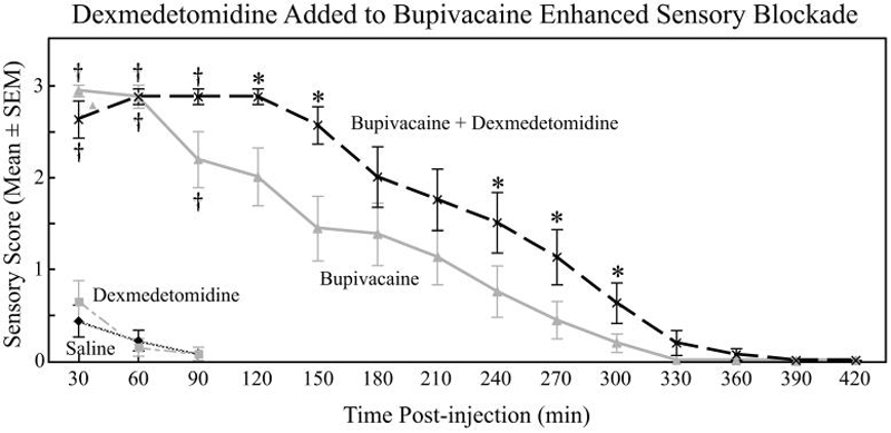
Sensory blockade- Dexmedetomidine added to bupivacaine enhanced the duration of sensory blockade in response to lateral paw pinch when compared with bupivacaine alone. Bupivacaine and bupivacaine plus dexmedetomidine showed improved sensory scores when compared with saline and dexmedetomidine. The time course demonstrates the progression from complete sensory blockade (score = 3) to recovery of normal sensory function (score = 0). Time course data for sensory testing were analyzed using a non-parametric model with ordinal logistic regression for repeated measures and generalized estimating equations. Asterisks (*) indicate significant differences for bupivacaine plus dexmedetomidine versus bupivacaine alone at specific times post-injection. † indicates significant differences between dexmedetomidine and saline when compared with bupivacaine plus dexmedetomidine and bupivacaine.
Table 2. Motor scores for bupivacaine and bupivacaine plus dexmedetomidine.
Motor scores (Complete blockade motor score = 3; Normal motor function = 0) were significantly improved at multiple time points when the bupivacaine plus dexmedetomidine group was compared with the bupivacaine group. Time course data for motor testing was analyzed using a non-parametric model with ordinal logistic regression for repeated measures and generalized estimating equations.
| Time (min) | Bupivacaine | Bupivacaine + DMET | P value | ||||||
|---|---|---|---|---|---|---|---|---|---|
| Mean ± SEM | Median | 25th IQR | 75th IQR | Mean ± SEM | Median | 25th IQR | 75th IQR | ||
| 30 | 2.69 ± 0.12 | 3 | 2 | 3 | 2.94 ± 0.06 | 3 | 3 | 3 | NS |
| 60 | 2.63 ± 0.15 | 3 | 2 | 3 | 2.94 ± 0.06 | 3 | 3 | 3 | NS |
| 90 | 2.31 ± 0.28 | 3 | 1 | 3 | 2.94 ± 0.06 | 3 | 3 | 3 | 0.042 |
| 120 | 2.06 ± 0.32 | 3 | 0 | 3 | 2.75 ± 0.11 | 3 | 2 | 3 | NS |
| 150 | 1.56 ± 0.32 | 2 | 0 | 3 | 2.56 ± 0.16 | 3 | 2 | 3 | 0.002 |
| 180 | 1.31 ± 0.30 | 1.5 | 0 | 2 | 2.19 ± 0.19 | 2 | 2 | 3 | 0.01 |
| 210 | 0.94 ± 0.28 | 0.5 | 0 | 2 | 1.75 ± 0.27 | 2 | 1 | 3 | 0.01 |
| 240 | 0.81 ± 0.28 | 0 | 0 | 2 | 1.31 ± 0.27 | 1 | 0 | 2 | 0.029 |
| 270 | 0.56 ± 0.24 | 0 | 0 | 1 | 1.00 ± 0.26 | 1 | 0 | 2 | 0.042 |
| 300 | 0.25 ± 0.11 | 0 | 0 | 0 | 0.63 ± 0.20 | 0 | 0 | 1 | 0.038 |
| 330 | 0 | 0 | 0 | 0 | 0.13 ± 0.09 | 0 | 0 | 0 | n/a |
| 360 | 0 | 0 | 0 | 0 | 0.13 ± 0.09 | 0 | 0 | 0 | n/a |
| 390 | 0 | 0 | 0 | 0 | 0.06 ± 0.06 | 0 | 0 | 0 | n/a |
| 420 | 0 | 0 | 0 | 0 | 0 | 0 | 0 | 0 | n/a |
DMET = dexmedetomidine; IQR = interquartile range; min = minutes; n/a = not applicable; NS = non-significant; p-value < 0.05 deemed significant; SEM = standard error of mean.
Figure 2.
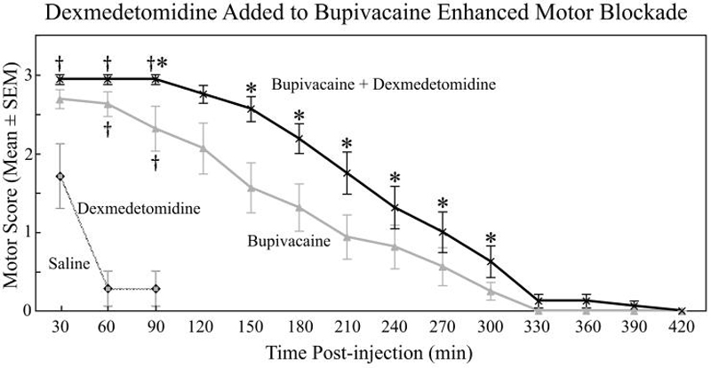
Motor blockade- Dexmedetomidine added to bupivacaine also extended the duration of motor blockade over time when compared to bupivacaine alone. Bupivacaine and bupivacaine plus dexmedetomidine showed improved motor scores when compared with saline and dexmedetomidine. The time course again shows the progression from complete motor blockade (score = 3) to the return of normal motor function (score = 0). Time course data for motor testing were analyzed using a non-parametric model with ordinal logistic regression for repeated measures and generalized estimating equations. Asterisks (*) indicate significant differences for bupivacaine plus dexmedetomidine versus bupivacaine alone at specific times post-injection. † indicates significant differences between dexmedetomidine and saline when compared with bupivacaine plus dexmedetomidine and bupivacaine.
The duration of complete sensory blockade (sensory score = 3) was significantly increased when bupivacaine plus dexmedetomidine was compared to bupivacaine. None of the rats in Group Saline-DMET ever had a complete sensory block. In addition, the time to recovery of normal sensory function (sensory score = 0) was increased when bupivacaine plus dexmedetomidine was compared to bupivacaine, dexmedetomidine, and saline (fig. 3).
Figure 3.
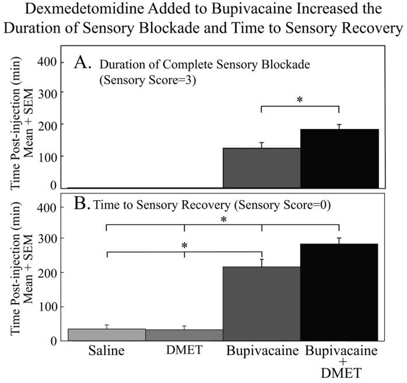
The duration of complete sensory blockade (A) was significantly increased when bupivacaine plus dexmedetomidine (182.0 ± 15.6 min) was compared to bupivacaine (123.8 ± 17.3 min; p < 0.01). None of the rats in the saline or dexmedetomidine groups had a complete sensory blockade. In addition, the time to recovery of normal sensory function (B) was increased when bupivacaine plus dexmedetomidine (282.0 ± 17.8 min) was compared to bupivacaine (215.6 ± 21.6 min, p < 0.05), dexmedetomidine (32.1 ± 11.1 min, p < 0.01), and saline (34.3 ± 11.7 min, p < 0.01). Asterisks (*) indicate significant differences. Analyses were completed using repeated measures analysis of variance (ANOVA) followed by Tukey-Kramer multiple comparisons test. The dependent measure for these analyses was time in minutes.
The duration of complete motor blockade (motor score = 3) was significantly lengthened when bupivacaine plus dexmedetomidine and bupivacaine were individually compared with dexmedetomidine and saline. The trend towards prolonged complete motor blockade when bupivacaine plus dexmedetomidine was compared to bupivacaine was not significant. The time to complete recovery of normal motor function (motor score = 0) was significantly longer when bupivacaine plus dexmedetomidine was compared with bupivacaine, dexmedetomidine, and saline. The increased time to motor recovery was also significant when bupivacaine was compared with dexmedetomidine and saline (fig. 4).
Figure 4.
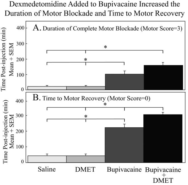
The duration of complete motor blockade (A) was significantly improved when bupivacaine plus dexmedetomidine (158.0 ± 19.1 min) and bupivacaine (101.3 ± 20.7 min) were individually compared with dexmedetomidine (21.4 ± 6.6 min, p < 0.01) and saline (21.4 ± 6.6 min, p < 0.01). The trend towards prolonged complete motor blockade when bupivacaine plus dexmedetomidine was compared to bupivacaine was not significant. The time to complete recovery of normal motor function (B) was significantly longer when bupivacaine plus dexmedetomidine (306.0 ± 14.4 min) was compared with bupivacaine (223.1 ± 22.4 min, p < 0.01), dexmedetomidine (42.9 ± 11.6 min, p < 0.01), and saline (42.9 ± 11.6 min, p < 0.01). The increased time to motor recovery was also significant when bupivacaine alone was compared with dexmedetomidine (p < 0.01) and saline (p < 0.01). Asterisks (*) indicate significant differences. Analyses were completed using repeated measures analysis of variance (ANOVA) followed by Tukey-Kramer multiple comparisons test. The dependent measure for these analyses was time in minutes.
In Group Saline-DMET, one rat was eliminated due to direct nerve damage during the dissection and was subsequently excluded and euthanized. All other animals underwent full neurobehavioral monitoring and subsequent nerve removal as described above in the Neurobehavioral Examination section of the Materials and Methods.
Histopathology
Sciatic nerve histopathology at 24 hours and 14 days showed normal axons and myelin in all nerves analyzed (histopathology score = 0). Figure 5 shows representative sciatic nerve histopathology. When compared with the saline control group, the bupivacaine group had significantly higher perineural inflammation scores at 24 hours. Nerves in the bupivacaine + dexmedetomidine group showed less perineural inflammation at 24 hours when compared to the bupivacaine group (table 3). There were no differences in perineural inflammation between the saline control, dexmedetomidine, and bupivacaine plus dexmedetomidine groups at 24 hours (table 3). None of the nerves analyzed at 14 days showed significant perineural inflammation.
Figure 5.
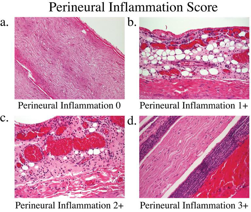
Nerves were sectioned and stained with hemotoxylin and eosin to assess perineural inflammation at 24 hours and 14 days. Nerves in the bupivacaine group had higher inflammation scores at 24 hours when compared with the saline control. Bupivacaine plus dexmedetomidine and dexmedetomidine alone had similar inflammation scores compared with normal saline at 24 hours. At 14 days, nerves in both groups were completely normal with inflammation scores of 0. (A) Inflammation score = 0: The perineural space is void of any significant inflammatory cells. (B) Inflammation score = 1: Focal portions of perineural inflammation involving 5-10% of the sections. (C) Inflammation score = 2: Moderate degree of perineural inflammation. (D) Inflammation score = 3: Severe inflammation is seen with large numbers of lymphocytes surrounding the nerve.
Table 3. Histopathological perineural inflammation scores at 24 hours.
Dexmedetomidine attenuated the acute bupivacaine-induced perineural inflammation. When compared with the saline control group, bupivacaine showed higher inflammation scores at 24 hours. The increased perineural inflammation at 24 hours when bupivacaine was compared with bupivacaine + dexmedetomidine was also statistically significant. There were no differences between the saline control, dexmedetomidine, and bupivacaine plus dexmedetomidine groups. Perineural inflammation scores were assessed by a blinded pathologist as follows: 0 = no inflammation, 1 = small focal areas of mild edema and/or cellular infiltrate, 2 = locally extensive areas of moderate edema/cellular infiltrate, 3 = diffuse areas of moderate to marked edema/cellular infiltrate.
| Drug | Perineural inflammation score | Comparison with saline (P value) | Bupivacaine vs Bupivacaine + DMET (P value) | ||
|---|---|---|---|---|---|
| Median | 25th IQR | 75th IQR | |||
| Saline | 2 | 1 | 2 | N/A | N/A |
| DMET | 1 | 0.5 | 2.5 | NS | N/A |
| Bupivacaine | 3 | 2 | 3 | < 0.04 * | < 0.04 ** |
| Bupivacaine + DMET | 1 | 1 | 2 | NS | N/A |
DMET = dexmedetomidine; IQR = interquartile range; NS = non-significant; p-value < 0.05 deemed significant; vs = versus.
Statistical significance determined based on comparison with saline placebo group (Wilcoxon Rank Sum/Mann-Whitney U Test).
Statistical significance when bupivacaine was compared with bupivacaine + DMET (Wilcoxon Rank Sum/Mann-Whitney U Test).
Discussion
Dexmedetomidine Enhancement of Sensory and Motor Blockade
In this placebo-controlled, randomized, blinded study, high-dose dexmedetomidine added to bupivacaine significantly enhanced sensory and motor blockade in sciatic nerve blocks in rat (fig. 1-4 and tables 1 and 2). The effect of dexmedetomidine was only significant when added to bupivacaine, thereby refuting the central analgesic effects of dexmedetomidine as the reason for increased duration of sensory blockade.
Dexmedetomidine alone did not produce significant sensory blockade nor sustained motor blockade, which is consistent with that which has been seen with clonidine in both laboratory and clinical work. In rabbit sciatic nerves, supraclinical doses of clonidine were found to inhibit C-fiber action potentials, however, doses almost 1000-fold lower prolonged the duration of lidocaine blocks.35 In humans, clonidine failed to provide adequate analgesia when used as the sole anesthetic in brachial plexus blockade.36 These data along with the laboratory and clinical clonidine data suggest a possible class effect for □2-adrenoceptor agonists in peripheral nerve blocks.
There have been four proposed mechanisms for the action of clonidine in peripheral nerve blocks. These mechanisms include centrally mediated analgesia, ∝2B-adrenoceptor mediated vasoconstrictive effects, attenuation of the inflammatory response, and direct action on the peripheral nerve.
Central analgesia, vasoconstriction and anti-inflammatory properties do not fully explain the efficacy of clonidine in peripheral nerve blocks. The duration of anesthesia and analgesia was prolonged with perineural clonidine compared with subcutaneous14 and intramuscular controls.9,37 Despite higher plasma levels of clonidine with intramuscular administration, perineural administration provided better analgesia, thereby refuting a central mechanism.9 Although ∝2B-adrenoceptors do mediate vasoconstriction in the periphery, the vasoconstrictive properties of clonidine are weaker than epinephrine.18 Unlike epinephrine, the enhancement of sensory blockade with clonidine is not attenuated by the co-administration of ∝-adrenoceptor antagonists.38,39 Recent work by Eisenach and colleagues has shown that □2-adrenoceptor agonists attenuate the inflammatory response in a nerve injury model in rats.40-45 Although these findings are extremely important in the study of neuropathic pain, they do not explain the immediate antinociceptive benefits of perineural clonidine added to local anesthetic in an acute pain model.
Laboratory work dating back as early as 197246 indicates that clonidine has a direct effect on the peripheral nerve that is not mediated via the ∝2-adrenoceptor. Clonidine produced a concentration-dependent, reversible blockade of compound action potentials in frog sciatic nerves,46 rat sciatic nerves,47 and desheathed rabbit vagus nerves.35,48 The effects were found to be greater on C-fibers than A∝-fibers.47 These local anesthetic effects at high concentrations, however, do not explain the apparent additive or synergistic effects of clonidine added to local anesthetics in peripheral nerve blocks in humans. In 1994, Gaumann et al.48 exhibited clonidine's ability to increase the hyperpolarizing afterpotential that follows a single compound action potential. Kroin et al.38 later found that lidocaine added to ZD 7288, a specific blocker of the Ih current, extended sensory blockade to pinprick in rat sciatic nerve blocks equivalent to the prolongation seen with the lidocaine and clonidine mixture. Dalle et al. later corroborated this finding.49 in a sucrose-gap method on the C-fibers of rabbit vagus nerves. The authors concluded that clonidine enhances activity-dependent hyperpolarization by inhibiting the Ih current. The Ih current plays a key role in cell excitability, especially the firing frequency, in both the central and peripheral nervous systems.50 The Ih current is activated during the hyperpolarization phase of an action potential and normally acts to reset a nerve for subsequent action potentials. Therefore, by blocking the Ih current clonidine enhances hyperpolarization and inhibits subsequent action potentials.
There are some recent studies investigating the mechanism of action of dexmedetomidine in the central nervous system. Dexmedetomidine was found to inhibit rat hypothalamic paraventricular nucleus neurons by activation of the G protein-coupled inwardly rectifying K+ current and paraventricular nucleus parvocellular neurons by suppression of Ih.51 An in vitro study of rat dorsal root ganglion neurons found that when combined with lidocaine both clonidine and dexmedetomidine produced an additive blockade-type interaction on tetrodotoxin-resistant sodium current.52 Although these studies investigated central mechanisms of action for dexmedetomidine, when combined with the above noted clonidine literature, hypotheses as to possible mechanisms of action for dexmedetomidine in peripheral nerves can be drawn.
The present study does not elucidate the mechanism by which dexmedetomidine enhances local anesthetics in peripheral nerve blocks. It is, however, the first study to report the peripheral perineural administration of dexmedetomidine. Given that dexmedetomidine and clonidine are both selective □2-adrenoceptor agonists, it is possible that they work in a similar manner and may indicate a class effect. The peripheral mechanism of clonidine, however, does not appear to be ∝2 mediated.
Histopathological Evaluation of Perineural Dexmedetomidine
To our knowledge, this is also the first reported histopathological evaluation of peripheral perineural administration of dexmedetomidine. Clonidine has long been used in clinical practice for oral, intravenous, subcutaneous, perineural, epidural, and intravenous administration without ill effect. In addition, previous studies of intrathecal administration of high doses of clonidine in rats53 and dogs54 did not find any toxicity to the spinal cord or nerve roots. A study published while the present results were being reviewed found demyelinization of the oligodendrocytes in the white matter of the spinal cord when dexmedetomidine 5 or 10 micrograms was injected into the epidural space in rabbits.55 The rabbits in the higher dose epidural dexmedetomidine group received between 6.06-6.25 mcg/kg, and the spinal cords were removed for histopathologic analysis 60 minutes after drug injection and only one day following the placement of the epidural catheter. A saline group was not included, and the injectate pH was neither adjusted nor reported. The neurotoxic effects of epidural dexmedetomidine reported could be due to a species effect, pH, vasoconstriction of the spinal cord vascular supply, or direct trauma from epidural placement.
In the present study, all of the nerves analyzed for histopathological changes were normal at 24 hours and 14 days. The concentration of dexmedetomidine was based on our proposed human clinical dosing of 2 mcg/kg for peripheral nerve blocks. This concentration was derived from previous human epidural and intravenous regional anesthesia studies. Estimating the average patient to be 75 kg, the concentration for a 30 ml brachial plexus block would be 5 mcg/ml. Thus, the present study used a concentration of 50 mcg/ml for a total dosing of between 28-40 mcg/kg to demonstrate a wider safety margin for both concentration and total dosing. Although there are significant differences between epidural and peripheral perineural administration, it is surprising that the 28-40 mcg/kg used in the present study did not affect the axons or myelin of the sciatic nerve, while the 6.25 mcg/kg in the epidural rabbit model led to significant myelin damage.55 Future studies in other animal species may help to clarify this discrepancy.
Consistent with the previously noted decrease in inflammatory mediators following perineural clonidine administration,40-45 the present study found a significant reduction in perineural inflammation at 24 hours when dexmedetomidine was added to bupivacaine as compared to bupivacaine alone. Bupivacaine alone had higher perineural inflammation scores at 24 hours compared with the saline control group (table 3). Both the bupivacaine plus dexmedetomidine and dexmedetomidine alone groups showed perineural inflammation scores similar to the saline control group. As previously discussed, the decrease in perineural inflammation is believed to be due to a decrease in proinflammatory products from immune cells recruited to the site of injury and an increase in anti-inflammatory cytokines.40-45
Local anesthetics are known to be myotoxic,21-24 however, the clinical significance of local anesthetic induced myotoxicity is still somewhat unclear as there are few reported cases of significant muscle pathology in the literature. This study did not investigate the myotoxic effects of local anesthetics combined with dexmedetomidine and may be an area of future research. Although the reports of clinically significant myotoxicity are limited to date,23 as regional anesthesia grows, the number of reported cases would be expected to increase. In addition, the increased use of peripheral nerve catheters56-58 and infusion over several days may make this issue more significant. As previous noted, some patients may be at a higher risk of post-procedural neurologic dysfunction due to comorbidities, such as diabetes or multiple sclerosis.25-27 In these patients, the proinflammatory, neurotoxic effects of local anesthetics may be contraindicated. The ability of □2-adrenoceptor agonists to attenuate the inflammatory response40-45,56 may improve safety in peripheral nerve catheters and single shot blocks.
Limitations
As noted above, the concentration of dexmedetomidine used far exceeds that which we propose as a potentially appropriate human dose, and the effects of more clinically relevant doses in this species are still unknown. In addition, it is unknown whether human responses to clinically relevant doses would be significant.
Clonidine is known to produce a dose dependent inhibition of A∝- and C-fibers with C-fibers having been shown to be more profoundly affected.35,47,48 C-fibers are known to mediate dull pain and burning sensations, however, the model used for neurobehavioral monitoring in the present study was equivalent to a surgical stimulus. Even though dexmedetomidine added to bupivacaine was shown to enhance both sensory and motor blockade, using a model to elicit dull pain may have shown more subtle sensory differences. This difference may better correlate with the postoperative sensory changes previously seen in human perineural clonidine studies.6-15
In addition, rats received high doses of dexmedetomidine, which is known to cause sedation. Whereas high doses, between 28-40 mcg/kg, were necessary to prove a satisfactory safety margin, the neurobehavioral monitoring was likely altered. Systemic dexmedetomidine is known to provide analgesia and sedation, which might also affect sensory and motor testing. Each rat received bilateral sciatic nerve blocks, however, and thereby acted as its own control. Furthermore, a blinded investigator conducted the neurobehavioral testing. Rats in the Saline-DMET group never had a complete sensory block, nor was their motor block as sustained as that which was seen in the Bupiv-DMET group. Although rats in the Saline-DMET group did have short durations of complete motor blockade, this was likely a product of centrally mediated sedation. However, the motor blockade seen in the Saline-DMET group was far less consistent than that in the Bupiv-DMET group and tended to quickly remit. Using a model in which separate rats received unilateral, single blocks with local anesthetic plus dexmedetomidine versus local anesthetic alone may be interesting. It would, however, be very difficult to blind due to the sedative effects of high dose dexmedetomidine. Future dose ranging studies will help to determine whether clinically relevant doses may have the same effect.
Conclusions
This study supports the hypothesis that large dose dexmedetomidine enhances the duration of bupivacaine anesthesia and analgesia of sciatic nerve block in rat. The results are consistent with laboratory clonidine data. The histopathological evaluation showed that nerve axon and myelin were normal in both groups at 24 hours and 14 days. To our knowledge, this is the first study to evaluate nerve histopathologic changes of a high dose □2-adrenoceptor agonist in peripheral nerve block in rat. Under the conditions of the study, high dose dexmedetomidine attenuates the acute bupivacaine-induced perineural inflammation without causing nerve damage. The finding that dexmedetomidine can safely improve the duration of bupivacaine-induced antinociception following sciatic nerve blockade in rat is an essential first step encouraging future studies in patients.
Acknowledgements
We thank Kevin K. Tremper, M.D., Ph.D. (Professor, Department of Anesthesiology, University of Michigan, Ann Arbor Michigan), Peter Gerner, M.D. (Assistant Professor, Department of Anesthesiology, Perioperative and Pain Medicine, Brigham and Women's Hospital, Harvard Medical School, Boston, Massachusetts), and Robert Hurley, M.D., Ph.D. (Assistant Professor, Pain Management Division, Department of Anesthesiology and Critical Care Medicine, Johns Hopkins School of Medicine, Baltimore, Maryland). For statistical support, we thank Kathy Welch, M.A., M.P.H. (Adjunct Lecturer, Biostatistics; Statistician Staff Specialist, Center for Statistical Consultation and Research, University of Michigan, Ann Arbor, Michigan. For technical support, we thank Diane Ignasiak, B.S. (Assistant Laboratory Manager), Christopher Watson, Ph.D. (Postdoctoral Fellow), Sarah Watson, B.S. (Research Associate), and Deborah Wagner, Pharm.D. (Associate Professor of Pharmacy, College of Pharmacy, Clinical Pharmacist, University of Michigan Hospital Pharmacy Services and Assistant Professor of Anesthesiology, University of Michigan School of Medicine).
Financial Support: Supported by the Department of Anesthesiology, University of Michigan, Ann Arbor, Michigan. Ralph Lydic, Ph.D. is supported by National Institutes of Health (Bethesda, Maryland) grants HL40881, HL57120, and HL65272. The authors have no conflict of interest.
Footnotes
National Academies Press, Washington D.C., 1996, www.nap.edu/readingroom/books/labrats, Last date accessed May 5, 2008
National Academies Press, Washington, D.C., 2003, www.national-academies.org/ilar, Last date accessed May 5, 2008
Preliminary data presented at the Association of University Anesthesiologists Annual Meeting, Chicago, Illnois, April 26-29, 2007.
Summary Statement: High dose dexmedetomidine added to bupivacaine significantly increased the duration of sensory and motor blockade in rat. Histopathology at 24 hours and 14 days revealed normal nerves.
References
- 1.Casati A, Fanelli G, Albertin A, Deni F, Anelati D, Antonino FA, Beccaria P. Interscalene brachial plexus anesthesia with either 0.5% ropivacaine or 0.5% bupivacaine. Minerva Anestesiol. 2000;66:39–44. [PubMed] [Google Scholar]
- 2.Hickey R, Hoffman J, Ramamurthy S. A comparison of ropivacaine 0.5% and bupivacaine 0.5% for brachial plexus block. Anesthesiology. 1991;74:639–42. doi: 10.1097/00000542-199104000-00002. [DOI] [PubMed] [Google Scholar]
- 3.Hickey R, Rowley CL, Candido KD, Hoffman J, Ramamurthy S, Winnie AP. A comparative study of 0.25% ropivacaine and 0.25% bupivacaine for brachial plexus block. Anesth Analg. 1992;75:602–6. doi: 10.1213/00000539-199210000-00024. [DOI] [PubMed] [Google Scholar]
- 4.Vaghadia H, Chan V, Ganapathy S, Lui A, McKenna J, Zimmer K. A multicentre trial of ropivacaine 7.5 mg x ml(-1) vs bupivacaine 5 mg x ml(-1) for supra clavicular brachial plexus anesthesia. Can J Anaesth. 1999;46:946–51. doi: 10.1007/BF03013129. [DOI] [PubMed] [Google Scholar]
- 5.Eisenach JC, De Kock M, Klimscha W. alpha(2)-adrenergic agonists for regional anesthesia. A clinical review of clonidine (1984-1995) Anesthesiology. 1996;85:655–74. doi: 10.1097/00000542-199609000-00026. [DOI] [PubMed] [Google Scholar]
- 6.Casati A, Magistris L, Fanelli G, Beccaria P, Cappelleri G, Aldegheri G, Torri G. Small-dose clonidine prolongs postoperative analgesia after sciatic-femoral nerve block with 0.75% ropivacaine for foot surgery. Anesth Analg. 2000;91:388–92. doi: 10.1097/00000539-200008000-00029. [DOI] [PubMed] [Google Scholar]
- 7.El Saied AH, Steyn MP, Ansermino JM. Clonidine prolongs the effect of ropivacaine for axillary brachial plexus blockade. Can J Anaesth. 2000;47:962–7. doi: 10.1007/BF03024866. [DOI] [PubMed] [Google Scholar]
- 8.Eledjam JJ, Deschodt J, Viel EJ, Lubrano JF, Charavel P, d'Athis F, du Cailar J. Brachial plexus block with bupivacaine: effects of added alpha-adrenergic agonists: comparison between clonidine and epinephrine. Can J Anaesth. 1991;38:870–5. doi: 10.1007/BF03036962. [DOI] [PubMed] [Google Scholar]
- 9.Hutschala D, Mascher H, Schmetterer L, Klimscha W, Fleck T, Eichler HG, Tschernko EM. Clonidine added to bupivacaine enhances and prolongs analgesia after brachial plexus block via a local mechanism in healthy volunteers. Eur J Anaesthesiol. 2004;21:198–204. doi: 10.1017/s0265021504003060. [DOI] [PubMed] [Google Scholar]
- 10.Iohom G, Machmachi A, Diarra DP, Khatouf M, Boileau S, Dap F, Boini S, Mertes PM, Bouaziz H. The effects of clonidine added to mepivacaine for paronychia surgery under axillary brachial plexus block. Anesth Analg. 2005;100:1179–83. doi: 10.1213/01.ANE.0000145239.17477.FC. [DOI] [PubMed] [Google Scholar]
- 11.Iskandar H, Benard A, Ruel-Raymond J, Cochard G, Manaud B. The analgesic effect of interscalene block using clonidine as an analgesic for shoulder arthroscopy. Anesth Analg. 2003;96:260–2. doi: 10.1097/00000539-200301000-00052. [DOI] [PubMed] [Google Scholar]
- 12.Iskandar H, Guillaume E, Dixmerias F, Binje B, Rakotondriamihary S, Thiebaut R, Maurette P. The enhancement of sensory blockade by clonidine selectively added to mepivacaine after midhumeral block. Anesth Analg. 2001;93:771–5. doi: 10.1097/00000539-200109000-00043. [DOI] [PubMed] [Google Scholar]
- 13.Reinhart DJ, Wang W, Stagg KS, Walker KG, Bailey PL, Walker EB, Zaugg SE. Postoperative analgesia after peripheral nerve block for podiatric surgery: clinical efficacy and chemical stability of lidocaine alone versus lidocaine plus clonidine. Anesth Analg. 1996;83:760–5. doi: 10.1097/00000539-199610000-00018. [DOI] [PubMed] [Google Scholar]
- 14.Singelyn FJ, Dangoisse M, Bartholomee S, Gouverneur JM. Adding clonidine to mepivacaine prolongs the duration of anesthesia and analgesia after axillary brachial plexus block. Reg Anesth. 1992;17:148–50. [PubMed] [Google Scholar]
- 15.Singelyn FJ, Gouverneur JM, Robert A. A minimum dose of clonidine added to mepivacaine prolongs the duration of anesthesia and analgesia after axillary brachial plexus block. Anesth Analg. 1996;83:1046–50. doi: 10.1097/00000539-199611000-00025. [DOI] [PubMed] [Google Scholar]
- 16.Culebras X, Van Gessel E, Hoffmeyer P, Gamulin Z. Clonidine combined with a long acting local anesthetic does not prolong postoperative analgesia after brachial plexus block but does induce hemodynamic changes. Anesth Analg. 2001;92:199–204. doi: 10.1097/00000539-200101000-00038. [DOI] [PubMed] [Google Scholar]
- 17.Duma A, Urbanek B, Sitzwohl C, Kreiger A, Zimpfer M, Kapral S. Clonidine as an adjuvant to local anaesthetic axillary brachial plexus block: a randomized, controlled study. Br J Anaesth. 2005;94:112–6. doi: 10.1093/bja/aei009. [DOI] [PubMed] [Google Scholar]
- 18.Gaumann D, Forster A, Griessen M, Habre W, Poinsot O, Della Santa D. Comparison between clonidine and epinephrine admixture to lidocaine in brachial plexus block. Anesth Analg. 1992;75:69–74. doi: 10.1213/00000539-199207000-00013. [DOI] [PubMed] [Google Scholar]
- 19.Kanazi GE, Aouad MT, Jabbour-Khoury SI, Al Jazzar MD, Alameddine MM, Al-Yaman R, Bulbul M, Baraka AS. Effect of low-dose dexmedetomidine or clonidine on the characteristics of bupivacaine spinal block. Acta Anaesthesiol Scand. 2006;50:222–7. doi: 10.1111/j.1399-6576.2006.00919.x. [DOI] [PubMed] [Google Scholar]
- 20.Memis D, Turan A, Karamanlioglu B, Pamukcu Z, Kurt I. Adding dexmedetomidine to lidocaine for intravenous regional anesthesia. Anesth Analg. 2004;98:835–40. doi: 10.1213/01.ane.0000100680.77978.66. [DOI] [PubMed] [Google Scholar]
- 21.Benoit PW, Belt WD. Destruction and regeneration of skeletal muscle after treatment with a local anaesthetic, bupivacaine (Marcaine) J Anat. 1970;107:547–56. [PMC free article] [PubMed] [Google Scholar]
- 22.Yagiela JA, Benoit PW, Buoncristiani RD, Peters MP, Fort NF. Comparison of myotoxic effects of lidocaine with epinephrine in rats and humans. Anesth Analg. 1981;60:471–80. [PubMed] [Google Scholar]
- 23.Zink W, Graf BM. Local anesthetic myotoxicity. Reg Anesth Pain Med. 2004;29:333–40. doi: 10.1016/j.rapm.2004.02.008. [DOI] [PubMed] [Google Scholar]
- 24.Zink W, Seif C, Bohl JR, Hacke N, Braun PM, Sinner B, Martin E, Fink RH, Graf BM. The acute myotoxic effects of bupivacaine and ropivacaine after continuous peripheral nerve blockades. Anesth Analg. 2003;97:1173–9. doi: 10.1213/01.ANE.0000080610.14265.C8. [DOI] [PubMed] [Google Scholar]
- 25.Hebl JR. Ultrasound-guided regional anesthesia and the prevention of neurologic injury: fact or fiction? Anesthesiology. 2008;108:186–8. doi: 10.1097/01.anes.0000299835.04104.02. [DOI] [PubMed] [Google Scholar]
- 26.Koff MD, Cohen JA, McIntyre JJ, Carr CF, Sites BD. Severe brachial plexopathy after an ultrasound-guided single-injection nerve block for total shoulder arthroplasty in a patient with multiple sclerosis. Anesthesiology. 2008;108:325–8. doi: 10.1097/01.anes.0000299833.73804.cd. [DOI] [PubMed] [Google Scholar]
- 27.Horlocker TT, O'Driscoll SW, Dinapoli RP. Recurring brachial plexus neuropathy in a diabetic patient after shoulder surgery and continuous interscalene block. Anesth Analg. 2000;91:688–90. doi: 10.1097/00000539-200009000-00035. [DOI] [PubMed] [Google Scholar]
- 28.Gerner P, Luo SH, Zhuang ZY, Djalali AG, Zizza AM, Myers RR, Wang GK. Differential block of N-propyl derivatives of amitriptyline and doxepin for sciatic nerve block in rats. Reg Anesth Pain Med. 2005;30:344–50. doi: 10.1016/j.rapm.2005.04.008. [DOI] [PubMed] [Google Scholar]
- 29.Hung YC, Kau YC, Zizza AM, Edrich T, Zurakowski D, Myers RR, Wang GK, Gerner P. Ephedrine blocks rat sciatic nerve in vivo and sodium channels in vitro. Anesthesiology. 2005;103:1246–52. doi: 10.1097/00000542-200512000-00021. [DOI] [PubMed] [Google Scholar]
- 30.Kau YC, Hung YC, Zizza AM, Zurakowski D, Greco WR, Wang GK, Gerner P. Efficacy of lidocaine or bupivacaine combined with ephedrine in rat sciatic nerve block. Reg Anesth Pain Med. 2006;31:14–8. doi: 10.1016/j.rapm.2005.08.008. [DOI] [PubMed] [Google Scholar]
- 31.Dyhre H, Soderberg L, Bjorkman S, Carlsson C. Local anesthetics in lipid-depot formulations--neurotoxicity in relation to duration of effect in a rat model. Reg Anesth Pain Med. 2006;31:401–8. doi: 10.1016/j.rapm.2006.05.008. [DOI] [PubMed] [Google Scholar]
- 32.Soderberg L, Dyhre H, Roth B, Bjorkman S. Ultralong peripheral nerve block by lidocaine:prilocaine 1:1 mixture in a lipid depot formulation: comparison of in vitro, in vivo, and effect kinetics. Anesthesiology. 2006;104:110–21. doi: 10.1097/00000542-200601000-00017. [DOI] [PubMed] [Google Scholar]
- 33.Kytta J, Heinonen E, Rosenberg PH, Wahlstrom T, Gripenberg J, Huopaniemi T. Effects of repeated bupivacaine administration on sciatic nerve and surrounding muscle tissue in rats. Acta Anaesthesiol Scand. 1986;30:625–9. doi: 10.1111/j.1399-6576.1986.tb02488.x. [DOI] [PubMed] [Google Scholar]
- 34.Pere P, Watanabe H, Pitkanen M, Wahlstrom T, Rosenberg PH. Local myotoxicity of bupivacaine in rabbits after continuous supraclavicular brachial plexus block. Reg Anesth. 1993;18:304–7. [PubMed] [Google Scholar]
- 35.Gaumann DM, Brunet PC, Jirounek P. Clonidine enhances the effects of lidocaine on C-fiber action potential. Anesth Analg. 1992;74:719–25. doi: 10.1213/00000539-199205000-00017. [DOI] [PubMed] [Google Scholar]
- 36.Sia S, Lepri A. Clonidine administered as an axillary block does not affect postoperative pain when given as the sole analgesic. Anesth Analg. 1999;88:1109–12. doi: 10.1097/00000539-199905000-00027. [DOI] [PubMed] [Google Scholar]
- 37.Iskandar H, Benard A, Ruel-Raymond J, Cochard G, Manaud B. The analgesic effect of interscalene block using clonidine as an analgesic for shoulder arthroscopy. Anesth Analg. 2003;96:260–2. doi: 10.1097/00000539-200301000-00052. [DOI] [PubMed] [Google Scholar]
- 38.Kroin JS, Buvanendran A, Beck DR, Topic JE, Watts DE, Tuman KJ. Clonidine prolongation of lidocaine analgesia after sciatic nerve block in rats Is mediated via the hyperpolarization-activated cation current, not by alpha-adrenoreceptors. Anesthesiology. 2004;101:488–94. doi: 10.1097/00000542-200408000-00031. [DOI] [PubMed] [Google Scholar]
- 39.Leem JW, Choi Y, Han SM, Yoon MJ, Sim JY, Leem SW. Conduction block by clonidine is not mediated by alpha2-adrenergic receptors in rat sciatic nerve fibers. Reg Anesth Pain Med. 2000;25:620–5. doi: 10.1053/rapm.2000.16160. [DOI] [PubMed] [Google Scholar]
- 40.Lavand'homme PM, Eisenach JC. Perioperative administration of the alpha2-adrenoceptor agonist clonidine at the site of nerve injury reduces the development of mechanical hypersensitivity and modulates local cytokine expression. Pain. 2003;105:247–54. doi: 10.1016/s0304-3959(03)00221-5. [DOI] [PubMed] [Google Scholar]
- 41.Lavand'homme PM, Ma W, De Kock M, Eisenach JC. Perineural alpha(2A)-adrenoceptor activation inhibits spinal cord neuroplasticity and tactile allodynia after nerve injury. Anesthesiology. 2002;97:972–80. doi: 10.1097/00000542-200210000-00033. [DOI] [PubMed] [Google Scholar]
- 42.Liu B, Eisenach JC. Perineural clonidine reduces p38 mitogen-activated protein kinase activation in sensory neurons. Neuroreport. 2006;17:1313–7. doi: 10.1097/01.wnr.0000227995.45917.f5. [DOI] [PubMed] [Google Scholar]
- 43.Romero-Sandoval A, Eisenach JC. Perineural clonidine reduces mechanical hypersensitivity and cytokine production in established nerve injury. Anesthesiology. 2006;104:351–5. doi: 10.1097/00000542-200602000-00022. [DOI] [PubMed] [Google Scholar]
- 44.Romero-Sandoval A, Eisenach JC. Clonidine reduces hypersensitivity and alters the balance of pro- and anti-inflammatory leukocytes after local injection at the site of inflammatory neuritis. Brain Behav Immun. 2007;21:569–80. doi: 10.1016/j.bbi.2006.09.001. [DOI] [PMC free article] [PubMed] [Google Scholar]
- 45.Romero-Sandoval EA, McCall C, Eisenach JC. Alpha2-adrenoceptor stimulation transforms immune responses in neuritis and blocks neuritis-induced pain. J Neurosci. 2005;25:8988–94. doi: 10.1523/JNEUROSCI.2995-05.2005. [DOI] [PMC free article] [PubMed] [Google Scholar]
- 46.Starke K, Wagner J, Schumann HJ. Adrenergic neuron blockade by clonidine: comparison with guanethidine and local anesthetics. Arch Int Pharmacodyn Ther. 1972;195:291–308. [PubMed] [Google Scholar]
- 47.Butterworth JFt, Strichartz GR. The alpha 2-adrenergic agonists clonidine and guanfacine produce tonic and phasic block of conduction in rat sciatic nerve fibers. Anesth Analg. 1993;76:295–301. [PubMed] [Google Scholar]
- 48.Gaumann DM, Brunet PC, Jirounek P. Hyperpolarizing afterpotentials in C fibers and local anesthetic effects of clonidine and lidocaine. Pharmacology. 1994;48:21–9. doi: 10.1159/000139158. [DOI] [PubMed] [Google Scholar]
- 49.Dalle C, Schneider M, Clergue F, Bretton C, Jirounek P. Inhibition of the I(h) current in isolated peripheral nerve: a novel mode of peripheral antinociception? Muscle Nerve. 2001;24:254–61. doi: 10.1002/1097-4598(200102)24:2<254::aid-mus110>3.0.co;2-#. [DOI] [PubMed] [Google Scholar]
- 50.Pape HC. Queer current and pacemaker: the hyperpolarization-activated cation current in neurons. Annu Rev Physiol. 1996;58:299–327. doi: 10.1146/annurev.ph.58.030196.001503. [DOI] [PubMed] [Google Scholar]
- 51.Shirasaka T, Kannan H, Takasaki M. Activation of a G protein-coupled inwardly rectifying K+ current and suppression of Ih contribute to dexmedetomidine-induced inhibition of rat hypothalamic paraventricular nucleus neurons. Anesthesiology. 2007;107:605–15. doi: 10.1097/01.anes.0000281916.65365.4e. [DOI] [PubMed] [Google Scholar]
- 52.Oda A, Iida H, Tanahashi S, Osawa Y, Yamaguchi S, Dohi S. Effects of alpha2-adrenoceptor agonists on tetrodotoxin-resistant Na+ channels in rat dorsal root ganglion neurons. Eur J Anaesthesiol. 2007;24:934–41. doi: 10.1017/S0265021507000543. [DOI] [PubMed] [Google Scholar]
- 53.Gordh T, Jr., Post C, Olsson Y. Evaluation of the toxicity of subarachnoid clonidine, guanfacine, and a substance P-antagonist on rat spinal cord and nerve roots: light and electron microscopic observations after chronic intrathecal administration. Anesth Analg. 1986;65:1303–11. [PubMed] [Google Scholar]
- 54.Gordh TE, Ekman S, Lagerstedt AS. Evaluation of possible spinal neurotoxicity of clonidine. Ups J Med Sci. 1984;89:266–73. doi: 10.3109/03009738409179507. [DOI] [PubMed] [Google Scholar]
- 55.Konakci S, Adanir T, Yilmaz G, Rezanko T. The efficacy and neurotoxicity of dexmedetomidine administered via the epidural route. Eur J Anaesthesiol. 2007:1–7. doi: 10.1017/S0265021507003079. [DOI] [PubMed] [Google Scholar]
- 56.Ilfeld BM, Enneking FK. Continuous peripheral nerve blocks at home: a review. Anesth Analg. 2005;100:1822–33. doi: 10.1213/01.ANE.0000151719.26785.86. [DOI] [PubMed] [Google Scholar]
- 57.Ilfeld BM, Le LT, Meyer RS, Mariano ER, Vandenborne K, Duncan PW, Sessler DI, Enneking FK, Shuster JJ, Theriaque DW, Berry LF, Spadoni EH, Gearen PF. Ambulatory continuous femoral nerve blocks decrease time to discharge readiness after tricompartment total knee arthroplasty: a randomized, triple-masked, placebo-controlled study. Anesthesiology. 2008;108:703–13. doi: 10.1097/ALN.0b013e318167af46. [DOI] [PMC free article] [PubMed] [Google Scholar]
- 58.Ilfeld BM, Morey TE, Wright TW, Chidgey LK, Enneking FK. Continuous interscalene brachial plexus block for postoperative pain control at home: a randomized, double-blinded, placebo-controlled study. Anesth Analg. 2003;96:1089–95. doi: 10.1213/01.ANE.0000049824.51036.EF. [DOI] [PubMed] [Google Scholar]


