

Abstract
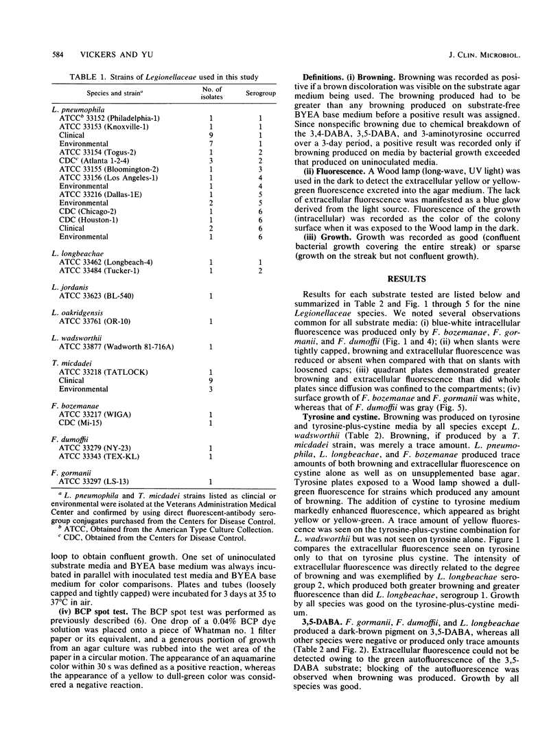
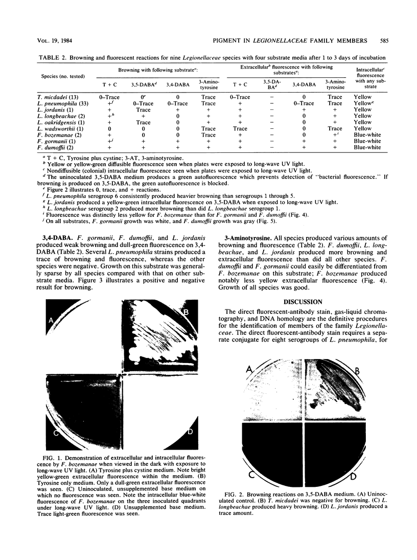
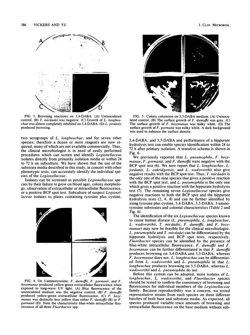
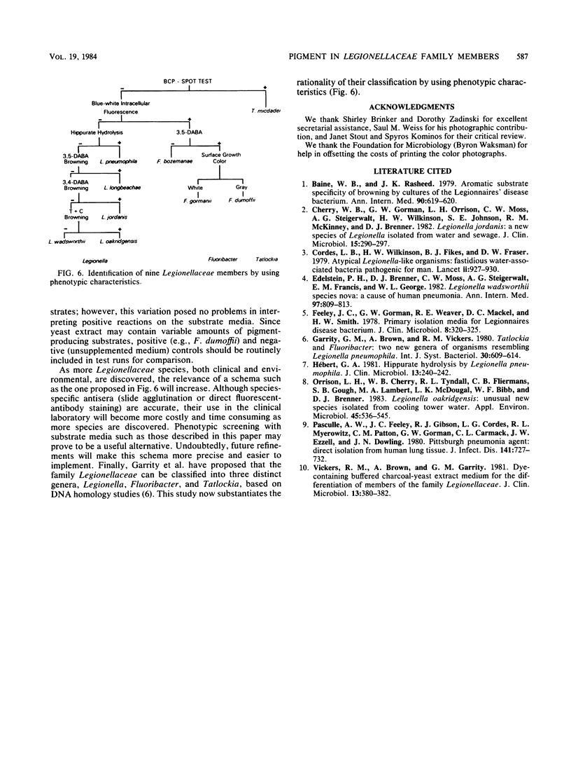
A systematic study of pigment production (browning) and fluorescence (extracellular yellow-green and intracellular blue-white) by nine Legionellaceae species was performed. A total of 56 strains representing Tatlockia micdadei (Pittsburgh pneumonia agent), Legionella pneumophila, Legionella jordanis, Legionella longbeachae, Legionella oakridgensis, Legionella wadsworthii, Fluoribacter bozemanae, Fluoribacter gormanii, and Fluoribacter dumoffii could be separated on media supplemented with tyrosine plus cystine, 3,4-diaminobenzoic acid, 3,5-diaminobenzoic acid, and 3-aminotyrosine. Parallel testing by hippurate hydrolysis and the bromocresol purple spot test enabled the identification of Legionellaceae species 24 to 72 h after primary isolation. This schema may be a practical alternative to species-specific antisera methods (slide agglutination or direct immunofluorescence) in the identification of members of the family Legionellaceae.
Full text
PDF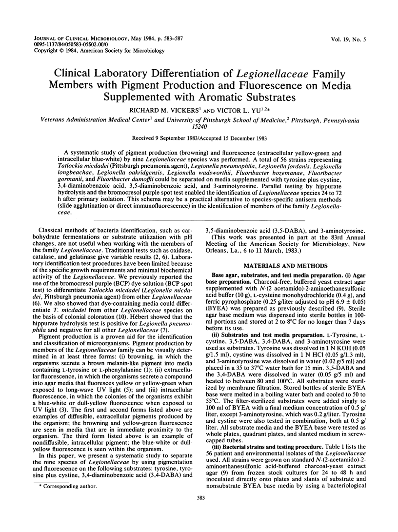
Images in this article
Selected References
These references are in PubMed. This may not be the complete list of references from this article.
- Baine W. B., Rasheed J. K. Aromatic substrate specificity of browning by cultures of the Legionnaires' disease bacterium. Ann Intern Med. 1979 Apr;90(4):619–620. doi: 10.7326/0003-4819-90-4-619. [DOI] [PubMed] [Google Scholar]
- Cherry W. B., Gorman G. W., Orrison L. H., Moss C. W., Steigerwalt A. G., Wilkinson H. W., Johnson S. E., McKinney R. M., Brenner D. J. Legionella jordanis: a new species of Legionella isolated from water and sewage. J Clin Microbiol. 1982 Feb;15(2):290–297. doi: 10.1128/jcm.15.2.290-297.1982. [DOI] [PMC free article] [PubMed] [Google Scholar]
- Cordes L. G., Wilkinson H. W., Gorman G. W., Fikes B. J., Fraser D. W. Atypical Legionella-like organisms: fastidious water-associated bacteria pathogenic for man. Lancet. 1979 Nov 3;2(8149):927–930. doi: 10.1016/s0140-6736(79)92623-0. [DOI] [PubMed] [Google Scholar]
- Edelstein P. H., Brenner D. J., Moss C. W., Steigerwalt A. G., Francis E. M., George W. L. Legionella wadsworthii species nova: a cause of human pneumonia. Ann Intern Med. 1982 Dec;97(6):809–813. doi: 10.7326/0003-4819-97-6-809. [DOI] [PubMed] [Google Scholar]
- Feeley J. C., Gorman G. W., Weaver R. E., Mackel D. C., Smith H. W. Primary isolation media for Legionnaires disease bacterium. J Clin Microbiol. 1978 Sep;8(3):320–325. doi: 10.1128/jcm.8.3.320-325.1978. [DOI] [PMC free article] [PubMed] [Google Scholar]
- Hébert G. A. Hippurate hydrolysis by Legionella pneumophila. J Clin Microbiol. 1981 Jan;13(1):240–242. doi: 10.1128/jcm.13.1.240-242.1981. [DOI] [PMC free article] [PubMed] [Google Scholar]
- Orrison L. H., Cherry W. B., Tyndall R. L., Fliermans C. B., Gough S. B., Lambert M. A., McDougal L. K., Bibb W. F., Brenner D. J. Legionella oakridgensis: unusual new species isolated from cooling tower water. Appl Environ Microbiol. 1983 Feb;45(2):536–545. doi: 10.1128/aem.45.2.536-545.1983. [DOI] [PMC free article] [PubMed] [Google Scholar]
- Pasculle A. W., Feeley J. C., Gibson R. J., Cordes L. G., Myerowitz R. L., Patton C. M., Gorman G. W., Carmack C. L., Ezzell J. W., Dowling J. N. Pittsburgh pneumonia agent: direct isolation from human lung tissue. J Infect Dis. 1980 Jun;141(6):727–732. doi: 10.1093/infdis/141.6.727. [DOI] [PubMed] [Google Scholar]
- Vickers R. M., Brown A., Garrity G. M. Dye-containing buffered charcoal-yeast extract medium for differentiation of members of the family Legionellaceae. J Clin Microbiol. 1981 Feb;13(2):380–382. doi: 10.1128/jcm.13.2.380-382.1981. [DOI] [PMC free article] [PubMed] [Google Scholar]