Abstract
The protein C pathway has an important function in regulating and modulating blood coagulation and ensuring patency of the microcirculation. Protein C deficiency leads to macro- and microvascular thrombosis. Congenital severe protein C deficiency is a life-threatening state with neonatal purpura fulminans and pronounced coagulopathy. Patients with heterozygous protein C deficiency have an increased risk for thromboembolic events or experience coumarin-induced skin necrosis during initiation of coumarin therapy. Replacement with protein C concentrates is an established therapy of congenital protein C deficiency, resulting in rapid resolving of coagulopathy and thrombosis without reasonable side effects. This article summarizes the current knowledge on protein C replacement therapy in congenital protein C deficiency.
Keywords: protein C, deficiency, replacement therapy, purpura fulminans, coagulopathy
Introduction
Protein C is part of a complex regulatory system, which has an important influence on the physiological function of hemostasis to ensure patency of the microcirculation. It was discovered in 1976 by Stenflo as the zymogen of an anticoagulant protein, which could be activated by thrombin (Stenlo 1976). The clinical importance of protein C became evident when Griffin et al demonstrated an association with thromboembolic events (Griffin et al 1981). Hereditary (congenital) protein C deficiency is rare, but a strong risk factor for thrombosis (Bertina et al 1982). At the beginning of the 1990s the resistance to activated protein C was discovered, a mutation in the gene of coagulation factor V, altering the inactivation of factor Va by protein Ca. This mutation occurs frequently in the general population and is also associated with an increased risk for thrombosis (Dahlbäck et al 1993; Bertina et al 1994). Acquired protein C deficiencies can occur in several other diseases. Sepsis and severe infections especially were the center of the scientific interest during the previous decade (Levi et al 1997; Esmon 2001, 2006; Faust et al 2001). It could be shown that the probability of survival of septic patients was correlated with their protein C activity (Fourrier et al 1992; Meesters et al 2000; Yan et al 2001; Macias et al 2004). Since 1988 larger amounts of protein C could be derived from human plasma and used for therapy of deficiency states.
This article summarizes the current knowledge on treating congenital protein C deficiency with concentrates of human protein C.
The physiology of the protein C pathway
The gene for protein C is located on chromosome 2q13-14. It is composed of 9 exons and 8 introns, 11 kilobases (kb) in size and homologous to the genes of other vitamin K dependent proteins (Esmon 2003). Until now more than 230 different mutations and polymorphisms in the protein C gene have been detected (Reitsma et al 1995; Reitsma 1997; http://www.hgmd.cf.ac.uk/ac/gene.php?gene=PROC).
The protein itself is synthesized in the liver as a precursor of 461 amino acids. In presence of vitamin K, gamma-carboxylation of 9 glutamic acid (gla) residues in the gla-domain is performed. The mature protein is composed of 419 amino acids and has a molecular weight of about 62 kD. Before secretion it is cleaved by a furin-like endoprotease at lys156 and arg157, resulting in a heavy and a light chain, connected by disulfide bonds. The gamma-carboxylated gla domain (amino acids 1–42) is necessary for the calcium-mediated binding of the protein to phospholipids, but also to thrombomodulin and the endothelial protein C receptor (EPCR) (Beckman et al 1985; Dahlbäck and Stenflo 1994; Esmon 2003).
Protein C zymogen is activated by thrombin. Once generated during the procoagulatoric cascades, thrombin can be bound to thrombomodulin on the surface of endothelial cells, and its procoagulatoric properties are blocked. In contrast, bound thrombin converts protein C zymogen into an active serine protease by cleavage of the heavy chain at position arg169-leu170. The localization of protein C activation to the surface of endothelial cells is an important mechanism to avoid the formation of thrombi in the microcirculation (Esmon 1989; Ye et al 1999; Van de Wouwer et al 2004).
The physiological plasma concentration of protein C zymogen is 63 nM or 4 μg/mL; the half-life under physiologic conditions is about 6–8 hours. In situations with increased activation of the coagulation pathways (ie, systemic response to inflammation, disseminated intravascular coagulation, consumption coagulopathy), this half-life can be considerably shortened, down to 2–3 hours, resulting in a relative deficiency of protein C and the development of a thrombogenic state. Activated protein C (protein Ca) can be found in the plasma in concentrations of about 40 pM. The half-life of protein Ca in plasma is short (about 20 minutes). Once formed, it is immediately degraded by specific and unspecific plasmatic inactivators. The protein C inhibitor, α-1-protease-inhibitor, and α-2-macroglobulin are the major inhibitors of activated protein C.
Activated protein C has a pronounced anticoagulatoric function, for which another vitamin K dependent protein, protein S, is necessary as a cofactor: it cleaves the activated coagulation factors V and VIII. These homologous proteins serve as cofactors during blood coagulation. The inhibition of factors Va and VIIIa is an important regulatory mechanism, modulating the time course and intensity of blood coagulation (Esmon 1987; 1989; 2003).
Activated protein C has an indirect profibrinolytic effect: it binds to the plasminogen-activator inhibitor 1 (PAI-1), resulting in an increase of the activity of tissue-type plasminogen activator (tPA). In addition, due to the reduced thrombin generation, the activation of TAFI (thrombin activatable fibrinolysis inhibitor) is diminished, thus resulting in an increased profibrinolytic potential (De Fouw et al 1988; Fouassier et al 2005).
Apart from its central role in regulation of coagulation activation, the protein C system also has an important function in modulating inflammation, and has antiapoptotic, cytoprotective, and barrier stabilizing effects (Okajima 2001; Joyce and Grinnell 2002; Feistritzer and Riewald 2005; Mosnier et al 2007). Most of these effects are mediated by the endothelial protein C receptor (EPCR), the protease-activated receptor-1 (PAR-1), or crossactivation of the sphingosine 1-phosphate receptor-1 (S1P1) (Esmon et al 1999; Riewald et al 2002; Brueckmann et al 2005; Feistritzer and Riewald 2005; Mosnier et al 2007). Binding of protein Ca to EPCR influences gene expression profiles by blocking NFκB nuclear translocation, which is necessary for the production of proinflammatory cytokines and adhesion molecules (White et al 2000a).
Congenital (hereditary) protein C deficiency
The hereditary protein C deficiency states are caused by mutations in the protein C gene located on chromosome 2(q13-14) (Online Mendelian Inheritance in Man, OMIM +176860) (www.ncbi.nlm.nih.gov/entrez/dispomim.cgi?id=176860). It may occur as heterozygous (only one of both chromosomes 2 affected), homozygous (both chromosomes 2 carry the same mutation), or mixed heterozygous (both chromosomes 2 are affected, but with different mutations) (Table 1) (Greengard et al 1994). Until now more than 230 different mutations causing protein C deficiency have been described and published in databases (http://www.hgmd.cf.ac.uk/ac/gene.php?gene=PROC). A review article of Reitsma summarized the main characteristics of these gene defects (Reitsma et al 1995; Reitsma 1997). Missense and nonsense mutations are most frequent, but also splicing abnormalities, small insertions or deletions, and regulatory defects have been described (Table 2). They may affect different parts of the gene, resulting in alterations of the various functions of protein C.
Table 1.
Different manifestations of congenital protein C deficiency
| Zygosity | Frequency |
|---|---|
| Type I heterozygous | 76% |
| Type II heterozygous | 12% |
| Type I homozygous | 5% |
| Type II homozygous | 0.6% |
| Type I mixed heterozygous | 3% |
| Type II mixed heterozygous | 0.6% |
| Type I/II mixed heterozygous | 1.5% |
| Unknown | 1.3% |
Note: Analysis of 320 patients.
Table 2.
Types of mutations in the protein C gene causing protein C deficiency
| Mutation | Frequency n (%) |
|---|---|
| Missense/nonsense | 170 (72.0) |
| Splicing | 22 (9.3) |
| Regulatory | 11 (4.7) |
| Small deletions | 20 (8.5) |
| Small insertions | 10 (4.2) |
| Small indels | 1 (0.4) |
| Gross deletions | 2 (0.8) |
| Gross insertions | 0 |
| Complex rearrangements | 0 |
| Repeat variations | 0 |
| Total | 236 |
According to the Human Gene Mutation Database at Institute of Medical Genetics in Cardiff (http://www.hgmd.cf.ac.uk/ac/gene.php?gene=PROC).
Type I protein C deficiency is characterized by a parallel reduction of both protein C activity and antigen concentration in the plasma. It is mainly caused by mutations in the hydrophobic core of protein C, causing large deletions, insertions, or frameshifts.
Type II deficiency is characterized by a reduced activity of protein C, but a near normal antigen concentration. Mutations affect the active site of the enzyme, or domains responsible for protein-protein or protein-phospholipid interactions. Most of the mutations were found in exon 3 (gla-domain), other mutations affected arg1 (cleavage of the propeptide), arg169 (site of activation by thrombin), his11 (in the active site), or other mutations of the active site near exon 9. Type II deficiencies may not only affect the function of protein C, but also other properties: activation of protein C, binding to phospholipids, to thrombomodulin, protein S, or EPCR (Greengard et al 1994).
Diagnosis of congenital protein C deficiency
For clinical purposes, determinations of protein C activity and/or antigen are useful and reliable for the diagnosis of protein C deficiency. Only in a few cases, especially with mutations affecting other than the anticoagulant function of protein C, diagnostic problems may occur: disturbances of protein C activation (by mutations of the thrombin binding site) may cause wrong results with certain activation assays, but not with others (Table 3). In such cases the much more elaborate molecular analysis of the protein C gene may be helpful. This analysis from amniotic fluid or chorion villus biopsy may also be considered in pregnancies with a high risk for homozygous protein C deficiency of the fetus (ie, both parents heterozygous or consanguineous) (Barnes et al 2002).
Table 3.
Types and diagnosis of protein C deficiency
| Deficiency | Protein C activity (function) | Protein C antigen (proteinconcentration) |
|---|---|---|
| Heterozygous | ||
| Type I | decreased (about 0.5 IU/mL) | decreased (about 0.5 IU/mL) |
| Type II | decreased (about 0.5 IU/mL) | normal |
| Homozygous | ||
| Type I | severely decreased (<0.02 IU/mL) | severely decreased (<0.02 IU/mL) |
| Type II | severely decreased (<0.02 IU/mL) | normal/moderately decreased |
In patients not on vitamin K antagonist therapy, the lower normal limit for protein C activity is reported to be about 0.67–0.72 IU/mL, but the levels are dependent on age. Protein C levels of newborns are around 30% of the adult levels and increase within the first years of life. However, every coagulation laboratory should establish a local normal collective. Patients with protein C deficiency can usually be discriminated well from normal subjects, but other possible reasons for decreased protein C levels (vitamin K deficiency, liver disease) have to be excluded (by the determination of a prothrombin time assay; Pabinger et al [1992]).
Homozygous protein C deficiency
Homozygous protein C deficiency is rare. The incidence is 1 per 500,000–750,000 live births. It occurs when on both chromosomes 2 the genes coding for protein C are affected. The same mutation on both chromosomes causes the classical homozygous deficiency state. It occurs mainly in populations with frequent consanguineous marriage. Different mutations on the both chromosomes 2 lead to a double heterozygous protein C deficiency state (Lane et al 1997).
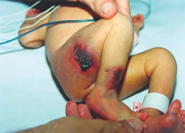
Plasma protein C activity levels are usually very low (below 0.01 IU/mL). Homozygous, severe protein C deficiency manifests in the first days after birth as neonatal purpura fulminans with necrosis of the skin, severe coagulopathy (disseminated intravascular coagulation), and arterial and venous thrombosis. These lesions have a characteristic morphology: they are irregularly formed, with a central hypo-perfused or necrotic area, and a red inflammatory border (Figure 1) The first reports on such a state were published in 1983 (Branson et al 1983). Mortality without therapy is almost 100%. However, in some patients a less dramatic clinical appearance may occur, resembling an early-onset heterozygous deficiency: such patients present with extensive venous thromboembolism. It seems that the severity of the clinical presentation is dependent on the residual amount of protein C; levels above 0.03–0.05 IU/mL are sufficient to ameliorate the symptoms of purpura fulminans, but also the type of mutation may be important (Branson et al 1983; Sharon et al 1986). Another remarkable finding is the observation that, in utero, retinal thrombosis and bleeding can occur, leading to blindness of the newborn (Hattenbach et al 1999). The fact that thrombosis occurs so late during pregnancy suggests the possibility of a transplacentar passage of some protein C, which is a rather small protein, from the mother to the fetus. It has further important impact on the management of mothers with a high probability of bearing a child with homozygous protein C deficiency (Manco-Johnson and Nuss 1992; Barnes et al 2002).
Figure 1a.
Five-day-old newborn with homozygous protein C deficiency and purpura fulminans. Irregularly formed hypoperfused or necrotic skin lesions surrounded by an inflammatory border.
Therapy of homozygous protein C deficiency
Treatment of homozygous protein C deficiency is performed primarily by substitution of the deficient protein (Marlar et al 1989; Salonvaara et al 2004) (Table 4). Until 1988, the only sources for protein C were fresh frozen plasma, and prothrombin complex concentrates (which also contain protein C). The first report of substitution therapy with a protein C concentrate was published in 1988 (Vukovich et al 1988). A 10-month-old daughter of a consanguineous Arab family, presenting with purpura fulminans due to homozygous protein C deficiency, was treated with a plasma-derived concentrate containing protein C and S. All skin lesions resolved and the child was treated with vitamin K antagonists thereafter. The in vivo half-life of protein C in this child was 8.3 hours, the recovery after infusion 44%. However, this concentrate never reached industrial production.
Table 4.
Therapeutic options for severe (homozygous or mixed heterozygous) protein C deficiency
| • Plasma-derived protein C zymogen concentrates |
| (Ceprotin®, Protexel®) |
| 60–80 IU/kg body weight slow i.v. bolus every 6 hours |
| (maximum rate 2 mL/min in adults or 0.2 ml/min in children <10 kg) |
| Target: post-infusion plasma protein C activity 1.0 IU/mL, pre-infusion plasma protein C activity >0.25 IU/mL |
| Alternative regimen: |
| 100 U/kg slow i.v. bolus, followed by 10 IU/kg/h continuous infusion |
| Target: plasma protein C activity 1.0 IU/mL |
| • Fresh frozen plasma |
| 20–30 mL/kg i.v. every 6 hours |
| • Activated human protein C – drotrecogin alpha activated (Xigris®) |
| 20 μg/kg/hour for >10 hours? |
| not approved for substitution therapy of congenital protein C deficiency |
| After symptoms have resolved: |
| Vitamin K antagonist therapy – warfarin |
| Slow initiation, overlapping with protein C substitution |
The next report of protein C substitution with concentrates was published in 1991 (Dreyfus et al 1991). This group used another plasma-derived protein C concentrate (Ceprotin®, described below) in a child with homozygous protein C deficiency and purpura fulminans. This concentrate was thereafter approved for the treatment of congenital protein C deficiency. Until now, several reports on the use of this concentrate in congenital protein C deficiency have been published (Auberger 1992; Conard et al 1993; DeStefano et al 1993; Alhenc-Gelas et al 1995; Baliga et al 1995; Dreyfus et al 1995; Muller et al 1996; Gatti et al 2003). In almost all treated children an impressive response with resolution of coagulopathy and skin lesions had been observed. Pharmacokinetic analysis demonstrated half-lives of 4.2–8.3 hours and recoveries of about 44% after infusion. Table 4 describes the modalities of dosing and monitoring this therapy. This concentrate may also be given as a subcutaneous injection (Minford et al 1996; Sanz-Rodriugez et al 1999; Mathias et al 2004) or continuous infusion, although it is not approved for these kinds of application.
Another series of patients were treated with a plasma-derived protein C concentrate of French origin (Protexel®) (Dreyfus et al 2007). Details of these patients are presented below.
Another possible source of protein C is the concentrate of recombinant activated protein C (drotrecogin alpha activated; Xigris®), which is approved only for the therapy of severe sepsis. However, several reports demonstrated a beneficial effect in congenital purpura fulminans, too (Manco-Johnson et al 2004).
A special situation is pregnancy with a high risk for homozygous protein C deficiency in the child (ie, heterozygous deficiency in both parents, or neonatal purpura fulminans in a previous pregnancy). In such a case, prophylactic protein C replacement after elective cesarian section in the 34th gestation week has been suggested, as fetal retinal vein thrombosis usually occurs late in pregnancy (Manco-Johnson et al 1992; Hattenbach et al 1999; Barnes et al 2002). Another theoretical approach may be a prophylactic protein C substitution of the (heterozygous) mother to high normal levels, assuming the possibility of a transplacentar crossing of protein C, but this has never been performed in practice.
Long-term therapy of patients with homozygous protein C deficiency after the initial symptoms have resolved is performed with oral anticoagulation, supplemented with protein C zymogen concentrates in high-risk situations (DeStefano et al 1993).
Heterozygous protein C deficiency
In patients with a heterozygous protein C deficiency only one chromosome 2 carries a mutation in the protein C gene, the other induces the synthesis of normal protein C (Griffin et al 1991; Lane et al 1997). Therefore, such patients have about half of the normal protein C activity in their plasma. The incidence in healthy blood donors is about 0.4% (Milletich et al 1987). In 5% of patients with venous thromboembolism a heterozygous protein C deficiency is found.
Venous thromboembolism is the major clinical manifestation of heterozygous protein C deficiency (Pabinger et al 1994; Pabinger and Schneider 1996; Sanson et al 1999). A prospective study on (primary asymptomatic) patients with heterozygous protein C deficiency found an elevated risk for thrombosis of 2.5% per year (5.9-year observation period) (Pabinger et al 1992). Other epidemiological studies found similar rates (Bovill 1989; Sanson 1999). Thrombosis occurs often spontaneously, most frequently in the veins of the lower extremity, but also in the brain, the mesenteric, or renal veins. The typical age of the first manifestation is between 25 and 40 years, earlier in women than in men (Griffin et al 1981; Bertina et al 1982; Bovill et al 1989; Allaart et al 1993; Goodwin et al 1995; Mustafa et al 1998).
Another manifestation of heterozygous protein C deficiency is coumarin–induced skin necrosis. This symptom occurs during the first days after initiation of a therapy with vitamin K antagonists, and its morphology resembles that of purpura fulminans: microvascular thrombosis and inflammation (Broekmans et al 1983). It was first described in 1943 (Flood et al 1943), and, untreated, it can lead to large necrosis needing surgical debridement. A report on the coincidences of coumarin-induced skin necrosis and heterozygous protein C deficiency in 1983 (Broekmans et al 1983) led to the description of the pathophysiology of this phenomenon: the reason is the short half-life of protein C in comparison with other procoagulatoric vitamin K dependent coagulation factors (ie, F II, IX, X) (Kelly and O'Malley 1979). Because of the lower initial protein C levels in such patients the protein C activity decreases faster and to lower levels than the procoagulatoric proteins, therefore causing a procoagulatoric state in the first days of such a therapy. It is unclear why this phenomenon occurs in the microcirculation of the skin. However, the clinical and histological manifestation resembles that of the lesions of neonatal purpura fulminans in homozygous protein C deficiency, and of infection-induced purpura fulminans (for example in meningococcal sepsis) (Shimamura et al 1983). All these states are associated with a localized breakdown of the protein C system in the microcirculation, and respond well to protein C replacement therapy.
Treatment of a heterozygous protein C deficiency state is usually performed with long-term anticoagulation in cases of thromboembolism. Vitamin K antagonist therapy should be initiated slowly and with low doses under ongoing heparin treatment to avoid aggravation of the hypercoagulable state. However, it can be expected that the new anticoagulants currently under development (oral thrombin or factor Xa inhibitors) will overcome these problems. In patients without thromboembolism no therapy is necessary, but in situations with additional prothrombotic risk (for example immobilization, hormone replacement, surgery, pregnancy) low-molecular-weight heparins in a prophylactic dose should be consequently given (Table 5).
Table 5.
Therapeutic options for heterozygous protein C deficiency
| Asymptomatic carrier |
| No long-term therapy |
| Prolonged / intensified prophylaxis of thromboembolic events (with LMWH) in risk situations |
| Patients with first thromboembolic event |
| Therapeutic (full-dose) anticoagulation with LMWH |
| Overlapping slow initiation of warfarin (target INR 2.0–3.0) for 6–12 months |
| Recurrent thromboembolic events |
| Therapeutic (full-dose) anticoagulation with LMWH |
| Overlapping slow initiation of long-term warfarin therapy (target INR 2.0–3.0) |
| Cases of coumarin-induced skin necrosis |
| Therapeutic (full-dose) anticoagulation with LMWH or UFH |
| Protein C concentrate (Ceprotin® initial dose 60 IU/kg every 6 h) |
| Target plasma protein C activity: post-infusion 1.0 IU/mL and pre-infusion |
| >0.25 IU/mL |
| Drotrecogin alpha activated (not approved) |
| overlapping slow initiation of warfarin therapy (target INR 2.0–3.0) |
Abbreviations: INL, international normalized ratio; LMWH = low molecular weight heparin; UFH = unfractionated (standard) heparin.
In cases complicated by endogenous or exogenous factors forbidding anticoagulant therapy, short-term substitution therapy with protein C should be considered: plasma-derived protein C zymogen concentrates have been used frequently for this purpose (Schramm et al 1993; Lewandowski and Zawilska 1994; Toupance et al 1994; Goodwin et al 1995; Richards et al 1997; Tardy-Poncet et al 2001), but there are also reports on the use of drotrecogin alpha (activated) (Sugimoto et al 1997; Sekiyama et al 2003).
Protein C concentrates for replacement therapy of protein C deficiency
Ceprotin®
Ceprotin® is a highly purified plasma-derived concentrate of human protein C zymogen. The product has been developed since 1990 by the pharmaceutical company Immuno Ag, Vienna, Austria, and introduced into clinical trials and international approval by Baxter Healthcare Corp. Today, it is produced by Baxter AG, Vienna. Ceprotin® is produced from frozen human plasma, fulfilling the highest levels of international quality standards, and tested negative for HIV, hepatitis A, B and C virus, and parvovirus B19 by serological and specific polymerase chain reactions. The production process includes several anion exchange chromatography steps, and final purification is performed with immunoadsorption on a murine monoclonal antibody to human protein C. Virus inactivation steps include vapor heating and treatment with polysorbate 80.
Ceprotin® is delivered as a sterile lyophilized powder, bottled in glass vials containing 500 or 1000 international units (IU) of protein C. One IU corresponds to the measured protein C activity in 1 mL of normal human plasma. It is reconstituted with sterile water for injections, the product contains human protein C with a high specific activity (>200 IU/mg protein). The content of activated protein C is negligible. The content of other vitamin K dependent factors is below 1 IU/100 IU protein C. Additionally, 1 vial with 500 IU protein C contains 40 mg human albumin, 44 mg sodium chloride, and 22 mg trisodiumcitrate. Ceprotin® is approved by the European and US authorities for the treatment of severe congenital protein C deficiency, ie, for neonatal purpura fulminans and for patients with coumarin-induced skin necrosis. It can be used in such patients for short-term prophylaxis when surgery is required, during the initiation of coumarin therapy, when coumarin therapy is not sufficient to resolve the symptoms or when coumarin therapy is not feasible for other reasons.
The lyophilized material has a shelf life of at least 2 years when stored at 4 °C. The dissolved concentrate should be injected immediately, the injection rate must not exceed 2 mL/min in adults or 0.2 mL/min in children less then 10 kg weight. An initial dose of 60–80 IU/kg body weight should be useful in most cases of severe congenital protein C deficiency. Plasma protein C activity should be measured before and after injections to determine recovery and half-life. Following doses should be adjusted to obtain post-infusion plasma protein C activity levels of about 1.0 IU/mL during the initial phase. Doses should be repeated every 6 hours during the acute phase of the symptoms, considering the shortened half-life of protein C in such states. Protein C plasma levels should be measured before every infusion, whenever possible, and the Ceprotin® dose should be modified to obtain pre-infusion plasma levels >0.25 IU/mL. After stabilization, protein C measurements can be reduced to twice daily, and Ceprotin® dose or interval can be modified accordingly. Ceprotin® therapy should be continued until skin lesions and coagulopathy have resolved and the patient is stable, and during an overlapping alternative therapy (warfarin) until international normalized ratio (INR) is in the desired range.
Even a subcutaneous application of this substance can be effective as a salvage or maintenance therapy in patients with poor venous access. Experimental data and clinical reports demonstrated the feasibility of Ceprotin® therapy as a continuous infusion. The resolved drug is stable for at least 32 hours at 30 °C in a syringe. Continuous infusion may have advantages in patients with sepsis and purpura fulminans, in whom the biological half-life of protein C is markedly reduced, and plasma protein C levels may drop between the injections of the concentrate. However, the substance is not yet approved for such an application. If the patient is switched to therapy with vitamin K antagonists, protein C replacement should be continued until stable anticoagulation is obtained. Dose modifications in patients with liver or renal dysfunction are not required, but coagulation parameters in such patients should be controlled frequently. No signs of overdosage have been noted until now.
Ceprotin® is identical with the physiological plasmatic protein C zymogen. The intravenous injection of Ceprotin® results in an immediate increase of the protein C concentration in the plasma. As studied in 12 asymptomatic patients with homozygous or double heterozygous protein C deficiency, the administration of 1 IU Ceprotin®/kg body weight resulted in a median increase of plasma protein C activity of 1.4 % (0.014 IU/mL; range 0.005–0.017 IU/mL). The individual half-lives of protein C vary between 4.4 and 15.9 hours (median 10–12 hours). The individual in vivo recovery varied between 20.4% and 83.2% (median 68.5%). In patients with acute thrombosis, with purpura fulminans, sepsis, or disseminated intravascular coagulopathy, the half-life and the recovery were profoundly lower (1.1 and 1.5 hours in 2 newborns with congenital purpura fulminans).
Ceprotin® has an excellent safety profile. No cases of transmission of microorganisms have been observed. Similar to other intravenous protein preparations, allergic or hypersensitive reactions cannot be excluded completely. Ceprotin® may contain trace amounts of heparin. Therefore, in patients with heparin hypersensitivity, allergic reactions or a drop in platelet count (heparin-associated thrombocytopenia) can be observed. When Ceprotin® is used in patients with severe protein C deficiency, the formation of inhibiting alloantibodies cannot be completely excluded (as in severe hemophiliacs treated with factor VIII concentrates), but has not yet been observed. Although Ceprotin® has been used in the treatment of pregnant women with protein C deficiency without any side effects, this has not been studied in controlled trials. No information is available on the possible excretion of Ceprotin® in the milk of lactating mothers. Therefore, the benefits of Ceprotin® in pregnant or breast-feeding women must carefully be balanced against potential risks.
Ceprotin® in congenital protein C deficiency
Early case reports on the treatment of newborns with severe protein C deficiency and neonatal purpura fulminans demonstrated an impressive response to substitution therapy with protein C concentrates. These reports were the basis for the approval of Ceprotin®. Since then, several case reports on the use of Ceprotin® for substitution therapy of protein C deficiency states (either neonatal purpura fulminans or coumarin-induced skin necrosis) have been published (Dreyfus et al 1991, 1995; Auberger 1992; Manco-Johnson et al 1992; Conard et al 1993; DeStefano et al 1993; Alhenc-Gelas et al 1995; Baliga et al 1995; Goodwin et al 1995; Muller et al 1996). Obtaining venous access can be a considerable problem of long-term protein C replacement therapy. Several groups reported that subcutaneous application of Ceprotin® led to sufficient pharmacokinetic results (Minford et al 1996; Sanz-Rodriugez et al 1999; Mathias et al 2004).
Coumarin-induced skin necrosis
Several reports on beneficial effects of protein C replacement in patients with coumarin-induced skin necrosis have been published (Schramm et al 1993; Lewandowski and Zawilska 1994; Gatti et al 2003). An analysis of 8 patients, who received in total 78 infusions with 88,476 IU Ceprotin®, demonstrated that the treatment was safe and effective with an improvement of thrombosis/skin necrosis (unpublished data).
A retrospective surveillance program observed more than 10 years of Ceprotin® use on 79 patients with protein C deficiency of different origins, treated for acute episodes, short-term and long-term prophylaxis, and confirmed the above mentioned safety and efficacy data (unpublished data).
Sepsis and purpura fulminans
Several infections, especially meningococcal or pneumococcal infections in some susceptible patients, may lead to severe coagulopathy and typical skin lesions, resembling the phenotype of neonatal purpura fulminans (Figure 1). Histological studies of such lesions demonstrated microvascular fibrin deposition and inflammation, resulting from a local breakdown of the protein C system (Shimamura et al 1983). Early reports from 1995 demonstrated an impressive improvement of such lesions and of coagulopathy after protein C replacement in such cases of infection-induced purpura fulminans (Rivard et al 1995). Several other publications have presented case series of pediatric and adult patients with severe sepsis, purpura fulminans, and consumption coagulopathy due to meningococcal infections (Gerson et al 1993; Rivard et al 1995; Smith et al 1997; Favier et al 1998; Kreuz et al 1998; Rintalaa et al 1998; Ettingshausen et al 1999; Clarke et al 2000; Leclerc et al 2000; Rintalaa et al 2000; White et al 2000b; Fourrier et al 2003; Vaccarella et al 2003; Schellongowski et al 2006). All of these reported in part impressive success in these severely ill patients with extremely high bleeding risk. The mortality of the protein C-treated patients in these reports was very low in comparison with the mortality predicted by scoring systems like the Glasgow Meningococcal Prognostic Score. All authors describe very low plasma protein C levels at presentation and a fast resolution of the coagulopathy and normalization of the protein C levels after initiation of protein C zymogen substitution. The signs of purpura fulminans on the skin resolved quickly, as well as organ dysfunction. However, a considerable “publication bias” has to be assumed.
A randomized trial on the use of Ceprotin® in 40 pediatric patients with severe meningococcal sepsis and septic shock served as a dose-finding study (De Kleijn et al 2003). Due to the small number of patients no analysis of survival rates could be performed, but it could be shown that the protein C zymogen, infused with the concentrate, was converted to activated protein C in vivo. No side effects occurred. The study demonstrated the safety of Ceprotin® in a small collective of severely ill patients.
In summary, substitution of protein C in clinical states of acquired pronounced deficiency, mostly presenting as purpura fulminans, consumption coagulopathy, or fulminant thrombosis/organ failure, can cause in part impressive improvement. Although the grade of scientific evidence is low due to the lack of controlled trials, and Ceprotin® is not approved for these indications, the off-label use could be considered in such patients, who otherwise have a high mortality.
Protexel®
Protexel® (LFB, Les Ulis, France) is a protein C zymogen concentrate, derived from human plasma (Radosevich et al 2003). Plasma from unpaid blood donors collected after accurate donor selection and donation qualification is used as the source. The purification process is performed on the supernatant after initial cryoprecipitation, and includes three anion exchange chromatography purification steps, the last coupled with heparin-sepharose affinity chromatography. Virus inactivation is performed with solvent/detergent treatment (1% polysorbate and 0.3% tri(n-butyl)phosphate). The process has been shown to effectively inactivate enveloped viruses such as hepatitis B virus, human immunodeficiency virus and hepatitis C virus. This process yields a high-purity product with a specific activity of 215 IU protein C/mg total protein, bottled in vials containing 500 IU protein C. The content of factors II, VII, IX, protein S, and activated protein C is less than 0.01 IU/mL. The total protein concentration is 0.27 g/L. No albumin is added for stabilization.
Animal studies revealed a favorable safety profile: no signs of thrombogenicity were observed, even at high doses. No adverse reactions, acute toxicity, or immunologic response were observed. The efficacy of the virus inactivation procedure was tested with model viruses. Reconstituted Protexel® was stable up to 24 hours at room temperature.
The recommended dosing regimen of Protexel® is similar to that of Ceprotin®: for neonatal purpura fulminans or acute thrombosis in severe protein C deficiency 60 IU/kg initial bolus, repeated 4 times daily, under monitoring of plasma protein C activity (aimed to minimal levels of 0.25 IU/mL), D-dimer, or prothrombin fragment 1 + 2.
A comprehensive report on the use of this concentrate in patients with inherited protein C deficiency was by Dreyfus et al (2007). The authors performed a retrospective workup of 9 patients (5 male, 4 female, age 3 days to 52 years), treated in 7 centers in France for protein C deficiency. Four patients had homozygous deficiency, 1 double heterozygous, 3 heterozygous, and 1 protein C deficiency of unknown origin. Indications for therapy were neonatal purpura fulminans (n = 1), necrotic hematomas (n = 19), venous thrombo-embolism (n = 2), and problems during vitamin K antagonist therapy (n = 5). Thirty courses of treatment were given, ranging from 1 day to 33 months. In total, 914 infusions/883,110 IU on 2006 cumulative exposure days to protein C concentrate from 26 different batches were performed. Recovery analysis demonstrated a post-infusion increase of protein C levels between 0.8 and 1.1% per kg body weight. Clinical response was sufficient in all cases, as justified by the investigators. The safety profile was excellent; no untoward side effects were observed, even not high doses (up to 209 IU/kg/day). No abnormal bleeding occurred, and no evidence of blood-born infection transmission.
Drotrecogin alpha activated (Xigris®)
Drotrecogin alpha (activated) (Xigris®, Eli Lilly Co.) is available as is a recombinant analogue to the physiologic human activated protein C. It is approved for the adjunctive therapy of adult patients with severe sepsis. The recommended dose of Xigris® in this indication is 24 μg/kg/hour, given as a continuous intravenous infusion for a total duration of 96 hours. Xigris is not approved for the treatment of congenital protein C deficiency, but single case reports suggest efficacy. However, the optimal dose and duration of applications are not defined.
One paper has reported report on the use of recombinant activated protein C to treat an episode of purpura fulminans in a teenage girl with severe protein C deficiency who had developed anaphylaxis to fresh-frozen plasma that was given in the past to treat recurrent episodes of purpura fulminans (Manco-Johnson et al 2004). Infusion of activated protein C (20 μg/kg/hour for 10 hours) reduced d-dimer levels from 6450 to 847 ng/mL, indicating a control of coagulopathy, and reduced skin lesions. The teenager was then treated with heparin overlapping with warfarin for 4 days until the INR was more than 3.5 and the d-dimer level <230 ng/mL. At the end of the activated protein C infusion, all skin lesions of PF were resolved. There were no adverse reactions to activated protein C.
Another report was published by a Japanese group, who used another concentrate of activated protein C to treat a female newborn who developed purpura fulminans on the third day after birth due to homozygous protein C deficiency (Nakayama et al 2000). Intravenous infusions of activated protein C markedly improved the necrotic skin lesions and enabled successful control of coagulopathy.
In conclusion, there is some evidence that activated protein C may also be effective in treating congenital protein C deficiency. It may be used in emergency cases when Ceprotin® is not available.
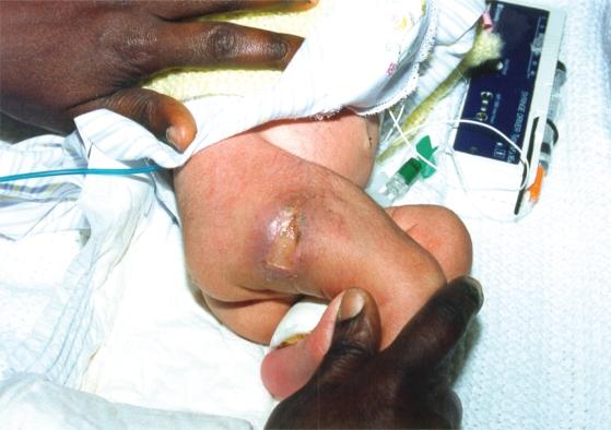
Figure 1b.
The same child 6 days after beginning of protein C replacement with Ceprotin®.
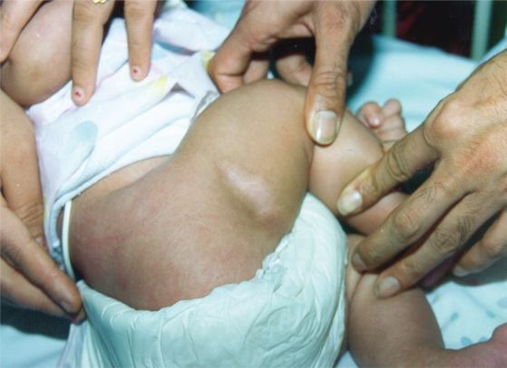
Figure 1c.
The same child 2 months later. All figures from Dreyfus M, Masterson M, David M, et al 1995. Replacement therapy with a monoclonal antibody purified protein C concentrate in newborns with severe congenital protein C deficiency. Semin Thromb Haemost, 21:371–81. Copyright © 1995. Reprinted by permission Thieme Publishers.
Acknowledgments
I gratefully acknowledge the assistance and positive input from Dr. Alex Veldman, Newborn Services, Monash Medical Centre, Monash University, Melbourne, Australia and Bruno Eberspaecher, Baxter Germany, Bioscience.
References
- Alhenc-Gelas M, Emmerich J, Gandrille S, et al. Protein C infusion in a patient with inherited protein C deficiency caused by two missense mutations: Arg 178 to Gln and Arg-1 to His. Blood Coagul Fibrinolysis. 1995;6:35–41. doi: 10.1097/00001721-199502000-00006. [DOI] [PubMed] [Google Scholar]
- Allaart CF, Poort SR, Rosendaal FR, et al. Increased risk of venous thrombosis in carriers of hereditary protein C deficiency defect. Lancet. 1993;341:134–8. doi: 10.1016/0140-6736(93)90003-y. [DOI] [PubMed] [Google Scholar]
- Auberger K. Evaluation of a new protein-C concentrate and comparison of protein-C assays in a child with congenital protein-C deficiency. Ann Hematol. 1992;64:146–51. doi: 10.1007/BF01697402. [DOI] [PubMed] [Google Scholar]
- Baliga V, Thwaites R, Tillyer ML, et al. Homozygous protein C deficiency-management with protein C concentrate. Eur J Pediatr. 1995;154:534–8. doi: 10.1007/BF02074829. [DOI] [PubMed] [Google Scholar]
- Barnes C, Newall F, Higgins S, et al. Perinatal management of patients at high risk of homozygous protein C deficiency. Thromb Haemost. 2002;88:370–1. [PubMed] [Google Scholar]
- Beckman RJ, Schmidt RJ, Santerre RF, et al. The structure and evolution of a 461 amino acid human protein C precursor and its messenger RNA, based upon the DNA sequence of cloned human liver cDNA’s. Nucl Acids Res. 1985;13:5233. doi: 10.1093/nar/13.14.5233. [DOI] [PMC free article] [PubMed] [Google Scholar]
- Bertina RM, Broekmans AW, van der Linden IK, et al. Protein C deficiency in a Dutch family with thrombotic disease. Thromb Haemost. 1982;48:1–5. [PubMed] [Google Scholar]
- Bertina RM, Koeleman BPC, Koster T, et al. Mutation in blood coagulation factor V associated with resistance to activated protein C. Nature. 1994;369:64–7. doi: 10.1038/369064a0. [DOI] [PubMed] [Google Scholar]
- Bovill EG, Bauer KA, Dickerman JD, et al. The clinical-spectrum of heterozygous protein C deficiency in a large new England Kindred. Blood. 1989;73:712–7. [PubMed] [Google Scholar]
- Branson HE, Katz K, Marble R, et al. Inherited protein C deficiency and coumarin-responsive chronic relapsing purpura fulminans in a newborn infant. Lancet. 1983;2:1165–8. doi: 10.1016/s0140-6736(83)91216-3. [DOI] [PubMed] [Google Scholar]
- Broekmans AW, Bertina RM, Loeliger EA, et al. Protein C and the development of skin necrosis during anticoagulant therapy. Thromb Haemost. 1983;49:244. [PubMed] [Google Scholar]
- Brueckmann M, Horn S, Lang S, et al. Recombinant human activated protein C upregulates cyclooxygenase-2 expression in endothelial cells via binding to endothelial cell protein C receptor and activation of protease-activated receptor-1. Thromb Haemost. 2005;93:743–50. doi: 10.1160/TH04-08-0511. [DOI] [PubMed] [Google Scholar]
- Clarke RC, Johnston JR, Mayne EE. Meningococcal septicaemia: treatment with protein C concentrate. Intensive Care Med. 2000;26:471–3. doi: 10.1007/s001340051184. [DOI] [PubMed] [Google Scholar]
- Conard J, Bauer KA, Gruber A, et al. Normalization of markers of coagulation activation with a purified protein C concentrate in adults with homozygous protein C deficiency. Blood. 1993;82(4):1159–64. [PubMed] [Google Scholar]
- Dahlbäck B, Carlsson M, Svensson PJ. Familial thrombophilia due to a previously unrecognized mechanism characterized by poor anticoagulant response to activated protein C: Prediction of a cofactor to activated protein C. Proc Natl Acad Sci USA. 1993;90:1004. doi: 10.1073/pnas.90.3.1004. [DOI] [PMC free article] [PubMed] [Google Scholar]
- Dahlbäck B, Stenflo J. Philadelphia: W.B. Saunders Company; 1994. The protein C anticoagulant system. In The Molecular Basis of Blood Diseases; p. 599. [Google Scholar]
- De Fouw NJ, de Jong YF, Haverkate F, et al. Activated protein C increases fibrin clot lysis by neutralization of plasminogen activator inhibitor – no evidence for a cofactor role of protein S. Thromb Haemost. 1988;60:328–33. [PubMed] [Google Scholar]
- De Kleijn ED, de Groot R, Hack CE, et al. Activation of protein C following infusion of protein C concentrate in children with severe meningococcal sepsis and purpura fulminans: a randomized, double-blinded, placebo-controlled, dose-finding study. Crit Care Med. 2003;31:1839–47. doi: 10.1097/01.CCM.0000072121.61120.D8. [DOI] [PubMed] [Google Scholar]
- De Stefano V, Mastrangelo S, Schwarz HP, et al. Replacement therapy with a purified protein C concentrate during initiation of oral anticoagulation in severe protein C congenital deficiency. Thromb Haemost. 1993;70:247–9. [PubMed] [Google Scholar]
- Dreyfus M, Magny JF, Bridey F, et al. Treatment of homozygous protein C deficiency and neonatal purpura fulminans with a purified protein C concentrate. N Engl J Med. 1991;325:1565–8. doi: 10.1056/NEJM199111283252207. [DOI] [PubMed] [Google Scholar]
- Dreyfus M, Masterson M, David M, et al. Replacement therapy with a monoclonal antibody purified protein C concentrate in newborns with severe congenital protein C deficiency. Semin Thromb Hemost. 1995;21:371–81. doi: 10.1055/s-2007-1000658. [DOI] [PubMed] [Google Scholar]
- Dreyfus M, Ladouzi A, Chambost H, et al. Treatment of inherited protein C deficiency by replacement therapy with the French purified plasma-derived protein C concentrate (PROTEXEL) Vox Sang. 2007;93:233–40. doi: 10.1111/j.1423-0410.2007.00953.x. [DOI] [PubMed] [Google Scholar]
- Esmon CT. The regulation of natural anticoagulant pathways. Science. 1987;235:1348–52. doi: 10.1126/science.3029867. [DOI] [PubMed] [Google Scholar]
- Esmon CT. The roles of protein C and thrombomodulin in the regulation of blood coagulation. J Biol Chem. 1989;264:4743–6. [PubMed] [Google Scholar]
- Esmon CT. Role of coagulation inhibitors in inflammation. Thromb Haemost. 2001;86:51–6. [PubMed] [Google Scholar]
- Esmon CT. The protein C pathway. Chest. 2003;124(3 Suppl):26S–32S. doi: 10.1378/chest.124.3_suppl.26s. [DOI] [PubMed] [Google Scholar]
- Esmon CT. Inflammation and the activated protein C anticoagulant pathway. Semin Thromb Hemost. 2006;32(Suppl 1):49–60. doi: 10.1055/s-2006-939554. [DOI] [PubMed] [Google Scholar]
- Esmon CT, Xu J, Gu JM, et al. Endothelial Protein C Receptor. Thromb Haemost. 1999;82:251–8. [PubMed] [Google Scholar]
- Ettingshausen CE, Veldman A, Beeg T, et al. Replacement therapy with protein C concentrate in infants and adolescents with meningococcal sepsis and purpura fulminans. Semin Thromb Hemost. 1999;25:537–41. doi: 10.1055/s-2007-994962. [DOI] [PubMed] [Google Scholar]
- Favier R, Deschamps A, Belhocine R, et al. Simultaneous administration of antithrombin III and protein C concentrates for the treatment of a devastating coagulopathy in a child. Hematol Cell Ther. 1998;40:67–70. [PubMed] [Google Scholar]
- Feistritzer C, Riewald M. Endothelial barrier protection by activated protein C through PAR1-dependent sphingosine 1–phosphate receptor-1 crossactivation. Blood. 2005;105:3178–84. doi: 10.1182/blood-2004-10-3985. [DOI] [PubMed] [Google Scholar]
- Flood EP, Redish MH, Bociek SJ, et al. Thrombophlebitis migrans disseminata : report of a case in which gangrene of a breast occured. N J State J Med. 1943;43:1121–4. [Google Scholar]
- Fouassier M, Moreau D, Thiolliere F, et al. Evolution of thrombin formation and fibrinolysis markers, including thrombin-activatable fibrinolysis inhibitor, during severe meningococcemia. Pathophysiol Haemost Thromb. 2005;34:284–7. doi: 10.1159/000093109. [DOI] [PubMed] [Google Scholar]
- Fourrier F, Chopin C, Goudemand J, et al. Septic shock, multiple organ failure, and disseminated intravascular coagulation. Compared patterns of antithrombin III, protein C, and protein S deficiencies. Chest. 1992;101:816–23. doi: 10.1378/chest.101.3.816. [DOI] [PubMed] [Google Scholar]
- Fourrier F, Leclerc F, Aidan K, et al. Combined antithrombin and protein C supplementation in meningococcal purpura fulminans: a pharmacokinetic study. Intensive Care Med. 2003;29:1081–7. doi: 10.1007/s00134-003-1784-1. [DOI] [PubMed] [Google Scholar]
- Gatti L, Carnelli V, Rusconi R, et al. Heparin-induced thrombocytopenia and warfarin-induced skin necrosis in a child with severe protein C deficiency: successful treatment with dermatan sulfate and protein C concentrate. J Thromb Haemost. 2003;1:387–8. doi: 10.1046/j.1538-7836.2003.00057.x. [DOI] [PubMed] [Google Scholar]
- Gerson WT, Dickerman JD, Bovill EG, et al. Severe acquired protein C deficiency in purpura fulminans associated with disseminated intra-vascular coagulation: treatment with protein C concentrate. Pediatrics. 1993;91:418–22. [PubMed] [Google Scholar]
- Goodwin TM, Gazit G, Gordon EM. Heterozygous protein C deficiency presenting as severe protein C deficiency and peripartum thrombosis: successful treatment with protein C concentrate. Obstet Gynecol. 1995;86:662–4. doi: 10.1016/0029-7844(95)00039-t. [DOI] [PubMed] [Google Scholar]
- Greengard JS, Fisher CL, Villoutreix B, et al. Structural basis for Type I and Type II deficiencies of antithrombotic plasma protein C: Patterns revealed by three-dirnensional molecular modelling of mutations of the protease domain. Proteins. 1994;18:367. doi: 10.1002/prot.340180407. [DOI] [PubMed] [Google Scholar]
- Griffin JH, Evatt B, Zimmerman TS, et al. Deficiency of protein C in congenital thrombotic disease. J Clin Invest. 1981;68:1370–3. doi: 10.1172/JCI110385. [DOI] [PMC free article] [PubMed] [Google Scholar]
- Hattenbach LO, Beeg T, Kreuz W, et al. Ophthalmic manifestation of congenital protein C deficiency. J AAPOS. 1999;3:188–90. doi: 10.1016/s1091-8531(99)70066-2. [DOI] [PubMed] [Google Scholar]
- Joyce DE, Grinnell BW. Recombinant human activated protein C attenuates the inflammatory response in endothelium and monocytes by modulating nuclear factor-kappaB. Crit Care Med. 2002;30(5 Suppl):S288–93. doi: 10.1097/00003246-200205001-00019. [DOI] [PubMed] [Google Scholar]
- Kelly JG, O’Malley K. Clinical pharmakokinetics of oral anticoagulants. Clin Pharmacokinet. 1979;4:1–11. doi: 10.2165/00003088-197904010-00001. [DOI] [PubMed] [Google Scholar]
- Kreuz W, Veldman A, Escuriola-Ettingshausen C, et al. Protein-C concentrate for meningococcal purpura fulminans. Lancet. 1998;351:986–7. doi: 10.1016/S0140-6736(05)60653-8. [DOI] [PubMed] [Google Scholar]
- Lane DA, Mannucci PM, Bauer KA, et al. Inherited thrombophilia: part 2. Thromb Haemost. 1997;76:824–34. [PubMed] [Google Scholar]
- Leclerc F, Cremer R, Leteurtre S, et al. Protein C concentrate and recombinant tissue plasminogen activator in meningococcal septic shock. Crit Care Med. 2000;28:1694–7. doi: 10.1097/00003246-200005000-00106. [DOI] [PubMed] [Google Scholar]
- Lewandowski K, Zawilska K. Protein C concentrate in the treatment of warfarin-induced skin necrosis in the protein C deficiency. Thromb Haemost. 1994;71:395. [PubMed] [Google Scholar]
- Macias WL, Nelson DR. Severe protein C deficiency predicts early death in severe sepsis. Crit Care Med. 2004;32(Suppl):S223–8. doi: 10.1097/01.ccm.0000126120.49367.ac. [DOI] [PubMed] [Google Scholar]
- Mesters RM, Helterbrand J, Utterback BG, et al. Prognostic value of protein C concentrations in neutropenic patients at high risk of severe septic complications. Crit Care Med. 2000;28:2209–16. doi: 10.1097/00003246-200007000-00005. [DOI] [PubMed] [Google Scholar]
- Manco-Johnson M, Nuss R. Protein C concentrate prevents peripartum thrombosis. Am J Hematol. 1992;40:69–70. doi: 10.1002/ajh.2830400116. [DOI] [PubMed] [Google Scholar]
- Manco-Johnson MJ, Knapp-Clevenger R. Activated protein C concentrate reverses purpura fulminans in severe genetic protein C deficiency. J Pediatr Hematol Oncol. 2004;26:25–7. doi: 10.1097/00043426-200401000-00008. [DOI] [PubMed] [Google Scholar]
- Marlar RA, Montgomery RR, Broekmans AW. Diagnosis and treatment of homozygous protein C deficiency. Report of the Working Party on Homozygous Protein C Deficiency of the Subcommittee on Protein C and Protein S, International Committee on Thrombosis and Haemostasis. J Pediatr. 1989;114:528–34. doi: 10.1016/s0022-3476(89)80688-2. [DOI] [PubMed] [Google Scholar]
- Mathias M, Khair K, Burgess C, et al. Subcutaneous administration of protein C concentrate. Pediatr Hematol Oncol. 2004;21:551–6. doi: 10.1080/08880010490477365. [DOI] [PubMed] [Google Scholar]
- Minford AM, Parapia LA, Stainforth C, Lee D. Treatment of homozygous protein C deficiency with subcutaneous protein C concentrate. Br J Haematol. 1996;93:215–6. doi: 10.1046/j.1365-2141.1996.4691021.x. [DOI] [PubMed] [Google Scholar]
- Mosnier LO, Zlokovic BV, Griffin JH. The cytoprotective protein C pathway. Blood. 2007;109:3161–72. doi: 10.1182/blood-2006-09-003004. [DOI] [PubMed] [Google Scholar]
- Muller FM, Ehrenthal W, Hafner G, et al. Purpura fulminans in severe congenital protein C deficiency: monitoring of treatment with protein C concentrate. Eur J Pediatr. 1996;155:20–5. doi: 10.1007/BF02115621. [DOI] [PubMed] [Google Scholar]
- Mustafa S, Mannhalter C, Rintelen C, et al. Clinical features of thrombophilia in families with gene defects in protein C or protein S combined with factor V Leiden. Blood Coagul Fibrinolysis. 1998;9:85–9. doi: 10.1097/00001721-199801000-00011. [DOI] [PubMed] [Google Scholar]
- Nakayama T, Matsushita T, Hidano H. A case of purpura fulminans is caused by homozygous delta8857 mutation (protein C-nagoya) and successfully treated with activated protein C concentrate. Br J Haematol. 2000;110:727–30. doi: 10.1046/j.1365-2141.2000.02230.x. [DOI] [PubMed] [Google Scholar]
- Okajima K. Regulation of inflammatory responses by natural anticoagulants. Immunol Rev. 2001;184:258–74. doi: 10.1034/j.1600-065x.2001.1840123.x. [DOI] [PubMed] [Google Scholar]
- Pabinger I, Allaart CF, Hermans J, et al. Hereditary protein C-deficiency. Laboratory values in transmitters and guidelines for the diagnostic procedure. Report on a study of the SSC Subcommittee on Protein C and Protein S. Thromb Haemost. 1992;68:470–4. [PubMed] [Google Scholar]
- Pabinger I, Kyrle PA, Heistinger M, et al. The risk of thrombosis in asymptomatic patients with hereditary protein C or protein S deficiency. A prospective cohort study. Thromb Haemost. 1994;71:441–5. [PubMed] [Google Scholar]
- Pabinger I, Schneider B. Thrombotic risk in hereditary antithrombin III, protein C, or protein S deficiency. A cooperative, retrospective study. Gesellschaft fuer Thrombose- und Haemostaseforschung (GTH) Study Group on Natural Inhibitors. Arterioscler Thromb Vasc Biol. 1996;16:742–8. doi: 10.1161/01.atv.16.6.742. [DOI] [PubMed] [Google Scholar]
- Radosevich M, Zhou1 F-L, Huart J-J, et al. Chromatographic purification and properties of a therapeutic human protein C concentrate. J Chromatography. 2003;790:199–207. doi: 10.1016/s1570-0232(03)00091-6. [DOI] [PubMed] [Google Scholar]
- Reitsma PH, Benuvdi F, Doig RG, et al. Protein C deficiency- a database of mutations, 1995 update. On behalf of the Subcommittee on Plasma Coagulation Inhibitors of the Scientifie and Standardization Committee of the ISTH. Thromb Haemost. 1995;73:876–89. [PubMed] [Google Scholar]
- Reitsma PH. Protein C deficiency: from gene defects to disease. Thromb Haemost. 1997;78:334–50. [PubMed] [Google Scholar]
- Richards EM, Makris M, Preston FE. The successful use of protein C concentrate during pregnancy in a patient with type 1 protein C deficiency, previous thrombosis and recurrent fetal loss. Br J Haematol. 1997;98:660–1. doi: 10.1046/j.1365-2141.1997.2873100.x. [DOI] [PubMed] [Google Scholar]
- Riewald M, Petrovan RJ, Donner A, et al. Activation of endothelial cell protease activated receptor 1 by the protein C pathway. Science. 2002;296:1880–2. doi: 10.1126/science.1071699. [DOI] [PubMed] [Google Scholar]
- Rintala E, Seppala OP, Kotilainen P, et al. Protein C in the treatment of coagulopathy in meningococcal disease. Crit Care Med. 1998;26:965–8. doi: 10.1097/00003246-199805000-00038. [DOI] [PubMed] [Google Scholar]
- Rintala E, Kauppila M, Seppala OP, et al. Protein C substitution in sepsis-associated purpura fulminans. Crit Care Med. 2000;28:2373–8. doi: 10.1097/00003246-200007000-00032. [DOI] [PubMed] [Google Scholar]
- Rivard GE, David M, Farrell C, et al. Treatment of purpura fulminans in meningococcemia with protein C concentrate. J Pediatr. 1995;126:646–52. doi: 10.1016/s0022-3476(95)70369-1. [DOI] [PubMed] [Google Scholar]
- Salonvaara M, Kuismanen K, Mononen T, et al. Diagnosis and treatment of a newborn with homozygous protein C deficiency. Acta Paediatr. 2004;93:137–9. doi: 10.1080/08035250310007411. [DOI] [PubMed] [Google Scholar]
- Sanson BJ, Simioni P, Tormene D, et al. The incidence of venous thromboembolism in asymptomatic carriers of a deficiency of anti-thrombin, protein C, or protein S: a prospective cohort study. Blood. 1999;94:3702–6. [PubMed] [Google Scholar]
- Sanz-Rodriguez C, Gil-Fernandez JJ, Zapater P, et al. Long-term management of homozygous protein C deficiency: replacement therapy with subcutaneous purified protein C concentrate. Thromb Haemost. 1999;81:887–90. [PubMed] [Google Scholar]
- Schramm W, Spannagl M, Bauer KA, et al. Treatment of coumarin-induced skin necrosis with a monoclonal antibody purified protein C concentrate. Arch Dermatol. 1993;129:753–6. [PubMed] [Google Scholar]
- Sekiyama K, Itoh H, Sagawa N, et al. Successful management of a pregnant woman with heterozygous protein C deficiency using activated protein C concentrate. J Obstet Gynaecol Res. 2003;29:412–5. doi: 10.1111/j.1341-8076.2003.00139.x. [DOI] [PubMed] [Google Scholar]
- Sharon C, Tirindelli MC, Mannucci PM, et al. Homozygous protein C deficiency with moderately severe clinical symptoms. Thromb Res. 1986;41:483–8. doi: 10.1016/0049-3848(86)91693-2. [DOI] [PubMed] [Google Scholar]
- Shimamura K, Oka K, Nakazawa M, et al. Distribution patterns of microthrombi in disseminated intravascular coagulation. Arch Path Lab Med. 1983;107:543–7. [PubMed] [Google Scholar]
- Smith OP, White B, Vaughan D, et al. Use of protein-C concentrate, heparin, and haemodiafiltration in meningococcus-induced purpura fulminans. Lancet. 1997;350:1590–3. doi: 10.1016/s0140-6736(97)06356-3. [DOI] [PubMed] [Google Scholar]
- Stenflo J. A new vitamin K-dependent protein. Purification from bovine plasma and preliminary characterization. J Biol Chem. 1976;251:355–63. [PubMed] [Google Scholar]
- Sugimoto M, Maruhashi Y, Kobayashi A, et al. Activated protein C concentrate: a new tool for the treatment of acute thromboembolism in patients with congenital protein C deficiency. Thromb Haemost. 1997;77:1223–4. [PubMed] [Google Scholar]
- Tardy-Poncet B, Rayet I, Damon G, et al. Protein C concentrates in a neonate with a cerebral venous thrombosis due to heterozygous type 1 protein C deficiency. Thromb Haemost. 2001;85:1118–9. [PubMed] [Google Scholar]
- Toupance O, Nguyen P, Brandt B, et al. Prevention of vascular thrombosis by human purified protein C concentrate in a patient with familial PC deficiency undergoing renal transplantation. Transpl Int. 1994;7:144–5. doi: 10.1007/BF00336478. [DOI] [PubMed] [Google Scholar]
- Vaccarella G, Pelella R. Replacement treatment with protein C in an 18-year-old man with meningococcal sepsis and purpura fulminans. Minerva Anestesiol. 2003;69:693–5. [PubMed] [Google Scholar]
- Van de Wouwer M, Collen D, Conway EM. Thrombomodulin-protein C-EPCR system: integrated to regulate coagulation and inflammation. Arterioscler Thromb Vasc Biol. 2004;24:1374–83. doi: 10.1161/01.ATV.0000134298.25489.92. [DOI] [PubMed] [Google Scholar]
- Vukovich T, Auberger K, Weil J, et al. Replacement therapy for a homozygous protein C deficiency-state using a concentrate of human protein C and S. Br J Haematol. 1988;70:435–40. doi: 10.1111/j.1365-2141.1988.tb02513.x. [DOI] [PubMed] [Google Scholar]
- White B, Schmidt M, Murphy C, et al. Activated protein C inhibits lipopolysaccharide-induced nuclear translocation of nuclear factor kappaB (NF-kappaB) and tumour necrosis factor alpha (TNF-alpha) production in the THP-1 monocytic cell line. Br J Haemato. 2000a;110:130–4. doi: 10.1046/j.1365-2141.2000.02128.x. [DOI] [PubMed] [Google Scholar]
- White B, Livingstone W, Murphy C, et al. An open-label study of the role of adjuvant hemostatic support with protein C replacement therapy in purpura fulminans-associated meningococcemia. Blood. 2000b;96:3719–24. [PubMed] [Google Scholar]
- Yan SB, Helterbrand JD, Hartman DL, et al. Low levels of protein C are associated with poor outcome in severe sepsis. Chest. 2001;120:915–22. doi: 10.1378/chest.120.3.915. [DOI] [PubMed] [Google Scholar]
- Ye X, Fukudome K, Tsuneyoshi N, et al. The endothelial cell protein C receptor (EPCR) functions as a primary receptor for protein C activation on endothelial cells in arteries, veins, and capillaries. Biochem Biophys Res Commun. 1999;259:671–7. doi: 10.1006/bbrc.1999.0846. [DOI] [PubMed] [Google Scholar]


