Abstract
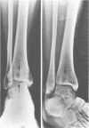
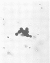
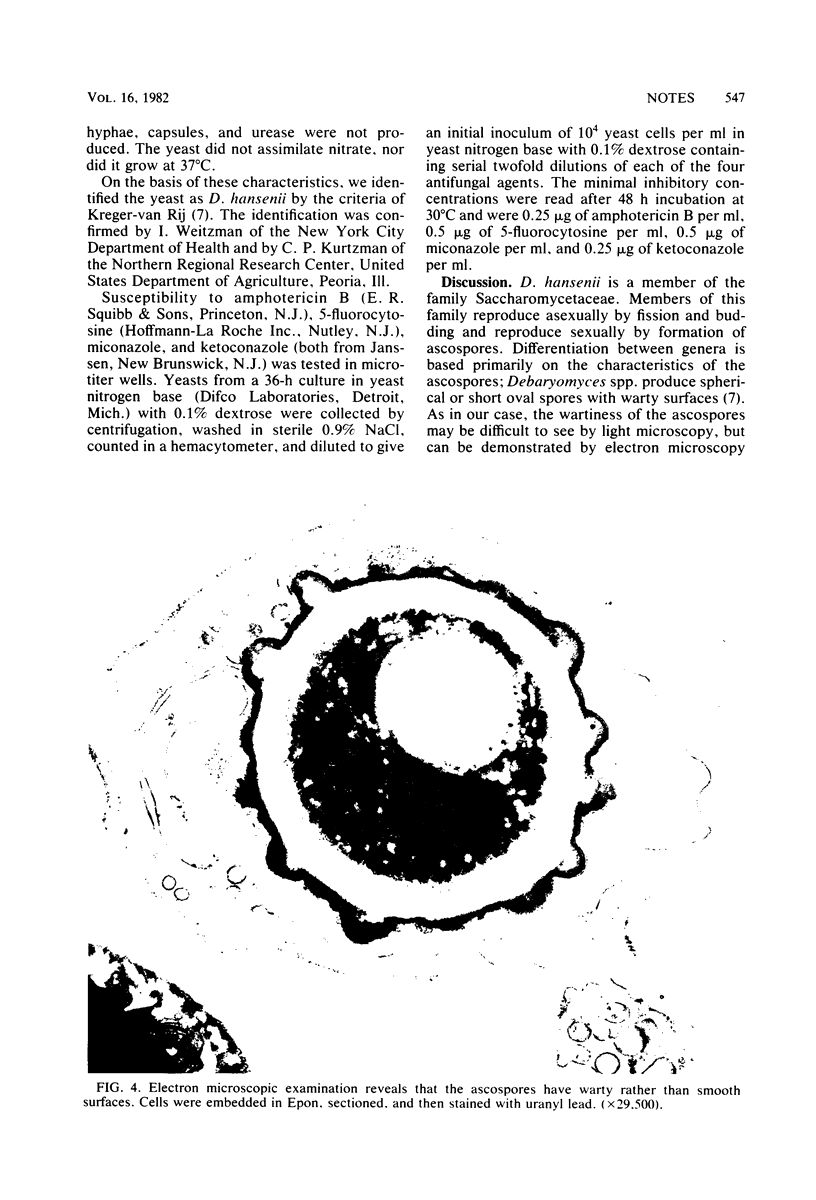
Debaryomyces hansenii was isolated from a cystic lesion in the distal tibia of a healthy 23-year-old woman. Ascospores were demonstrated when the organism was incubated at 25 but not at 30 degrees C. Electron microscopy was necessary to demonstrate the warty surface of the ascospore wall. Growth was inhibited in vitro by low concentrations of amphotericin B, 5-fluorocytosine, miconazole, and ketoconazole. Four previous cases of infection caused by Debaryomyces spp. and one caused by the related fungus Torulopsis candida were reviewed.
Full text
PDF
Images in this article
Selected References
These references are in PubMed. This may not be the complete list of references from this article.
- AJELLO L. Comments on the laboratory diagnosis of opportunistic fungus diseases. Lab Invest. 1962 Nov;11:1033–1034. [PubMed] [Google Scholar]
- BARNEY M., DODGE C. W., SIMMERS R. DEBARYOMYCES INFECTION IN VERMONT. Mycopathol Mycol Appl. 1963 Jun 15;19:246–254. doi: 10.1007/BF02051254. [DOI] [PubMed] [Google Scholar]
- Golubev V. I., Bab'eva I. P. Yeasts of the genus Debaryomyces Klöck in the nests of ants of the group Formica rufa L. Sov J Ecol. 1972 Jan-Feb;3(1):59–62. [PubMed] [Google Scholar]
- Joly S. Observaçes sôbre uma cepa de Torulopsis candida isolada de uma levedurose. Mycopathol Mycol Appl. 1969 May 28;37(4):369–371. doi: 10.1007/BF02129884. [DOI] [PubMed] [Google Scholar]
- KLIEWE H., HOFER J. Uber die Systematik eines pathogenen Sprosspilzes. Frankf Z Pathol. 1952 Jan;63(1):88–94. [PubMed] [Google Scholar]
- Kreger van Rij N. J., Veenhuis M. Electron microscopy of ascus formation in the yeast debaryomyces hansenii. J Gen Microbiol. 1975 Aug;89(2):256–264. doi: 10.1099/00221287-89-2-256. [DOI] [PubMed] [Google Scholar]


