Abstract
A homologue of the mammalian bilirubin transporter bilitranslocase (BTL) (TCDB 2.A.65.1.1), able to perform an apparent secondary active transport of flavonoids, has previously been found in carnation petals and red grape berries. In the present work, a BTL homologue was also shown in white berries from Vitis vinifera L. cv. Tocai/Friulano, using anti-sequence antibodies specific for rat liver BTL. This transporter, similarly to what found in red grape, was localized in the first layers of the epidermal tissue and in the vascular bundle cells of the mesocarp. In addition, a strong immunochemical reaction was detected in the placental tissue and particularly in peripheral integuments of the seed. The protein was expressed during the last maturation stages in both skin and pulp tissues and exhibited an apparent molecular mass of c. 31 kDa. Furthermore, the transport activity of such a carrier, measured as bromosulphophthalein (BSP) uptake, was detected in berry pulp microsomes, where it was inhibited by specific anti-BTL antibodies. The BTL homologue activity exhibited higher values, for both Km and Vmax, than those found in the red cultivar. Moreover, two non-pigmented flavonoids, such as quercetin (a flavonol) and eriodictyol (a flavanone), inhibited the uptake of BSP in an uncompetitive manner. Such results strengthen the hypothesis that this BTL homologue acts as a carrier involved also in the membrane transport of colourless flavonoids and demonstrate the presence of such a carrier in different organs and tissues.
Keywords: Bilitranslocase, flavonoid transport, immunohistochemistry, seed, Vitis vinifera L., white berry cultivar
Introduction
Flavonoids, among secondary metabolites, represent a noteworthy source of antioxidants in grapevine (Vitis vinifera L.) (Conde et al., 2007). These compounds include the classes of anthocyanins, flavonols, and flavan-3-ols, which subsequently polymerize to condensed tannins (or proanthocyanidins, PAs) (Adams, 2006).
Regarding flavonoid composition, red grape berry tissues differ from the white ones in the presence of anthocyanins and the hydroxylation pattern of flavonols and PAs. In fact, white grape cultivars are unable to synthesize anthocyanins and 3′,4′,5′-hydroxylated flavonoids, due to the absence of a specific expression of genes encoding for the UDP glucose:flavonoid 3-O-glucosyltransferase (UFGT) and the flavonoid 3′5′-hydroxylase (F3′5′H) (Bogs et al., 2006). In particular, pigmentation in red cultivars depends, exclusively, on the last step of anthocyanin biosynthesis, catalysed by UFGT, whose expression undergoes genetic control exerted by MYB-type transcription factors (Boss et al., 1996; Walker et al., 2007; Matus et al., 2009). Similarly, VvCytob5 is suggested to be a regulation factor involved in flavonol and PA biosynthesis (Bogs et al., 2006), by modulating the oxidative state of F3′5′H. These regulation mechanisms are correlated with the accumulation of the hydroxylated flavonols and PAs, as well as anthocyanins, occurring at different berry developmental stages. Regulation takes place early, before véraison in the case of PAs and flavonols, while for anthocyanins it occurs at the final maturation steps (Adams, 2006).
Grape berry flavonoids are mainly detected in the hypodermal layers of the skin and in the outer integument of the seed testa (Adams, 2006; Cadot et al., 2006). Moreover, flavonoids are absent from the white berry pulp, which contains only phenolic hydroxycinnamates (Adams, 2006). In particular, anthocyanins are found only in the berry skin of the red cultivars; genes responsible for colour variation have been extensively studied and their expression related to maturation stages or environmental conditions (Castellarin et al., 2006, 2007; Ageorges et al., 2006; Cutanda-Perez et al., 2009). Glycosylated flavonols are mainly accumulated in the skin of both red and white varieties. The latter contain a unique set of flavonol aglycone derivatives, namely quercetin, kaempferol, and isorhamnetin (3′-methylether of quercetin), with quercetin derivatives being the most representative (Mattivi et al., 2006). Conversely, they do not have the delphinidin-like flavonols myricetin, laricitrin, and syringetin, owing to, as mentioned above, the lack of expression of F3′5′H (Bogs et al., 2006). Their presence is cultivar-dependent and, therefore, the flavonol profile may be used to distinguish different varieties (Masa et al., 2007).
Tannins are another class of flavonoids, whose subunit composition changes in relation to seed or skin localization, while their degree of polymerization increases throughout the ripening process (He et al., 2008). The higher tannin condensation of grape skin is responsible for its organoleptic properties, since astringency and bitterness of PAs are known to be inversely correlated to the degree of polymerization (Downey et al., 2003).
The subcellular compartmentation of flavonoids in red berries has been demonstrated to involve the cell wall and vacuole, while less attention has been paid to white cultivars. Anthocyanins are known to be stored only in the vacuole as spherical pigmented inclusions, named anthocyanic vacuolar inclusions (AVIs) (Conn et al., 2003), whereas flavanols and tannins are also located in the cell wall (Gény et al., 2003; Gagné et al., 2006), where they exert a defensive role (Grotewold, 2004; Mayer et al., 2008). At the subcellular level, therefore, flavonoids are delivered from the cytoplasm, the site of their synthesis, to the accumulation targets.
Similarly, at the organ level, a long-distance transport could also be performed from ‘source’ to ‘sink’ tissues (Yazaki et al., 2008). The translocation of secondary metabolites, like flavonoids, across membranes has been suggested to occur by active transport processes, although the specific transporters involved have not yet been completely elucidated (Kutchan et al., 2005; Braidot et al., 2008a). In particular, it has been suggested that the flavonoid transfer from the cytoplasmic face of the endoplasmic reticulum to the vacuole involves primary and secondary active transporters (Kitamura, 2006). The first kind of carrier belongs to the ATP-binding cassette (ABC) protein family. These proteins, recently identified in grape (Zhang et al., 2008), are responsible for vacuolar sequestration or secretion of flavonoids, chlorophyll catabolites, and xenobiotic molecules (Klein et al., 2006; Yazaki et al., 2008). Conversely, secondary active transport (Law et al., 2008) may be mediated by multi-drug and toxic compound extrusion (MATE) proteins, as reported for PAs in Arabidopsis (Debeaujon et al., 2001) and tomato (Mathews et al., 2003), and for saponarin in barley (Marinova et al., 2007). Recent evidence shows the presence of a MATE transporter even in grape, as described by Cutanda-Perez et al. (2009) and Gomez et al. (2009).
Another secondary active transporter, homologue to mammalian bilitranslocase (BTL), has been described in carnation petals (Passamonti et al., 2005) and, more recently, in red grape berries (Braidot et al., 2008b). In the latter, this translocator is a membrane protein of c. 31 kDa located, at the subcellular level, on the microsomal membranes, while, at tissue level, it may be detected in teguments and vascular bundles. Its expression varies during the developmental stages (starting from véraison) and is strongly correlated with spatial and temporal anthocyanin accumulation. In addition, it is modulated by water deficit. Kinetic studies of the transport activity in red berry pulp microsomes (Braidot et al., 2008b) showed that this protein mediates an electrogenic transport of bromosulphophthalein (BSP). In particular, the transporter exhibits a Michaelis–Menten constant (Km=2.39 μM) similar to that of the mammalian homologue. Its activity is inhibited by an antibody raised against a specific BTL sequence and by quercetin, a colourless flavonol. These features, together with the protein localization in the vascular tissue, suggest the participation of the BTL homologue in the transport of colourless flavonoids and precursors, besides pigmented anthocyanins.
Considering these findings related to red grape, the present study aimed at demonstrating the presence of the BTL homologue in a white berry cultivar, where the synthesis and accumulation of some flavonoids (other than anthocyanins) occur early in berry maturation. According to this aim, BTL homologue transport activity and its inhibition by flavonoids were also characterized. Moreover, its expression was examined at different maturation stages, in order to compare this pattern to that observed in the red cultivar. Finally, immunohistochemical analysis was extended to seeds, to verify the presence of the BTL homologue in an organ representing a major source of tannins and PAs in grape.
Materials and methods
Plant material
Berries from Vitis vinifera L. cv. Tocai/Friulano were collected during the 2006 and 2007 growing seasons from a commercial vineyard ‘Tenuta Villanova di Farra d'Isonzo’, Italy, at different developmental stages. Developmental stages were: fruit set (27 d after anthesis); pre-véraison (40 d after anthesis); véraison (55 d after anthesis); maturation (74 d after anthesis); and harvest (95 d after anthesis). Random samples of at least 40 bunches from about 20 different plants were collected. For immunological assays, skin, pulp, and seed samples were obtained from grape berries and immediately stored at –80 °C.
Fresh grape berries, at the harvest stage, were used for transport assays.
Isolation of microsomes from berry skin and pulp
For immunoblot analysis, 50 g of grape berry skin and pulp were blended in 150 ml of 20 mM HEPES-TRIS (pH 7.6), 0.4 M sucrose, 5 mM EDTA, 1 mM DTE, and 1 mM phenylmethylsulphonyl fluoride at 4 °C. The homogenization process was performed as described in Braidot et al. (2008b). The supernatant was collected and considered as the cytosolic fraction, while the microsomal membrane fraction was resuspended in 0.25 M sucrose and 20 mM TRIS-HCl (pH 7.5).
For transport assays, 50 g of grape berry skin and pulp, only at the harvest stage, were blended in 150 ml of 0.5 M glucose (pH 8.2 with ascorbic acid), 80 mM PPi, 0.2% (w/v) bovine serum albumin (BSA), 1% (w/v) defatted casein, 1% (w/v) polyvinylpolypyrrolidone, 5 mM EDTA, 5 mM dithioerythritol, 20 mM β-mercaptoethanol, and 1 mM phenylmethylsulphonyl fluoride at 4 °C. The homogenization was performed as described in Terrier et al. (2001) and the microsomal fraction was resuspended in 20% (v/v) glycerol, 0.25 M sucrose, 20 mM HEPES-TRIS (pH 7.0) and 0.025% (w/v) BSA.
Immunoblotting
Electrophoresis of 25 μg protein from cytosolic and microsomal fractions isolated as previously described, for each different developmental stage, was carried out in 12% (w/v) polyacrylamide gel containing 0.1% (w/v) SDS. Gel was layered onto a nitrocellulose membrane to transfer the proteins by electroblotting. The nitrocellulose membrane was incubated with anti-BTL antibody (0.66 μg ml IgG-1), raised against a synthetic peptide corresponding to segment 235–246 of the primary structure of rat liver bilitranslocase as described by Passamonti et al. (2005). The immune reaction was developed by the activity of alkaline phosphatase, conjugated to anti-rabbit IgG (dilution of 1/2500) and densitometry analysis was carried out using the software Quantity One®, version 4.2.3. (Bio-Rad, Hercules, CA, USA). BTL levels are expressed relative to its homologue in skin of red cv. Merlot sampled at the maturation stage and processed as in Braidot et al. (2008b). Four independent immunoblots of different extracts were used for the calculation of the mean average ±SE. Incubation with preimmune serum was used as a negative control. Furthermore, a serial dilution of microsomal and cytosolic protein was performed to ensure the specificity of the antibody and a linear relationship between the amount of protein loaded and densitometry results.
Measurement of PPi-dependent proton gradient (ΔpH)
Acridine orange was used to monitor spectrofluorimetrically (Perkin Elmer spectrofluorimeter, model LS50B, MA, USA) proton-pumping in microsomal vesicles obtained from berry skin and pulp (Braidot et al., 2008b).
Transport activity assay
Transport activity of the BTL homologue was assayed spectrophotometrically by Agilent 1200 diode array and multiple wavelength detector (Agilent, Santa Clara, CA, USA). Microsomal protein (20 μg ml−1), obtained from fresh berry pulp at the harvest stage, and BSP (from 2–48 μM) were added to a stirred cuvette containing 2 ml of assay medium (0.2 M KCl, 20 mM HEPES-TRIS, pH 8.0). The reaction was started by the addition of 60 μg of valinomycin; the initial rate of the absorbance drop (wavelength pair 580–514 nm) was calculated by Agilent Instrument 1 software (Santa Clara, CA, USA) and referred to as electrogenic BSP uptake. Upon the addition of valinomycin, K+ ions diffuse into the microsomal vesicles, thus building up a membrane potential that provides the driving force for the secondary active transport of anionic BSP, as previously shown (Passamonti et al., 2005). Indeed, in mammalian cells it has been demonstrated that BSP uptake mediated by BTL is an uniport transport (Sottocasa et al., 1982; Miccio et al., 1990), activated by the transmembrane electrical potential.
Inhibition of the electrogenic BSP uptake was examined by adding 50 μM of different flavonoids (Extrasynthese, Lyon Nord, Genay Cedex, France) to the assay medium. Inhibition of the electrogenic BSP uptake by an anti-BTL antibody was examined after preincubation of the microsomal vesicles with the antibody (240, 300, 360, and 480 ng) at 25 °C for 5 min.
Data of the electrogenic BSP uptake and inhibition by flavonoids or the anti-BTL antibody were expressed as a mean value of independent experiments, performed on four different microsomal preparations.
Microscopy analysis
Immunofluorescence microscopy analysis was carried out on grape berries collected during the harvest stage. The samples were processed as described in Braidot et al. (2008b). Sections were incubated with the primary anti-BTL antibody (3.3 μg ml−1 IgG), whereas control sections were incubated with preimmune serum. The immune reaction was shown under UV light by the activity of the fluorescein isothiocyanate (FITC)-conjugated secondary antibody (dilution of 1/2500).
Immunohistochemical microscopy analysis was carried out on grape berries, collected at the pre-véraison, maturation, and harvest stages, and on seeds, collected at the harvest stage. The samples were processed as described in Braidot et al. (2008b). Sections were incubated with the primary anti-BTL antibody (3.3 μg ml−1 IgG), whereas control sections were incubated with preimmune serum. The immune reaction was developed by the activity of alkaline phosphatase, conjugated to anti-rabbit IgG (dilution of 1/2500).
Reagents and chemicals
All reagents were purchased from Sigma-Aldrich (St Louis, MO, USA), if not differently stated.
Results
Immunochemical analysis of the BTL homologue in microsomal fractions isolated from skin and pulp during the development of white grape berry
An immunoblot analysis of microsomal proteins, isolated from both skin and pulp of grape berries of V. vinifera cv. Tocai/Friulano, was carried out to detect the BTL homologue and to estimate its expression at different berry developmental stages. The antibody used was an affinity-purified polyclonal antibody raised against the segment 235–246 of the amino-acidic sequence of rat liver BTL (Passamonti et al., 2005).
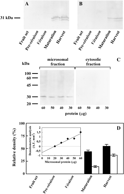
The results showed a cross-reactivity, involving a protein of c. 31 kDa, in the microsomal fraction isolated from both skin (Fig. 1A) and pulp (Fig. 1B) tissues. As previously reported by Braidot et al. (2008b), two very close bands were detected. The specificity of the antibody was also tested in both the microsomal and cytosolic fractions (Fig. 1C). This assay allowed the absence of other immunoreactive bands to be demonstrated and confirmed that the BTL homologue is localized only in the membrane fractions. Moreover, BTL homologue expression was undetectable in the negative controls (results not shown). BTL homologue expression began from the maturation stage until harvest. The densitometric analysis of the immunoblot staining (Fig. 1D) showed that its expression increased throughout the last two developmental stages in both skin and pulp. However, the signal associated with the microsomal fraction of the epicarp (black bars) was stronger than that of the mesocarp (white bars), particularly during the maturation stage. The accuracy of this approach was confirmed by the linear relationship among the amounts of the microsomal protein and the densitometry measurements detected in the pulp at the harvest stage (inset of Fig. 1D).
Fig. 1.
Immunoblotting of the BTL homologue carrier, during different developmental stages of grape berries. Proteins of microsomal fractions, obtained from skin (A) and pulp (B) of grape fruits, were probed against the anti-BTL antibody. Different amounts of microsomal and cytosolic proteins isolated from pulp at the maturation stage (C) were also probed against the same antibody. The value on the left represents the apparent molecular mass. (D) Densitometric analysis of the immunoblot. Data (n=4) are mean values ±SE, calculated from immunoblotting experiments performed on four independent microsomal preparations. Vertical bars represent: black, skin; white, pulp. Inset: correlation among increasing amounts of pulp microsomal protein from grape berries, collected at the harvest stage, and densitometry records.
Characterization of BTL homologue transport activity
In the plant tissues so far investigated, the BTL antibody, used in the above immunoblot, had been shown to inhibit the activity of a membrane transporter catalysing electrogenic BSP transport into microsomal vesicles (Passamonti et al., 2005; Braidot et al., 2008b).
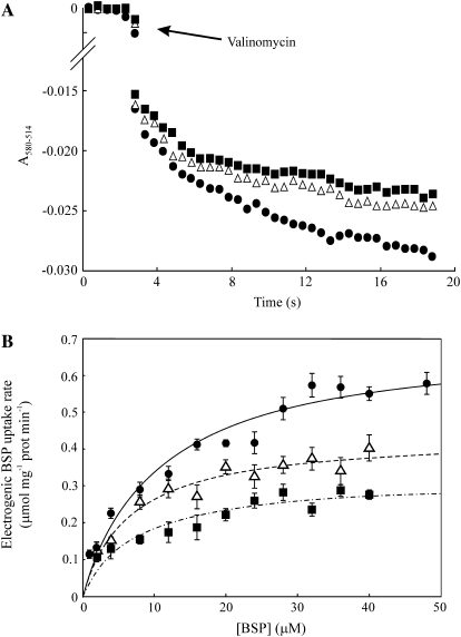
With the aim of investigating such specific transport activity in microsomal vesicles isolated from both skin and pulp of fresh berry at the harvest stage, preliminary tests of PPi-dependent H+-pumping were carried out in order to assess their compartmental intactness, a prerequisite for detecting any transport activity. The assay was identical to that described earlier by Braidot et al. (2008b) and the results were similar. Despite the higher expression of the BTL homologue protein found in berry skin, microsomal vesicles isolated from this tissue were not able to generate and maintain a membrane H+ gradient and to catalyse an electrogenic BSP transport (results not shown). Therefore, only vesicles obtained from berry pulp were used for the enzymatic characterization. As described previously (Passamonti et al., 2005; Braidot et al., 2008b), the transport assay is based on the generation of a K+ diffusion into the vesicles, as a consequence of the addition of valinomycin. The electrogenic potassium flux generates a transmembrane gradient. Such a gradient induces the movement of BSP (a pH-indicator phthalein substrate) from the assay medium to the vesicles, mediated by the BTL homologue. As shown in Fig. 2A (closed circles), the transport activity, measured as a decrease of BSP absorbance, was detected in pulp microsomes. In order to evaluate the kinetic properties of this transport activity, the effects of compounds belonging to different flavonoid classes, such as eriodictyol (a flavanone) and quercetin (a flavonol), on BSP uptake were tested. In both cases, the initial rate of the BSP uptake differed in comparison to the control (Fig. 2A, closed squares and open triangles, respectively). These inhibitory effects were further investigated under increasing BSP concentrations (Fig. 2B). The initial rate values could fit the Michaelis–Menten equation, whose parameters were Vmax=0.70±0.05 μmol BSP min−1 mg−1 protein and Km=11.12±2.35 μM BSP in the control assay (closed circles); Vmax=0.32±0.03 μmol BSP min−1 mg−1 protein and apparent Km (Km App)=7.35±2.22 μM BSP in the presence of quercetin (closed squares); Vmax=0.44±0.02 μmol BSP min−1 mg−1 protein and Km App=6.70±1.33 μM BSP in the presence of eriodictyol (open triangles). The flavonoids, by decreasing the values of both Km and Vmax, exerted an uncompetitive inhibition on electrogenic BSP uptake. Inhibition constants, calculated as Ki=[I]/(α′–1), where α′=Km/Km App was 97.48 μM and 75.76 μM for quercetin and eriodictyol, respectively.
Fig. 2.
Electrogenic BSP uptake into grape pulp microsomes and effect of two flavonoids. (A) Continuous spectrophotometric recording of electrogenic BSP uptake into microsomes. Each test was carried out by the addition of 60 μg valinomycin to a stirred cuvette containing 20 μg ml−1 microsomal protein in 2 ml of transport medium supplemented with 20 μM BSP without (closed circles) or with 50 μM of quercetin (closed squares) or eriodictyol (open triangles). Change of A580–514=0.005 corresponds to 1.04 nmol BSP. Traces are representative of experiments performed on four independent microsomal preparations. Other details are described in the Materials and methods. (B) Dependence of the initial rate of electrogenic BSP uptake on BSP concentration and the effect of two flavonoids. The assay was carried out as described in the Materials and methods, without (closed circles) and with 50 μM of either quercetin (closed squares) or eriodictyol (open triangles). Data (n=4) are means ±SE of the initial rate, fitted to the equation V=Vmax[BSP]/(Km+[BSP]).
Inhibition of electrogenic BSP uptake by anti-sequence BTL antibodies
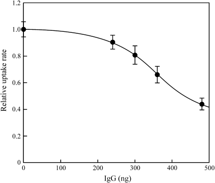
To test whether electrogenic BSP uptake into berry pulp microsomal vesicles was mediated by the protein identified in the above immunoblot, microsomes were preincubated with the same antibody and then assayed for electrogenic BSP uptake activity. Figure 3 shows the dependence of electrogenic BSP uptake upon increasing concentration of the antibody; the residual activity was about 30%, a value similar to that observed previously (Passamonti et al., 2005; Braidot et al., 2008b). Moreover, the transport activity was unaffected by bovine IgG or IgG purified from the rabbit preimmune serum (data not shown).
Fig. 3.
Effect of different amounts of anti-BTL antibody on BSP uptake in grape pulp microsomes. Each experiment was performed by preincubating 40 μg microsomal protein at 25 °C for 5 min with different amounts of anti-BTL antibody. BSP uptake was then measured as described in the Materials and methods. Data (n=4) are means ±SE of the initial rate expressed as a percentage with respect to the control (without anti-BTL antibody). Data were fitted to the sigmoid, four parameter equation y=y0+a/1+e–[(x–x0)/b], where y is relative transport activity, y0 is the residual relative transport activity (bottom level), a is the fraction of relative transport activity that is amenable to inhibition by the antibody, e=2.7183, x is the IgG dose, x0 is the IgG dose causing half-maximal inhibition, and b is the difference between the x values at 75% and 25% of the inhibition amplitude. The parameters found were: a=0.74±0.01; b=–65.3±1.6 ng; x0=350.5±1.9 ng; y0=0.36±0.01; r2=0.999.
Immunohistochemical analysis of the BTL homologue in white grape berries and seeds at the harvest stage
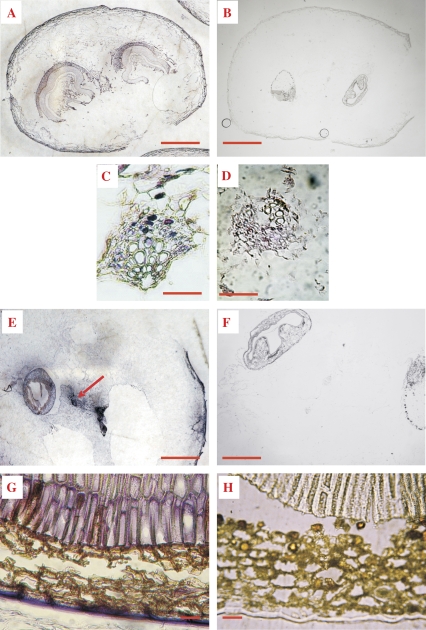
The above-described immunochemical results corroborate the use of this antibody specifically to localize the BTL homologue, by immunohistochemistry, in intact white berries at the harvest stage. Sections of the berry with seeds (Fig. 4A) showed that the BTL homologue was expressed not only in the epicarp and hypodermal layers, but also in seeds. Higher magnification images of the mesocarp showed a cross-reaction, indicated by violet staining, corresponding to the vascular bundle cells (Fig. 4C), especially in the phloem, and also in the placental tissue (Fig. 4E), localized in the centre of the berry. Furthermore, the distribution of the BTL homologue in seed tissues was investigated. In particular, the immunohistochemical analysis of the seed section exhibited a higher cross-reaction in some peripheral zones of the testa, namely the epidermis and palisade layer (Fig. 4G). The specificity of the cross-reaction in these experiments was confirmed probing the corresponding controls with preimmune serum (Fig. 4B, D, F, H).
Fig. 4.
Immunohistochemical microscopy analysis of the BTL homologue in grape berry and seed. Sections of grape fruits, collected during the maturation stage, were fixed and incubated with anti-BTL Ab (A, C, E, G) or preimmune serum (B, D, F, H) as described in the Materials and methods. The immune reaction was visualized under visible light with BCIP/NBT. Fruit berry with seeds (A, B; scale bar=2 mm); vascular bundles of the berry mesocarp (C, D; scale bar=100 μm); seed and maternal tissue of the berry (E, F; scale bar=1 mm); seed peripheral integuments (G, H; scale bar=25 μm). Arrow: cross-reaction in placenta tissue.
Immunohistochemical analysis of the BTL homologue during development of white grape berry
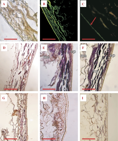
To characterize the localization of the BTL homologue in grape fruit better, an immunohistochemical analysis was performed in the peripheral tissues during berry development. Sections of grape berries, sampled at the maturation stage and examined in detail at the level of the pericarp, showed, under visible light, the typical parenchymatic and epidermal tissues (Fig. 5A). Under UV light, in the presence of the BTL antibody, a strong fluorescence signal was observed, mainly localized in the first layers of the epidermal tissue (Fig. 5B). In addition, the omission of the primary antibody (not shown) or the presence of preimmune serum (Fig. 5C), caused only a weak fluorescence signal, probably due to waxes localized in the epicuticular tissue.
Fig. 5.
Immunofluorescence and immunohistochemical microscopy analysis of the BTL homologue during development of grape berry. Sections of grape fruits, collected during the harvest stage, were fixed and incubated with the anti-BTL antibody (A, B) or preimmune serum (C). Sections were observed under visible light (A) or visualized with FITC under UV light (B, C). Sections of grape fruits, collected during pre-véraison (D, G), maturation (E, H), and harvest stage (F, I), were fixed and incubated with anti-BTL antibody (D, E, F) or preimmune serum (G, H, I). The immune reaction was visualized under visible light with BCIP/NBT. Scale bar=100 μm. Abbreviations: ep, epicarp; me, mesocarp; hy, hypodermal layers. Arrow: cuticular layer, unspecific reaction.
The expression of the BTL homologue, during different developmental stages, was further studied under visible light, using an alkaline phosphatase-catalysed colorimetric reaction. At the pre-véraison stage (Fig. 5D), the immunoreaction (violet staining) showed a faint signal. However, starting from maturation (Fig. 5E) up to harvest (Fig. 5F), the staining was clearly detectable, and the BTL homologue was localized in both the epicarp and the hypodermal layers. The highest cross-reactivity was associated with the harvest stage, consistently with the immunoblot data shown above (Fig. 1). The absence of unspecific reactions was confirmed by analysing the corresponding controls (with preimmune serum) at the pre-véraison, maturation and harvest stages, respectively (Fig. 5G–I).
Discussion
The flavonoid end-product transport in plant cells, from their site of biosynthesis towards vacuolar and other subcellular compartments, cannot be explained by a single model or mechanism. Much evidence shows, mainly by a genetic approach, that various carriers can mediate the transport of the most important flavonoids in plant cells. On the contrary, in grapevine, few reports, if any, have studied the transporters involved in flavonoid uptake at a subcellular level, by measuring transport activity. Nevertheless, some recent work has shown an overexpression of the MATE gene in relation to the synthesis of anthocyanins in the fruit (Cutanda-Perez et al., 2009; Gomez et al., 2009).
Recently, a BTL homologue has been identified in carnation petal teguments by immunohistochemical analysis, and a role for this carrier as a potential flavonoid transporter has been suggested by studying its transport activity in plasma membrane and tonoplast fractions (Passamonti et al., 2005). These results were then extended to red grape berry, where it has been demonstrated that the BTL homologue is responsible for an ATP-independent transport, which is inhibited by an anti-BTL antibody and by a colourless flavonol, quercetin (Braidot et al., 2008b). This protein is mainly present in tegumental cells, while, in the mesocarp, it is limited to the vascular tissues. In addition, its expression depends on the developmental stages of berry ripening.
Evidence is now supplied on the presence of the BTL homologue in a white grape berry (cv. Tocai/Friulano), in agreement with the above reported findings obtained in red grape berry (cv. Merlot). Although this cultivar, as well as the other white grapes, does not accumulate anthocyanins in the skin, the expression of the BTL homologue was similar to that shown in cv. Merlot. Immunofluorescence analysis on sections of grape berry, at the maturation stage, showed that the BTL homologue was mainly expressed in the hypodermal layers of the berry (Fig. 5B). The peripheral location of the homologue overlapped with the well-known pattern of flavonol and PAs accumulation in the skin of white varieties (Adams, 2006; Mattivi et al., 2006). In parallel, immunolabelling with alkaline phosphatase allowed BTL homologue expression during the development of berry fruit to be characterized better (Fig. 5D–F). Similarly to what found in the red grape berry, the presence of the BTL homologue in the skin tissues was larger in the berries sampled during the final period of maturation. This observation was confirmed by immunochemical analysis, which allowed a cross-reacting microsomal protein to be identified in both skin and pulp only from ripened grape berries (Fig. 1). The molecular size of the protein, reacting with the anti-BTL antibody, was also similar to that found in red grape (c. 31 kDa). Nevertheless, the BTL homologue expression in cv. Tocai/Friulano showed some peculiarities. The protein was expressed to a lesser extent in both skin and pulp, if compared to the strong signal found in red grape (Braidot et al., 2008b); its presence was confined just to the last two developmental stages (maturation and harvest), whereas in cv. Merlot it increased throughout the maturation process. A physiological explanation for this different behaviour could be the absence of anthocyanin accumulation in the white skin of cv. Tocai/Friulano. Conversely, even if flavonol synthesis in the skin of white varieties occurs early during maturation, a second period of accumulation of these compounds takes place from véraison until the final stages of berry maturation (Downey et al., 2006). Consequently, it could be suggested that the BTL homologue might function in the intracellular transport of colourless flavonoids, besides anthocyanins in red varieties, and that its contribution might be predominant only in late berry maturation.
The presence of the BTL homologue in microsomal membranes isolated from grape pulp was further confirmed by immunohistochemical microscope observations (Fig. 4C), showing that the BTL homologue localized in the phloem cells in the vascular bundles. These observations, already reported for red grape berry, suggest that this transporter could also be involved in the intercellular phloem translocation of secondary metabolites, such as colourless flavonoids and their precursors, towards the sites of accumulation in the berry, by analogy to similar pathways already proposed by Yazaki et al. (2008). Interestingly, recent work in transgenic tomatoes expressing the AtMYB75 gene showed anthocyanin production in different organs (leaves, stems, roots, flowers, and fruits) and also accumulation in cells close to the vascular bundles (Zuluaga et al., 2008).
This assumption could be justified by the observation that, during the last developmental stages of the berry, a shift from xylem (Wada et al., 2008) to phloem translocation occurs (Bondada et al., 2005). Thus, transport along the phloem tissue seems to be the preferential pathway after grape berry véraison. Furthermore, another change during the late maturation stages has been observed concerning phloem unloading, which is no longer symplastic but apoplastic (Zhang et al., 2006); this behaviour could be a general feature, since it has also been observed in apple fruit (Zhang et al., 2004).
The study of BTL homologue localization was extended to seeds, where flavonoids are accumulated in the outer integuments of the testa (Fig. 4E, G). Interestingly, immunohistochemical observations, performed on sections of the whole berry at the maturation stage, showed that the BTL homologue was also present in the maternal tissues in the proximity of the seeds. This finding leads to the supposition that this translocator could be implicated in the import of flavonoid precursors into developing seeds, which represent strong sink compartments during berry ripening. At the same time, some species possess modified uptake seed cells, enriched in plasmodesmata, supporting the symplasmic transfer of nutrients and/or secondary metabolites (Patrick and Offler, 2001). This mechanism also appears to be shared by grape seed, which exhibits this type of transfer cells in the palisade layer of the seed coat (the inner layer of the outer integument) (Walker et al., 1999). Different from berry skin, the immunoreactive signal in grape seed of cv. Tocai/Friulano was found in these layers, while it was absent from the layers, where a major accumulation of flavonoids occurs, as reported by Cadot et al. (2006). This result therefore supports a putative role of the BTL homologue, at least in white grapes, in delivering PA precursors from the external placenta to the seed coat layers devoted to their final accumulation.
The putative involvement of the BTL homologue in the transport of flavonoid precursors into the seeds, as well as its participation in phloematic transport, requires additional studies. In particular, it would be interesting to confirm the presence of the carrier in the seeds of red cultivar or teinturier varieties and to verify its localization in the phloem tissues of stalk or other organs.
To confirm further that the BTL homologue, identified by immunochemical analysis, could function as a flavonoid carrier, BSP uptake was followed in microsomal vesicles isolated from fresh grape pulp (Fig. 2). The unexpected lack of competence in skin microsomes and the consequent failure to detect BSP uptake, may be justified by the complex organic composition of this tissue (presence of cutin, waxes, and tannins), which might prevent the formation of sealed vesicles during the isolation procedure. In addition, in this tissue, the transporter might be lesser active owing to membrane leakage, which may be expected in senescing tissues (Thompson, 1988; Braidot et al., 1993).
BSP uptake was specifically inhibited by the anti-BTL antibody (Fig. 3). By analogy with red grape, the existence of a secondary active transport of this phthalein, which possesses similarity in its molecular structure with flavonoid compounds, was confirmed. This transport was mediated by a carrier-like enzyme, as shown by the curve obtained by plotting BSP uptake rate as a function of the substrate, and by calculation of its specific kinetic parameters, Vmax and Km. These values, compared with those measured in the red variety, show a lower affinity of the carrier for the transport substrate BSP, whereas the Vmax of electrogenic BSP transport is slightly higher.
The effect of non-pigmented flavonoids, such as quercetin (a flavonol) and eriodictyol (a flavanone), on the BSP electrogenic transport (Fig. 2) was assayed. Different from red grape, where it displayed a competitive inhibition (Ki=4.2 μM, Braidot et al., 2008b), quercetin, the more representative flavonol in white berries, showed a much lower affinity for the carrier. The same considerations are true for eriodictyol. The different affinity displayed by the two BTL homologues for artificial and natural substrates could be related to the different composition of the transported secondary metabolites present in the two cultivars studied.
Both flavonoids had Ki values for the BTL homologue larger by one order of magnitude and induced uncompetitive inhibition. Hence, the inhibitor binding site appears to be different from the active site for BSP and flavonoids seem to be able to bind only to the enzyme–substrate complex. Consequently, these observations suggest the existence in the BTL homologue of multiple binding sites for different kinds of secondary metabolites. These results are in agreement with observations reporting that flavonoids act as uncompetitive inhibitors of the carriers/enzymes involved in both transport and detoxification of several metabolites or xenobiotics (Hayeshi et al., 2007; Wang et al., 2008). Nevertheless, it should also be considered that the electrogenic BSP transport assay conditions do not exactly reflect the physiological intracellular conditions. The in vitro conditions might induce some unusual conformational change in the carrier, so that a competitive inhibitor, under physiological conditions, might act as an uncompetitive one under different milieu conditions. A temperature shift from 27 °C to 37 °C was shown to be enough for such a change on inhibition modality (Ajloo et al., 2007). Such distinctive features are also shared by mammalian BTL, whose ability to transport several metabolites is well-known, ranging from biline pigments to flavonoids (Karawajczyk et al., 2007; Passamonti et al., 2009) and possessing a minimal common structure (that of nicotinic acid). In a previous work, dealing with carnation, it has been shown that the plant homologue also exhibits multiple surface-accessible binding sites, specific for the various substrates and distinct from the catalytic (transport) site (Passamonti et al., 2005).
On the basis of the results presented above, it is suggested that white grape berries could represent a suitable model to investigate the secondary transport of colourless flavonoids, avoiding any interference due to high pigment concentration as happens in the red cultivar. Further research on the expression and function of the BTL homologue could involve other grape varieties, to confirm that only fine functional differences (kinetics and temporal pattern of expression) of the BTL homologues occur across the red/white cultivar division.
Acknowledgments
This research was supported by the ‘Tenuta Villanova’ Vineyard and Winery at Farra d'Isonzo, Italy, by the Regione Autonoma Friuli-Venezia Giulia (LR 26/2005 art. 17) and by the Ministero dell'Istruzione, Università e Ricerca (PRIN project 2004070118). We are grateful to Dr Michela Terdoslavich (University of Trieste) for the preparation of the anti-BTL antibody.
Glossary
Abbreviations
- ABC
ATP binding cassette
- AVIs
anthocyanic vacuolar inclusions
- BCIP/NBT
5-bromo-4-chloro-3-indolyl phosphate/nitroblue tetrazolium
- BSA
bovine serum albumin
- BSP
bromosulphophthalein
- BTL
bilitranslocase
- ΔpH
transmembrane proton gradient
- F3′5′H
flavonoid 3′5′-hydroxylase
- FITC
fluorescein isothiocyanate
- IgG
immunoglobulin G
- MATE
multi-drug and toxic compound extrusion
- PAs
proanthocyanidins
- PPi
inorganic pyrophosphate
- UFGT
UDP-glucose:flavonoid 3-O-glucosyltransferase
References
- Adams DO. Phenolics and ripening in grape berries. American Journal of Enology and Viticulture. 2006;57:249–256. [Google Scholar]
- Ageorges A, Fernandez L, Vialet S, Merdinoglu D, Terrier N, Romieu C. Four specific isogenes of the anthocyanin metabolic pathway are systematically co-expressed with the red colour of grape berries. Plant Science. 2006;170:372–383. [Google Scholar]
- Ajloo D, Saboury AA, Haghi-Asli N, Ataei-Jafarai G, Moosavi-Movahedi AA, Ahmadi M, Mahnam K, Namaki S. Kinetic, thermodynamic and statistical studies on the inhibition of adenosine deaminase by aspirin and diclofenac. Journal of Enzyme Inhibition and Medical Chemistry. 2007;22:395–406. doi: 10.1080/14756360701229085. [DOI] [PubMed] [Google Scholar]
- Bogs J, Ebadi A, McDavid D, Robinson SP. Identification of the flavonoid hydroxylases from grapevine and their regulation during fruit development. Plant Physiology. 2006;140:279–291. doi: 10.1104/pp.105.073262. [DOI] [PMC free article] [PubMed] [Google Scholar]
- Bondada BR, Matthews MA, Shackel KA. Functional xylem in the post-véraison grape berry. Journal of Experimental Botany. 2005;56:2949–2957. doi: 10.1093/jxb/eri291. [DOI] [PubMed] [Google Scholar]
- Boss PK, Davies C, Robinson SP. Expression of anthocyanin biosynthesis pathway genes in red and white grapes. Plant Molecular Biology. 1996;32:565–569. doi: 10.1007/BF00019111. [DOI] [PubMed] [Google Scholar]
- Braidot E, Zancani M, Petrussa E, Peresson C, Bertolini A, Patui S, Macrì F, Vianello A. Transport and accumulation of flavonoids in grapevine (Vitis vinifera L.) Plant Signaling and Behavior. 2008a;3:626–632. doi: 10.4161/psb.3.9.6686. [DOI] [PMC free article] [PubMed] [Google Scholar]
- Braidot E, Petrussa E, Bertolini A, Peresson C, Ermacora P, Loi N, Passamonti S, Terdoslavich M, Macrì F, Vianello A. Evidence for a putative flavonoid translocator similar to mammalian bilitranslocase in grape berries (Vitis vinifera L.) during ripening. Planta. 2008b;228:203–213. doi: 10.1007/s00425-008-0730-4. [DOI] [PubMed] [Google Scholar]
- Braidot E, Vianello A, Petrussa E, Macri F. Dissipation of the electrochemical proton gradient in phospholipase-induced degradation of plant mitochondria and microsomes. Plant Science. 1993;90:31–39. [Google Scholar]
- Cadot Y, Miñana-Castelló MT, Chevalier M. Anatomical, histological, and histochemical changes in grape seeds from Vitis vinifera L. cv. Cabernet franc during fruit development. Journal of Agricultural and Food Chemistry. 2006;54:9206–9215. doi: 10.1021/jf061326f. [DOI] [PubMed] [Google Scholar]
- Castellarin SD, Di Gaspero G, Marconi R, Nonis A, Peterlunger E, Paillard S, Adam-Blondon AF, Testolin R. Colour variation in red grapevines (Vitis vinifera L.): genomic organization, expression of flavonoid 3′-hydroxylase, flavonoid 3′,5′-hydroxylase genes and related metabolite profiling of red cyanidin-/blue delphinidin-based anthocyanins in berry skin. BMC Genomics. 2006;7:17–29. doi: 10.1186/1471-2164-7-12. [DOI] [PMC free article] [PubMed] [Google Scholar]
- Castellarin SD, Pfeiffer A, Sivilotti P, Degan M, Peterlunger E, Di Gaspero G. Transcriptional regulation of anthocyanin biosynthesis in ripening fruits of grapevine under seasonal water deficit. Plant. Cell and Environment. 2007;30:1381–1399. doi: 10.1111/j.1365-3040.2007.01716.x. [DOI] [PubMed] [Google Scholar]
- Conde C, Silva P, Fontes N, Dias ACP, Tavares RM, Sousa MJ, Agasse A, Delrot S, Gerós H. Biochemical changes throughout grape berry development and fruit and wine quality. Food. 2007;1:1–22. [Google Scholar]
- Conn S, Zhang W, Franco C. Anthocyanic vacuolar inclusions (AVIs) selectively bind acylated anthocyanins in Vitis vinifera L. (grapevine) suspension culture. Biotechnology Letters. 2003;25:835–839. doi: 10.1023/a:1024028603089. [DOI] [PubMed] [Google Scholar]
- Cutanda-Perez MC, Ageorges A, Gomez C, Vialet S, Terrier N, Romieu C, Torregrosa L. Ectopic expression of VlmybA1 in grapevine activates a narrow set of genes involved in anthocyanin synthesis and transport. Plant Molecular Biology. 2009;69:633–648. doi: 10.1007/s11103-008-9446-x. [DOI] [PubMed] [Google Scholar]
- Debeaujon I, Peeters AJ, Léon-Kloosterziel KM, Koornneef M. The TRANSPARENT TESTA12 gene of Arabidopsis encodes a multidrug secondary transporter-like protein required for flavonoid sequestration in vacuoles of the seed coat endothelium. The Plant Cell. 2001;13:853–871. doi: 10.1105/tpc.13.4.853. [DOI] [PMC free article] [PubMed] [Google Scholar]
- Downey MO, Harvey JS, Robinson SP. Analysis of tannins in seeds and skins of Shiraz grapes throughout berry development. Australian Journal of Grape Wine Research. 2003;9:15–27. [Google Scholar]
- Downey MO, Dokoozlian NK, Krstic MP. Cultural practice and environmental impacts on the flavonoid composition of grapes and wine: a review of recent research. American Journal of Enology and Viticulture. 2006;57:257–268. [Google Scholar]
- Gagné S, Saucier C, Gény L. Composition and cellular localization of tannins in Cabernet Sauvignon skins during growth. Journal of Agricultural and Food Chemistry. 2006;54:9465–9471. doi: 10.1021/jf061946g. [DOI] [PubMed] [Google Scholar]
- Gény L, Saucier C, Bracco S, Daviaud F, Glories Y. Composition and cellular localization of tannins in grape seeds during maturation. Journal of Agricultural and Food Chemistry. 2003;51:8051–8054. doi: 10.1021/jf030418r. [DOI] [PubMed] [Google Scholar]
- Gomez C, Terrier N, Torregrosa L, et al. Grapevine MATE-type proteins act as vacuolar H+-dependent acylated anthocyanin transporters. Plant Physiology. 2009;150:402–415. doi: 10.1104/pp.109.135624. [DOI] [PMC free article] [PubMed] [Google Scholar]
- Grotewold E. The challenges of moving chemicals within and out of cells: insights into the transport of plant natural products. Planta. 2004;219:906–909. doi: 10.1007/s00425-004-1336-0. [DOI] [PubMed] [Google Scholar]
- Hayeshi R, Mutingwende I, Mavengere W, Masiyanise V, Mukanganyama S. The inhibition of human glutathione S-transferases activity by plant polyphenolic compounds ellagic acid and curcumin. Food and Chemical Toxicology. 2007;45:286–295. doi: 10.1016/j.fct.2006.07.027. [DOI] [PubMed] [Google Scholar]
- He F, Pan Q-H, Shi Y, Duan C-Q. Biosynthesis and genetic regulation of proanthocyanidins in plants. Molecules. 2008;13:2674–2703. doi: 10.3390/molecules13102674. [DOI] [PMC free article] [PubMed] [Google Scholar]
- Karawajczyk A, Drgan V, Medic N, Oboh G, Passamonti S, Novic M. Properties of flavonoids influencing the binding to bilitranslocase investigated by neural network modelling. Biochemical Pharmacology. 2007;73:308–320. doi: 10.1016/j.bcp.2006.09.024. [DOI] [PubMed] [Google Scholar]
- Kitamura S. Transport of flavonoids: from cytosolic synthesis to vacuolar accumulation. In: Grotewold E, editor. Science of flavonoids. Berlin, Germany: Springer; 2006. pp. 123–146. [Google Scholar]
- Klein M, Burla B, Martinoia E. The multidrug resistance-associated protein (MRP/ABCC) subfamily of ATP-binding cassette transporters in plants. FEBS Letters. 2006;580:1112–1122. doi: 10.1016/j.febslet.2005.11.056. [DOI] [PubMed] [Google Scholar]
- Kutchan TM. A role for intra- and intercellular translocation in natural product biosynthesis. Current Opinion in Plant Biology. 2005;8:292–300. doi: 10.1016/j.pbi.2005.03.009. [DOI] [PubMed] [Google Scholar]
- Law CJ, Maloney PC, Wang DN. Ins and outs of major facilitator superfamily antiporters. Annual Review of Microbiology. 2008;62:289–305. doi: 10.1146/annurev.micro.61.080706.093329. [DOI] [PMC free article] [PubMed] [Google Scholar]
- Marinova K, Pourcel L, Weder B, Schwarz M, Barron D, Routaboul JM, Debeaujon I, Klein M. The Arabidopsis MATE transporter TT12 acts as a vacuolar flavonoid/H+-antiporter active in proanthocyanidin-accumulating cells of the seed coat. The Plant Cell. 2007;6:2023–2038. doi: 10.1105/tpc.106.046029. [DOI] [PMC free article] [PubMed] [Google Scholar]
- Masa A, Vilanova A, Pomar F. Varietal differences among the flavonoid profiles of white grape cultivars studied by high-performance liquid chromatography. Journal of Chromatography. 2007;1164:291–297. doi: 10.1016/j.chroma.2007.06.058. [DOI] [PubMed] [Google Scholar]
- Mathews H, Clendennen SK, Caldwell CG, et al. Activation tagging in tomato identifies a transcriptional regulator of anthocyanin biosynthesis, modification, and transport. The Plant Cell. 2003;15:1689–1703. doi: 10.1105/tpc.012963. [DOI] [PMC free article] [PubMed] [Google Scholar]
- Matus JT, Loyola R, Vega A, Peña-Neira A, Bordeu E, Arce-Johnson P, Alcalde JA. Post-véraison sunlight exposure induces MYB-mediated transcriptional regulation of anthocyanin and flavonol synthesis in berry skins of Vitis vinifera. Journal of Experimental Botany. 2009;60:853–867. doi: 10.1093/jxb/ern336. [DOI] [PMC free article] [PubMed] [Google Scholar]
- Mattivi F, Guzzon R, Vrhovsek U Stefanini M, Velasco R. Metabolite profiling of grape: flavonols and anthocyanins. Journal of Agricultural and Food Chemistry. 2006;54:7692–7702. doi: 10.1021/jf061538c. [DOI] [PubMed] [Google Scholar]
- Mayer R, Stecher G, Wurzner R, et al. Proanthocyanidins: target compounds as antibacterial agents. Journal of Agricultural and Food Chemistry. 2008;56:6959–6966. doi: 10.1021/jf800832r. [DOI] [PubMed] [Google Scholar]
- Miccio M, Lunazzi GC, Gazzin B, Sottocasa GL. Reconstitution of sulphobromophthalein transport in erythrocyte membranes induced by bilitranslocase. Biochimica et Biophysica Acta. 1990;1023:140–142. doi: 10.1016/0005-2736(90)90019-k. [DOI] [PubMed] [Google Scholar]
- Passamonti S, Cocolo A, Braidot E, Petrussa E, Peresson C, Medic N, Macri F, Vianello A. Characterization of electrogenic bromosulphophthalein transport in carnation petal microsomes and its inhibition by antibodies against bilitranslocase. FEBS Journal. 2005;272:3282–3296. doi: 10.1111/j.1742-4658.2005.04751.x. [DOI] [PubMed] [Google Scholar]
- Passamonti S, Terdoslavich M, Franca R, Vanzo A, Tramer F, Braidot E, Petrussa E, Vianello A. Bioavailability of flavonoids: a review of their membrane transport and the function of bilitranslocase in animal and plant organisms. Current Drug Metabolism. 2009;10:369–394. doi: 10.2174/138920009788498950. [DOI] [PubMed] [Google Scholar]
- Patrick JW, Offler CE. Compartmentation of transport and transfer events in developing seeds. Journal of Experimental Botany. 2001;52:551–564. [PubMed] [Google Scholar]
- Sottocasa GL, Baldini G, Sandri G, Lunazzi GC, Tiribelli C. Reconstitution in vitro of sulphobromophthalein transport by bilitranslocase. Biochimica et Biophysica Acta. 1982;685:123–128. doi: 10.1016/0005-2736(82)90088-8. [DOI] [PubMed] [Google Scholar]
- Terrier N, Sauvage FX, Ageorges A, Romieu C. Changes in acidity and in proton transport at the tonoplast of grape berries during development. Planta. 2001;213:20–28. doi: 10.1007/s004250000472. [DOI] [PubMed] [Google Scholar]
- Thompson JE. The molecular basis for membrane deterioration during senescence. In: Noodén LD, Leopold AC, editors. Senescence and aging in plants. San Diego, USA: Academic Press; 1988. pp. 52–84. [Google Scholar]
- Wada H, Shackel KA, Matthews MA. Fruit ripening in Vitis vinifera: apoplastic solute accumulation accounts for pre-véraison turgor loss in berries. Planta. 2008;227:1351–1361. doi: 10.1007/s00425-008-0707-3. [DOI] [PubMed] [Google Scholar]
- Walker AR, Lee E, Bogs J, McDavid DAJ, Thomas MR, Robinson SP. White grapes arose through the mutation of two similar and adjacent regulatory genes. The Plant Journal. 2007;49:772–785. doi: 10.1111/j.1365-313X.2006.02997.x. [DOI] [PubMed] [Google Scholar]
- Walker RP, Chen Z-H, Técsi L, Famiani F, Lea PJ, Leegood RC. Phosphoenolpyruvate carboxykinase plays a role in interactions of carbon and nitrogen metabolism during grape seed development. Planta. 1999;210:9–18. doi: 10.1007/s004250050648. [DOI] [PubMed] [Google Scholar]
- Wang X, Wang Q, Morris ME. Pharmacokinetic interaction between the flavonoid luteolin and γ-hydroxybutyrate in rats: potential involvement of monocarboxylate transporters. APPS Journal. 2008;10:47–55. doi: 10.1208/s12248-007-9001-8. [DOI] [PMC free article] [PubMed] [Google Scholar]
- Yazaki K, Sugiyama A, Morita M, Shitan N. Secondary transport as an efficient membrane transport mechanism for plant secondary metabolites. Phytochemistry Reviews. 2008;7:513–524. [Google Scholar]
- Zhang LY, Peng YB, Pelleschi-Travier S, Fan Y, Lu YF, Lu YM, Gao XP, Shen YY, Delrot S, Zhang DP. Evidence for apoplasmic phloem unloading in developing apple fruit. Plant Physiology. 2004;135:574–586. doi: 10.1104/pp.103.036632. [DOI] [PMC free article] [PubMed] [Google Scholar]
- Zhang XY, Wang XL, Wang XF, Xia GH, Pan QH, Fan RC, Wu FQ, Yu XC, Zhang DP. A shift of phloem unloading from symplasmic to apoplasmic pathway is involved in developmental onset of ripening in grape berry. Plant Physiology. 2006;142:220–232. doi: 10.1104/pp.106.081430. [DOI] [PMC free article] [PubMed] [Google Scholar]
- Zhang J, Ma H, Feng J, Zeng L, Wang Z, Chen S. Grape berry plasma membrane proteome analysis and its differential expression during ripening. Journal of Experimental Botany. 2008;59:2979–2990. doi: 10.1093/jxb/ern156. [DOI] [PubMed] [Google Scholar]
- Zuluaga DL, Gonzali S, Loreti E, Pucciariello C, Degl'Innocenti E, Guidi L, Alpi A, Perata P. Arabidopsis thaliana MYB75/PAP1 transcription factor induces anthocyanin production in transgenic tomato plants. Functional Plant Biology. 2008;35:606–618. doi: 10.1071/FP08021. [DOI] [PubMed] [Google Scholar]


