Abstract
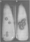
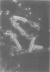
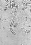
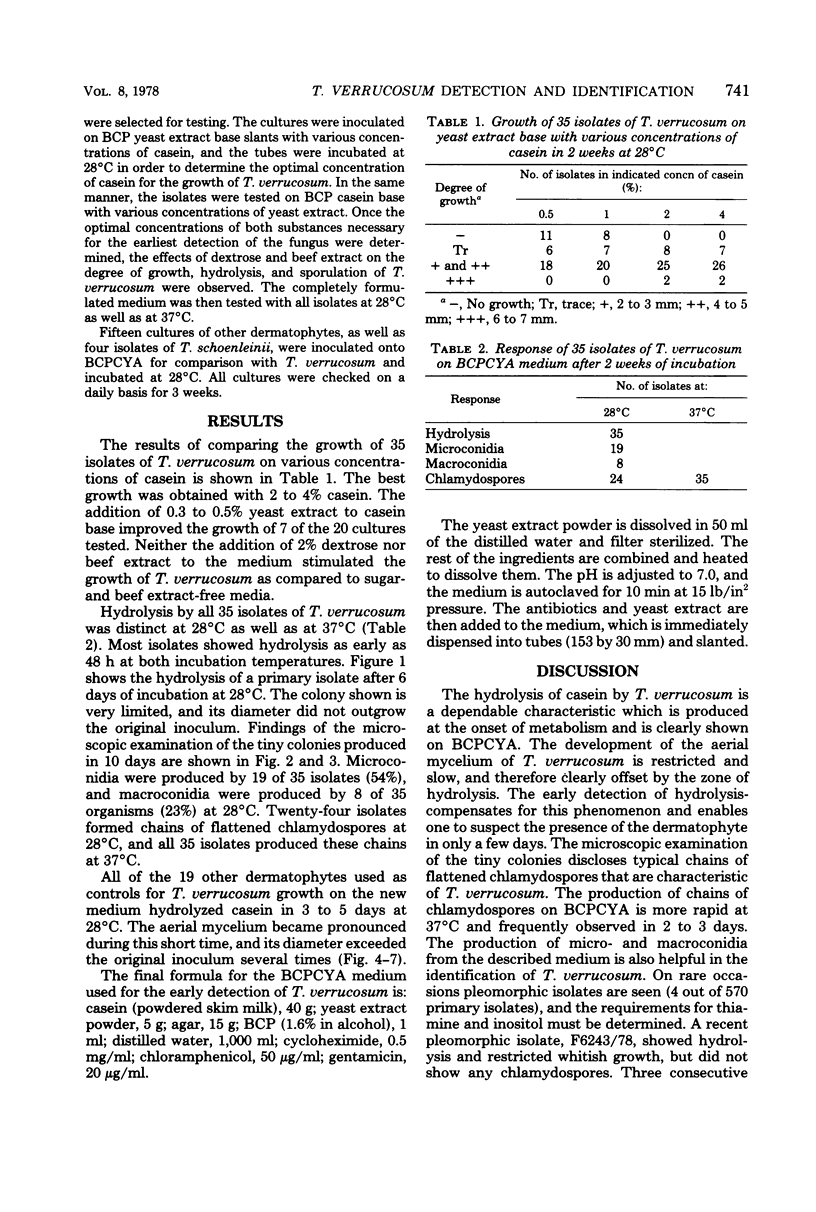
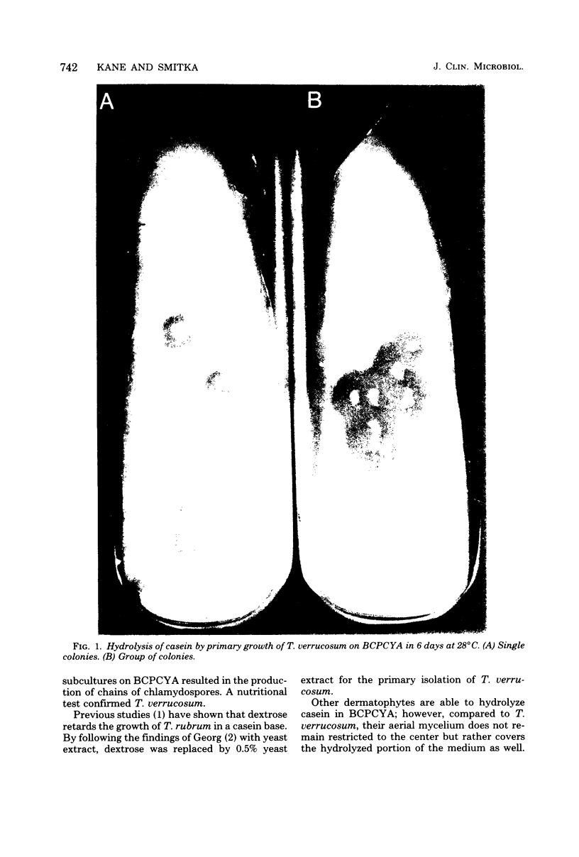
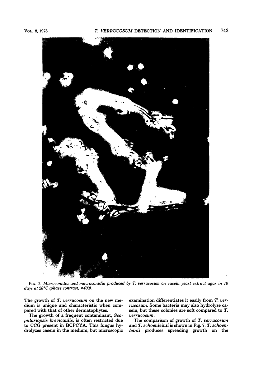
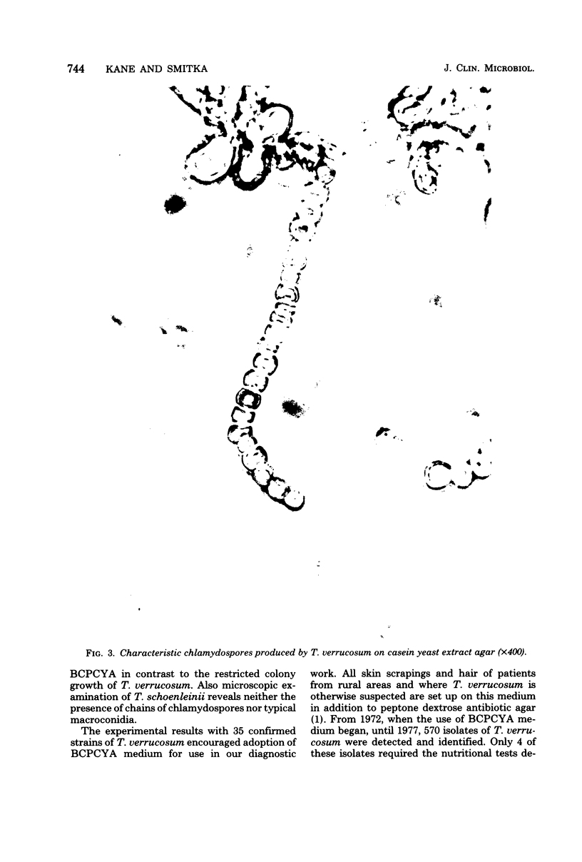
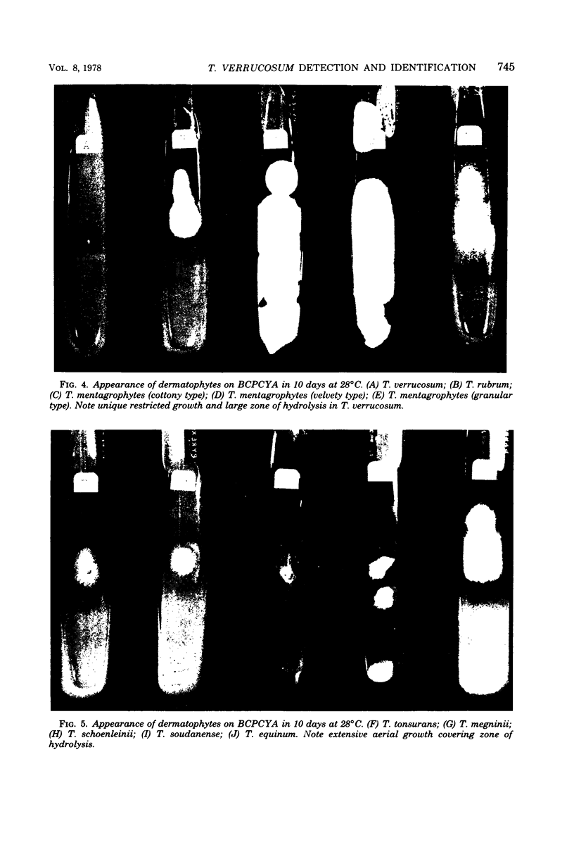
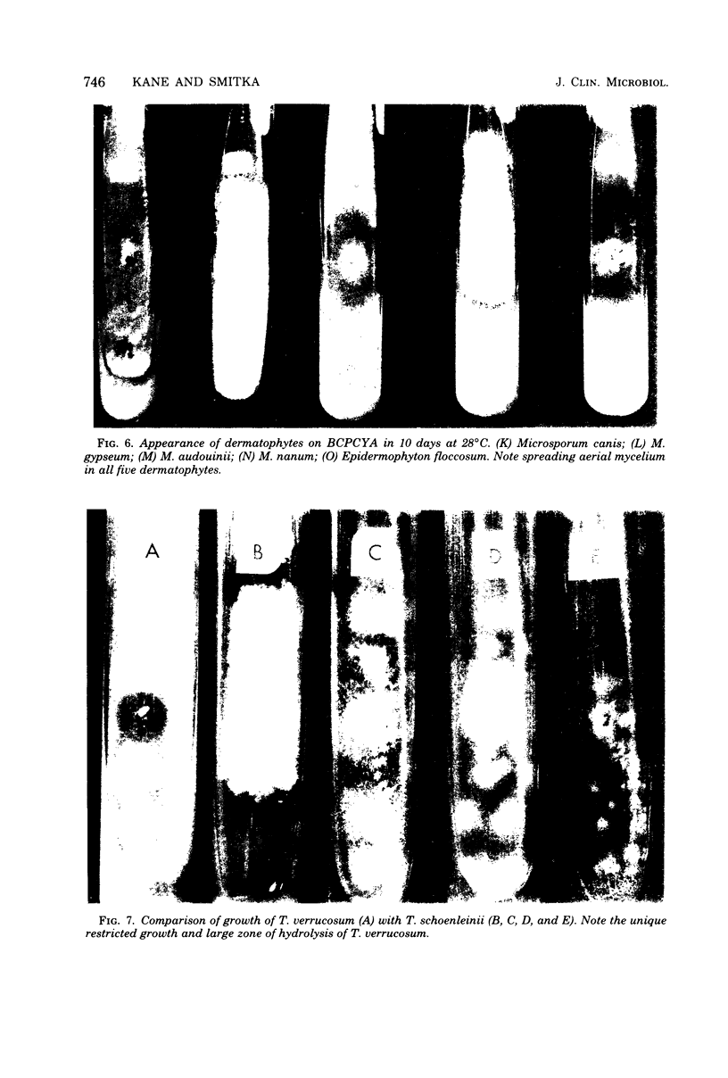
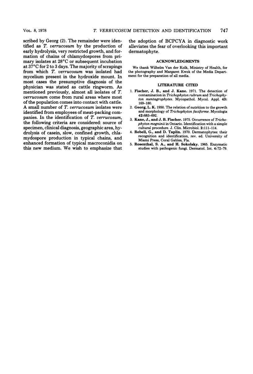
A new medium for the early detection and identification of Trichophyton verrucosum has been formulated. The key ingredients of the medium are 4% casein and 0.5% yeast extract. T. verrucosum is recognized by its early hydrolysis of casein and very slow growth. Microconidia were produced by 19 out of 35 isolates (54%), and macroconidia were produced by 8 out of 35 isolates (23%). All isolates formed chains of chlamydospores at 37 degrees C, and 24 out of 35 isolates formed chains at 28 degrees C. Nutritional requirements of all 35 strains of T. verrucosum were confirmed. The medium was evaluated by isolating 570 suspected T. verrucosum from skin scrapings. The early detection of hydrolysis, formation of characteristic chains of chlamydospores, and restricted slow growth of this dermatophyte differentiate it from T. schoenleinii.
Full text
PDF
Images in this article
Selected References
These references are in PubMed. This may not be the complete list of references from this article.
- Fischer J. B., Kane J. The detection of contamination in Trichophyton rubrum and Trichophyton mentagrophytes. Mycopathol Mycol Appl. 1971 Feb 19;43(2):169–180. doi: 10.1007/BF02051718. [DOI] [PubMed] [Google Scholar]
- Kane J., Fischer J. B. Occurrence of Trichophyton megninii in Ontario. Identification with a simple cultural procedure. J Clin Microbiol. 1976 Aug;2(2):111–114. [PMC free article] [PubMed] [Google Scholar]
- Rosenthal S. A., Sokolsky H. Enzymatic studies with pathogenic fungi. Dermatol Int. 1965 Apr-Jun;4(2):72–78. doi: 10.1111/j.1365-4362.1965.tb05127.x. [DOI] [PubMed] [Google Scholar]


