

Abstract
This work explores using self-assembled DNA nanostructures as carriers for drug delivery. We have recently developed an organic nanotube system that is assembled from a single component: a 52-base-long DNA single strand. In this work, functional agents (folate as a cancer cell target agent and Cy3 as a fluorescence imaging agent) are conjugated with the DNA strands; the conjugates self-assemble into micrometers-long nanotubes (NTs). The conjugated DNA-NTs can be effectively taken by cancer cells as demonstrated by fluorescence imaging and fluorescence activated cell sorting (FACS). No obvious toxicity has been observed under current experimental conditions.
Keywords: DNA, drug delivery, nanotubes, cancer cells
Introduction
A key research effort in nanomedicine is to develop universal drug carriers that can transport multiple functional agents: guiding agents for targeting to specific cells/tissues, imaging agents for diagnosis and monitoring treatment effects, drugs for disease treatment, and nucleic acids for gene therapy. Previously reported drug delivery systems include synthetic polymers (such as dendrimers),1-3 lipids and liposomes,4-6 carbon nanotubes,7-11 nanoparticles,12-15 RNAs,16, 17 and viral capsids.18 Their effectiveness has been clearly demonstrated. However, some of them are non-natural or foreign molecules for cells. Their long-term effects on human bodies, including immune response and toxicity, are not clear and remain to be examined. It is desirable to develop alternative systems, especially from degradable biomacromolecules that naturally exist in human bodies. They might provide more choices, and more importantly might have biocompatibility. This work is an effort along this line. Here, the delivery vehicle is based on short, synthetic DNA molecules. DNA is relatively stable when compared with RNA and proteins, but still be biodegradable by both spontaneous hydrolysis and nuclease-mediated digestion. DNA is also a natural component in human body and short DNA molecules are non-immunogenic.
This work is built upon structural DNA nanotechnology.19-21 Recently we have developed a DNA nanotube (DNA-NT) system that requires only one single component: a 52-base-long, single DNA strand, which consists of four palindromic segments.22 Many copies of the DNA strands self-assemble into DNA-NTs with diameters of 50 – 200 nm and lengths up to 40 μm. The DNA-NTs are robust and can tolerate chemical modifications. Herein, the DNA-NTs have been exploited as carriers to deliver chemicals into cells.
We have tested the strategy by using DNA-NTs as carriers to deliver Cy3 into human cancer cells (nasopharyngeal epidermal carcinoma KB cells, Figure 1). Cy3 is chosen as a model drug because its red fluorescence allows easy visualization. For targeting the Cy3-DNA-NTs to cancer cells, the NTs are also chemically modified with folate because folate receptors (FRs) are over-expressed on the cell surfaces of various cancer cells (including KB cells) and have been commonly used as biomarkers for cancer cells.2, 23-26 Cy3 and folate are separately conjugated to some DNA strands at their 5′ ends through a well-established NHS chemistry. DNA strands with and without chemical modification (Cy3 and folate) are mixed and assembled into Cy3 and folate modified DNA-NTs upon slowly cooling down from 95°C to 20°C. Cy3 and folate are randomly distributed along the DNA-NTs. The contents of Cy3 and folate in the DNA-NTs can be easily varied. When incubated with KB cells, the dual functionalized NTs will bind to the cell surfaces through specific folate – FR interaction and be further taken into the cells. Note that Cy3 and folate are not covalently linked to the same DNA strands.
Figure 1.

Scheme of the overall strategy. (a) Self-assembled DNA nanotubes (DNA-NTs) contain both Cy3 (a fluorescence dye) and folate (a targeting agent). (b) Dual-functionalized DNA-NTs specifically bind to cancer cells through folate-folate receptor (FR) interaction.
Experimental Section
DNA oligonucleotides
The DNA sequence is adapted from our previous work,22 5′-CCA AGC TTG GAC TTC AGG CCT GAA GTG GTC ATT CGA ATG ACC TGA GCG CTC A-3′. Oligonucleotides without any modification and with a cy3 label at the 5′ end were purchased from IDT, Inc. An oligonucleotide, with the same sequence as above oligos but containing an amino group and an extra T at its 5′ end, was purchased from Sigma GenoSys. The amino DNA was conjugated with folate by a reaction between NHS ester of folic acid and the amino DNA. All the chemicals were purchased from Aldrich except triethylamine from EMD. In a typical procedure, 100 mg folic acid (0.23 mmol), 250 μL triethylamine were added 1 mL DMSO, after dissolving all the solid, dicyclohexylcarbodiimide (DCC) (94mg, 0.45 mmol) and N-hydroxysuccinimide (NHS) (52 mg, 0.45 mmol) were added, the mixture was then agitated at room temperature for 12 hours. After brief centrifugation using a bench top centrifuge, the folate-NHS was used without further purification. 110 nmol amine-modified oligonucleotide was dissolved in 100 μL 0.2 M sodium phosphate buffer (100 mmol NaCl, pH 7.5), to this solution was added 45 μL 10 mg/mL folate-NHS ester dissolved in DMSO. The mixture was agitated for 4-10 hours at room temperature. After passing the solution through a gel filtration column (Sephadex G-25), the conjugate product was purified by denaturing polyacrylamide gel electrophoresis (PAGE, 20%).
Preparation of DNA-NTs
45% and 10% out of the strands were substituted with identical, cy3-labeled and folate-conjugated strand at 5′-ends, respectively. Three different DNA strands (without labels, with Cy3, with folate) were mixed to form dual-functionalized DNA tubes by the same annealing procedure as we previously reported.17 In brief, 5 μM DNA was dissolved in TAE/Mg2+ buffer composed of tris(hydroxymethyl) aminomethane (Tris) base (40 mm, pH 8.0), acetic acid (20 mm), ethylenediaminetetraacetate (EDTA; 2 mm), and MgAc2 (12.5 mm) and slowly cooled from 95°C to 4°C over 48 hours. It was then diluted to 500nM with PBS/10 mM MgCl2 for incubation with cells unless otherwise stated.
Denaturing Polyacrylamide Gel Electrophoresis
Gels contained 20% polyacrylamide (acrylamide/bisacrylamide, 19:1) and 8.3 M urea and were run at 55°C. Tris-borate-EDTA (TBE) was used as the separation buffer and consisted of Tris base (89 mM, pH 8.0), boric acid (89 mM), and EDTA (2 mM). Gels were run on a Hoefer SE 600 electrophoresis unit at 600 V (constant voltage).
Cell Culture
KB cells were grown as monolayer in 12 ml folate-deficient RPMI-1640 cell culture medium (Invitrogen) supplemented with 10% heat inactivated (60°C for 30 minutes) fetal bovine serum (Atlanta Biologicals), 50 units/ml penicillin (Cellgro), and 50 μg/ml streptomycin (Cellgro) at 37°C in a 5% CO2 / 95% air humidified atmosphere. A T-75 flask (75 cm2 effective growth area) was used for cell culture. When the cells grew to ∼80 % confluency (usually in 3-4 days), the culture medium was discarded. Then the cells were washed with 10 ml PBS (Phosphate Buffered Saline, Cellgro) and trypsinized with 1.5 ml 0.25% trypsin − 2.21 mM EDTA (Cellgro) for 2-3 minutes at room temperature. In order to detach the cells, the side of the flask was gently whacked several times. Then 5 ml fresh medium was added into the flask and the solutions are pipetted up-and-down several times to separate the cells from each other. 1 ml of the cell suspension (out of 6.5 ml) was quickly redispersed into another T-75 flask that contained 12 ml fresh medium for further cell culture.
Incubation of Living Cells in DNA Solutions
It was done by direct deposition of 100μl of 500nM DNA nanotube solution onto cells. Prior to incubation, the human nasopharyngeal epidermal carcinoma KB cells were seeded on a glass cover slide loaded in a well of 6 well-plate at a cell density of 3×10⁶ cells/mL and precultured in folate-deficient RPMI-1640 cell culture medium (Invitrogen) supplemented with 10% fetal bovine serum (FBS), 50 unit/ml penicillin, and 50 μg/ml streptomycin for one day. After 1 hour incubation in PBS/MgCl2 of DNA nanotubes, the cells were extensively washed with PBS/MgCl2 to remove excess DNA and further cultured for different periods in fresh cell medium, unless otherwise stated, in 5% CO2 atmosphere at 37°C.
Fluorescence Imaging
The cover slide with cells was placed onto a glass slide and images were obtained with a Nikon Fluorescence Microscope E800. KB cells were stained by Hoechst 33342 (Invitrogen) for dual color detection of DNA transportation. Prior to Hoechst 33342 staining, cells were washed and equilibrated in PBS. 1μM Hoechst 33342 solution was loaded onto cells and kept for ∼10 min and washed with PBS.
Confocal Microscopy Imaging of Cells
All confocal images were taken after the incubation/culture and washing with PBS. For confocal imaging, KB cells were seeded at 4 × 10⁴ cells/ml in tissue culture treated μ-Slide 8 well (Ibidi) and further cultured at 37°C and 5% CO2 as usual. Images were acquired using a Radiance 2100 MP Rainbow (Bio-Rad, Hemel Hempstead, England) on a TE2000 (Nikon, Tokoyo, Japan) inverted microscope using a 60× oil 1.4 NA lens. Cy3-labeled DNA-NTs and Hoechst 33342 fluorescence images were collected sequentially. The Cy3-labeled DNA-NTs were excited at 543 nm using a green HeNe laser and the fluorescence emission greater than 560 nm in wavelength was collected. Multi-photon excitation for Hoechst 33342 was provided by the Mai Tai laser (Spectra-Physics, Mountain View, CA) at 750 nm and the emission between 420 and 480 nm was collected.
Flow Cytometry
KB cells were precultured and trypsinized for 1 hr incubation in various concentrations of DNA solution at a concentration of ∼3 × 10⁴ cells per mixture. They were extensively washed twice with PBS/MgCl2 and resuspended in ice-cold PBS. The mean red fluorescence of cells was measured for 10,000 cells by flow cytometry (FACSCalibur, Becton Dickinson). For investigation of longer period culturing following 1 hour incubation, cells were also collected after a certain desired period and washed twice for flow cytometry. For each sample, a geometric mean was used as “mean fluorescence”.
YOYO-1 Staining
KB cells were seeded on a glass cover slide loaded in a well of a 24 well plate at a cell density of 1×10⁵ cells/500μL. After two days's culturing in folate-deficient RPMI-1640 cell culture medium (Invitrogen) supplemented with 10% fetal bovine serum (FBS), 100 unit/ml penicillin, and 100 μg/ml streptomycin, the cells were sequentially incubated with 500nM DNA NTs in PBS/Mg2+ buffer for 1 hour, washed with PBS buffer, and further cultured in a fresh medium for one day. Then, 100 μL of 100 mM YOYO-1 (Molecular Probes) solution (in PBS buffer) was directly loaded onto cells and incubated for 20 min at room temperature. Finally the cells were washed with PBS buffer and imaged by fluorescence microscopy.
DNase I treatment
Similar to YOYO-1 staining, the cells were precultured for two days. KB cells were incubated with DNase I (New England BioLab, 2 units) in 100 μL DNase I reaction buffer for 1 hour at 37°C. The cells were then washed with PBS buffer and imaged by fluorescence microscopy.
Results and Discussion
Formation of DNA-NTs
Dual functionalized DNA-NTs are readily formed through cooling-induced DNA self-assembly. Cy3 is a red fluorescent dye, which allows direct visualization of the DNA-NTs. Figure 2 shows a fluorescence image of the DNA samples. DNA-NTs appear 10 to 40 μm long, similar to the DNA-NTs without chemical modifications.22 They are stable in PBS buffer and cell medium when supplemented with 10mM MgCl2.
Figure 2.
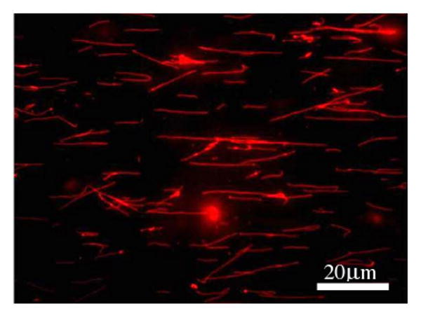
A fluorescence image of DNA-NTs containing both Cy3 and folate (45% and 10% of the DNA strands are labeled with Cy3 and folate, respectively.)
Cellular Uptake of DNA-NTs
Folate modified DNA-NTs can efficiently transport Cy3 into cancer cells (Figure 3). After incubation with FR-overexpressing KB cells for one hour, folate modified DNA-NTs are significantly concentrated onto the cell surfaces as shown by florescence imaging (Figures 3a-c). The red color indicates the Cy3 moieties on DNA-NTs. This observation suggests that the dual functionalized DNA-NTs strongly bind to cell surfaces. Figures 3a-c are images of cells without washing. The DNA-NTs (red) and KB cells are clearly colocalized, which suggests that the DNA-NTs are either in the cells or adsorbed onto the cell surfaces. Some DNA-NTs are still recognizable (indicated by white arrows).
Figure 3.
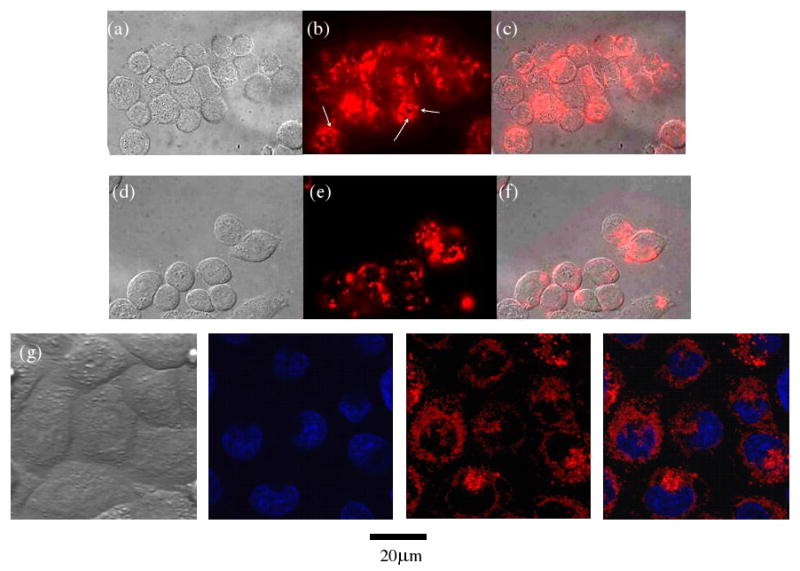
Fluorescence images of KB cells incubated with dual-functionalized DNA-NTs for one hour. (a-c) before and (d-f) after extensive washing. (a) & (d): brightfield images; (b) & (e): fluorescence images; (c) & (f): the superimposed images of (a) & (b) and (d) & (e), respectively. White arrows in (b) indicate some recognizable DNA-NTs. (g) Confocal fluorescence images of washed cells. From left to right: brightfield image, Hoechst stain (indicating the nuclei) image, Cy3 (indicating DNA-NTs) image, superimposed nuclei and DNA-NT image. Hoechst 33342: a blue fluorescence dye that specifically stains nuclei.
To remove the bound but not internalized DNA-NTs, we have extensively washed the cells. The red fluorescence on the cells generally decreases (Figures 3d-f), but some fluorescence remains, which suggests that the Cy3 moieties are internalized into the cells. Internalization is more clearly shown by confocal fluorescence microscopy imaging (Figure 3g). To further confirm the internalization, we have used DNase I to treat the cells. If the DNA-NTs are inside of the cells, they would not be accessible by DNase I and the red fluorescence will remain. In contrast, if the DNA-NTs only adsorb onto the cell surfaces and are not internalized, they will be degraded by DNase I and the red fluorescence will disappear. The experimental data (see Supporting Information, Figure S1) support that the the DNA-NTs are internalized as the red fluorescence remains in the cells.
After internalization, the morphologies of the DNA-NTs are not recognizable any more. Presumably, the internalization process changes the supramolecular structures of the DNA-NTs. It might be possible that shortening the DNA-NTs is necessary for its internalization. However, it remains speculation at this stage. Note that the nanotube morphology will become non-recognizable under an optical microscope when NTs are shorter than 1 μm due to an optics limitation. After up to 4-days incubation, no significant cell death has been observed; indicating that DNA-NTs are biocompatible and their toxicity is low. After incubating the cells with DNA-NTs, we have investigated whether the cell membranes are intact by staining the cells with a green fluorescence dye, YOYO-1. The dye can not effectively cross the cell membranes to stain the cells as the green fluorescence of the cells did not increase after staining (see Supporting Information, Figure S1). This experiment suggests that the cell membranes are intact.
The self-assembled DNA nanostructure is important for delivering Cy3 into the cancer cells as confirmed by a control experiment. We have performed a thermal denaturation: the DNA-NTs were heated at 95°C for 5 minutes and then quenched onto ice. The cooling process is fast that prevents DNA strands from meeting and hybridizing with each other to properly assemble into nanotube structures. Instead, the DNA strands fold into some metastable, single stranded structures, or small aggregates. The process destroys the NT structures into separated single DNA strands. Thus, Cy3 and folate moieties are not in the same complexes. When incubating cells with the denatured DNA samples, no fluorescence is observed to be colocalized with the cells, which suggests that the self-assembled DNA nanostructure is essential for the cellular uptake of Cy3.
Quantification of Cellular Uptake of DNA-NTs
Fluorescence activated cell sorting (FACS) is used for quantitative study of cellular uptakes of DNA-NTs (Figure 4). After one hour incubation with dually functionalized DNA-NTs, KB cells show increased fluorescence in comparison with the untreated cells. The fluorescence intensity is positively correlated with the DNA concentration. We have also performed a series of control experiments. There is no significant fluorescence increase when incubating KB cells with control DNA samples, including denatured DNA-NTs, DNA-NTs that contain only Cy3 but without folate, and Cy3-labeled DNA single strands that have the same length but cannot self-assemble into NTs. This observation further suggests that the DNA complexes are important for cellular uptakes of Cy3. When the DNA concentration is high, there is a small fluorescence increase for the control samples. It is presumably caused by small DNA aggregates because at such a high concentration, even a quenching process can not completely avoid inter-strand interactions.
Figure 4.
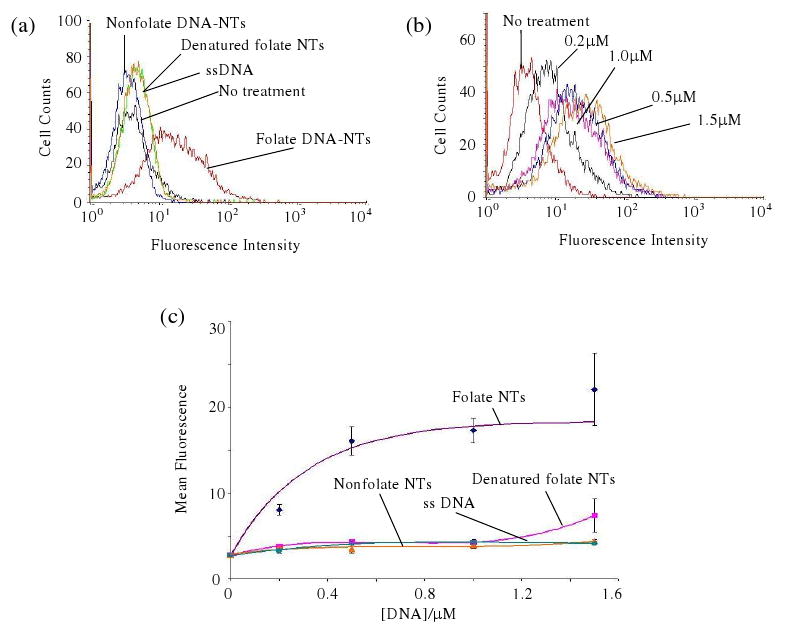
Flow cytometry analysis after 1 hour incubation. (a) Fluorescence histogram of cells incubated with different DNA samples. (b) Fluorescence histogram of cells incubated with different concentrations of DNA-NTs. (c) Mean fluorescence intensity of cells versus DNA concentrations.
Effect of Incubation Duration on DNA Uptake
We have varied the incubation duration of cells with DNA-NT solution to examine how it affects the DNA uptake. Mean fluorescence of cells increased as the incubation duration increased and reached a plateau over ∼ 1 hour (Figure 5a). It seems that there is a limitation on the amount of DNA that can be taken into cells.
Figure 5.
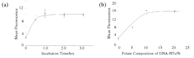
Effects of incubation time and folate content on DNA uptake. (a) Mean fluorescence of cells versus incubation time with DNA solution (45% of Cy3, 10% of folate, 500 nM DNA). (b) Mean fluorescence of cells versus folate content of DNA-NTs. The data were taken after 1 hour incubation in the presence of DNA-NTs (500 nM DNA).
Effect of Folate Composition on DNA Uptake
We have systematically changed the folate composition in DNA-NTs to study how folate affects the DNA uptake (Figure 5b). A higher folate content leads to higher cellular DNA uptake. When folate increases to 10%, the uptake reaches a plateau. Increasing folate content higher than 10% does not further increase the cellular uptake of DNA (Figure 5b). Considering the limited amount of FR on cell surface, it is reasonable that the FR-dependent DNA uptake has an upper limit.
To further confirm that the internalization of DNA-NTs is through the FR pathway, we have performed a control experiment. Immediately before introducing folate-functionalized DNA-NTs, incubating the cells with folate-saturated culture medium dramatically impairs the DNA-NT internalization (Figure 6).
Figure 6.
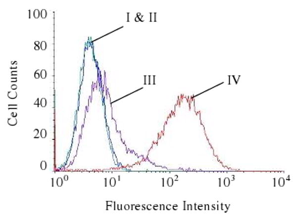
The effect of folate pre-incubation on DNA-NTs internalization as analyzed flow cytometry. (I) cells without any treatment; (II) cells incubated with folate-saturated medium for 1 hour; (III) cells incubated with folate-saturated medium for 1 hour and then with folate-functionalized DNA-NTs for 1 hour; (IV) cells incubated with folate-functionalized DNA-NTs for 1 hour.
Conclusion
We have successfully used DNA-NTs to deliver chemicals into cancer cells. It would be possible to further develop this system to become a platform for multifunctional delivery vehicle, which is under investigation. Surprisingly, the DNA accumulates at specific cellular locations, perinuclear regions. The mechanism of intracellular transportation is not clear at this stage and requires further study.
Supplementary Material
Acknowledgments
This work was supported by the National Science Foundation (CCF-0622093) and the National Institute of Health (R21EB007472). The cell culture works were performed in the Center for Combinatorial Chemical Biology at Purdue University. We thank Jennifer Sturgis for help with confocal microscopy imaging.
References
- 1.Shukla R, Thomas TP, Peters J, Kotlyar A, Myc A, Baker JR. Chem Commun. 2005;(46):5739–5741. doi: 10.1039/b507350b. [DOI] [PubMed] [Google Scholar]
- 2.Quintana A, Raczka E, Piehler L, Lee I, Myc A, Majoros I, Patri AK, Thomas T, Mule J, Baker JR. Pharmaceut Res. 2002;19:1310–1316. doi: 10.1023/a:1020398624602. [DOI] [PubMed] [Google Scholar]
- 3.Oupicky D, Konak C, Ulbrich K, Wolfert MA, Seymour LW. J Control Release. 2000;65:149–171. doi: 10.1016/s0168-3659(99)00249-7. [DOI] [PubMed] [Google Scholar]
- 4.Felgner PL, Gadek TR, Holm M, Roman R, Chan HW, Wenz M, Northrop JP, Ringold GM, Danielsen M. Proc Natl Acad Sci USA. 1987;84:7413–7417. doi: 10.1073/pnas.84.21.7413. [DOI] [PMC free article] [PubMed] [Google Scholar]
- 5.Voinea M, Simionescu M. J Cell Mol Med. 2002;6:465–474. doi: 10.1111/j.1582-4934.2002.tb00450.x. [DOI] [PMC free article] [PubMed] [Google Scholar]
- 6.Luo D, Saltzman WM. Nat Biotechnol. 2000;18:33–37. doi: 10.1038/71889. [DOI] [PubMed] [Google Scholar]
- 7.Kam NWS, O'Connell M, Wisdom JA, Dai HJ. Proc Natl Acad Sci USA. 2005;102:11600–11605. doi: 10.1073/pnas.0502680102. [DOI] [PMC free article] [PubMed] [Google Scholar]
- 8.Kam NWS, Liu ZA, Dai HJ. Angew Chem Intl Ed. 2006;45:577–581. doi: 10.1002/anie.200503389. [DOI] [PubMed] [Google Scholar]
- 9.Kam NWS, Liu Z, Dai HJ. J Am Chem Soc. 2005;127:12492–12493. doi: 10.1021/ja053962k. [DOI] [PubMed] [Google Scholar]
- 10.Kam NWS, Jessop TC, Wender PA, Dai HJ. J Am Chem Soc. 2004;126:6850–6851. doi: 10.1021/ja0486059. [DOI] [PubMed] [Google Scholar]
- 11.Kam NWS, Dai HJ. J Am Chem Soc. 2005;127:6021–6026. doi: 10.1021/ja050062v. [DOI] [PubMed] [Google Scholar]
- 12.Rosi NL, Giljohann DA, Thaxton CS, Lytton-Jean AKR, Han MS, Mirkin CA. Science. 2006;312:1027–1030. doi: 10.1126/science.1125559. [DOI] [PubMed] [Google Scholar]
- 13.Khaled A, Guo SC, Li F, Guo PX. Nano Lett. 2005;5:1797–1808. doi: 10.1021/nl051264s. [DOI] [PMC free article] [PubMed] [Google Scholar]
- 14.Yih TC, Al-Fandi M. 2006;97:1184–1190. doi: 10.1002/jcb.20796. [DOI] [PubMed] [Google Scholar]
- 15.Bertin PA, Gibbs JM, Shen CKF, Thaxton CS, Russin WA, Mirkin CA, Nguyen ST. J Am Chem Soc. 2006;128:4168–4169. doi: 10.1021/ja056378k. [DOI] [PubMed] [Google Scholar]
- 16.Guo SC, Tschammer N, Mohammed S, Guo PX. Hum Gene Ther. 2005;16(9):1097–1109. doi: 10.1089/hum.2005.16.1097. [DOI] [PMC free article] [PubMed] [Google Scholar]
- 17.Guo S, Huang F, Guo P. Gene Ther. 2006;13:814–820. doi: 10.1038/sj.gt.3302716. [DOI] [PMC free article] [PubMed] [Google Scholar]
- 18.Brown WL, Mastico RA, Wu M, Heal KG, Adams CJ, Murray JB, Simpson JC, Lord JM, Taylor-Robinson AW, Stockley PG. Intervirology. 2002;45:371–380. doi: 10.1159/000067930. [DOI] [PubMed] [Google Scholar]
- 19.Seeman NC. Biochemistry. 2003;42:7259–7269. doi: 10.1021/bi030079v. [DOI] [PubMed] [Google Scholar]
- 20.Samori B, Zuccheri G. Angew Chem Intl Ed. 2005;44:1166–1181. doi: 10.1002/anie.200400652. [DOI] [PubMed] [Google Scholar]
- 21.Feldkamp U, Niemeyer CM. Angew Chem Intl Ed. 2006;45(12):1856–1876. doi: 10.1002/anie.200502358. [DOI] [PubMed] [Google Scholar]
- 22.Liu HP, Chen Y, He Y, Ribbe AE, Mao CD. Ang Chem Intl Ed. 2006;45:1942–1945. doi: 10.1002/anie.200504022. [DOI] [PubMed] [Google Scholar]
- 23.Hilgenbrink AR, Low PS. J Pharm Sci. 2005;94:2135–2146. doi: 10.1002/jps.20457. [DOI] [PubMed] [Google Scholar]
- 24.Lu YJ, Sega E, Leamon CP, Low PS. Adv Drug Deliver Rev. 2004;56:1161–1176. doi: 10.1016/j.addr.2004.01.009. [DOI] [PubMed] [Google Scholar]
- 25.Sudimack J, Lee RJ. Targeted drug delivery via the folate receptor. Adv Drug Deliver Rev. 2000;41:147–162. doi: 10.1016/s0169-409x(99)00062-9. [DOI] [PubMed] [Google Scholar]
- 26.Lee RJ, Low PS. J Biol Chem. 1994;269:3198–3204. [PubMed] [Google Scholar]
Associated Data
This section collects any data citations, data availability statements, or supplementary materials included in this article.