Abstract
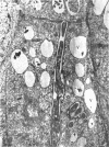
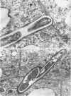
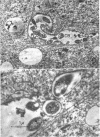
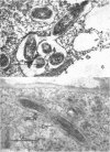
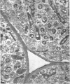
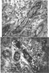
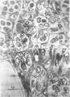
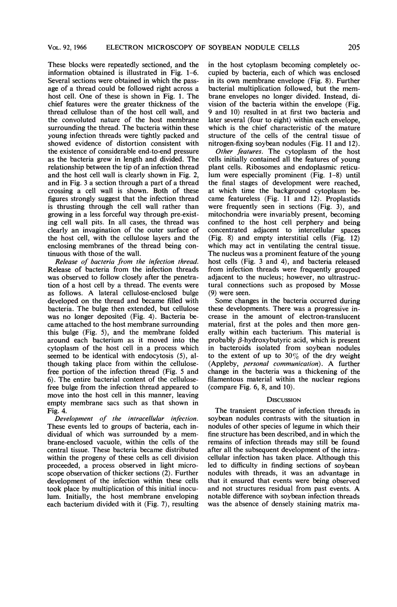
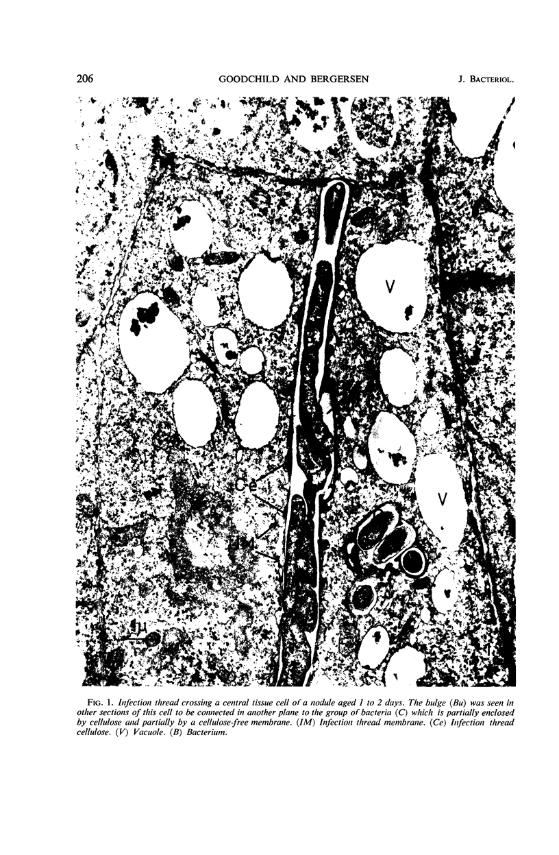
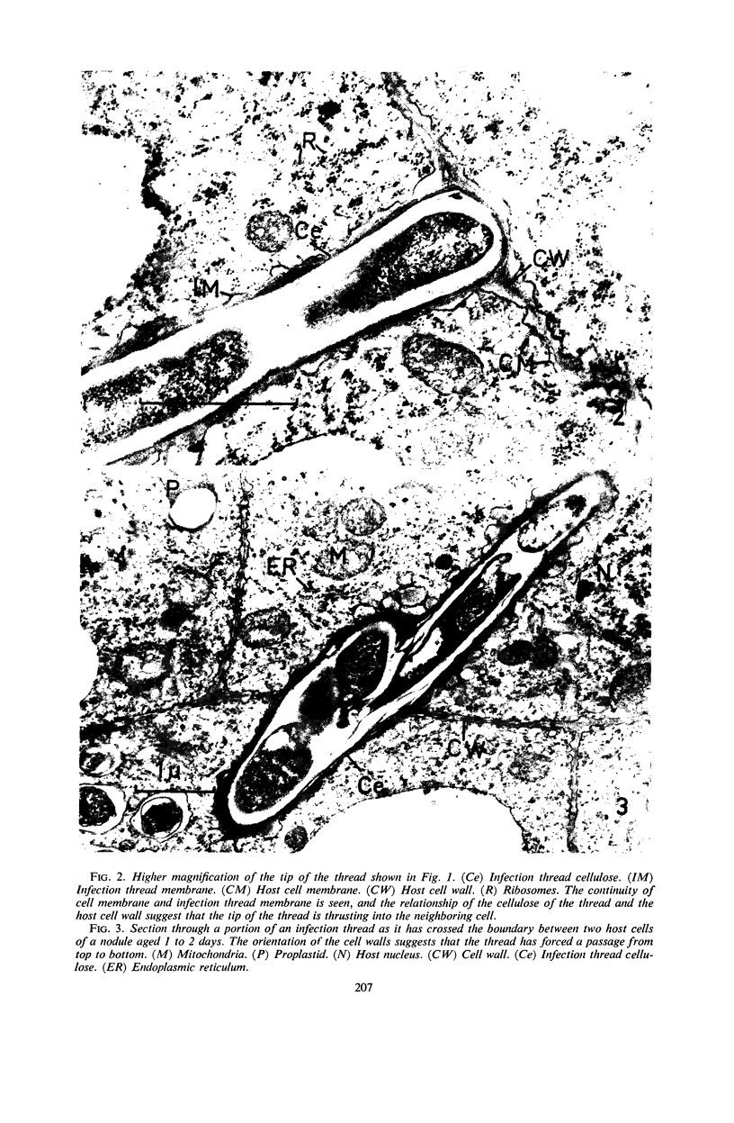
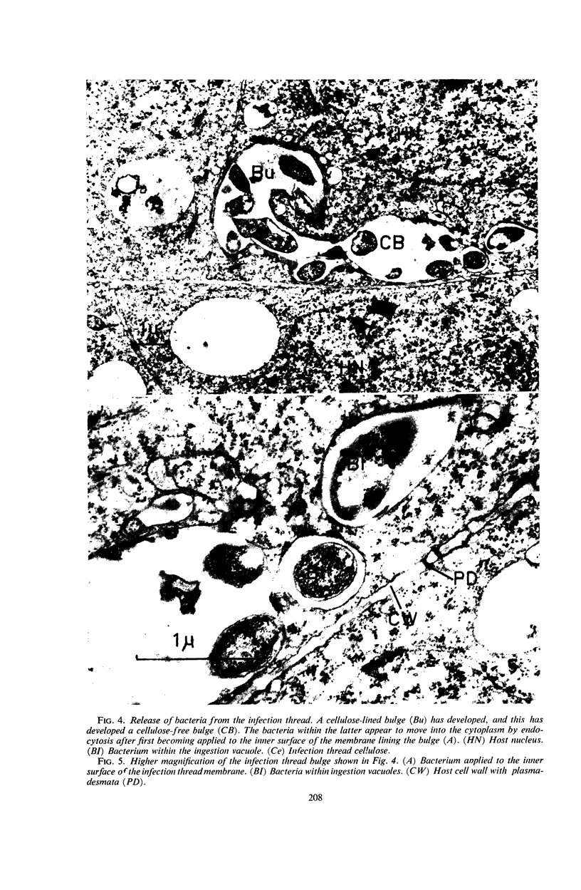
Goodchild, D. J. (Commonwealth Scientific and Industrial Research Organization, Canberra, Australia), and F. J. Bergersen. Electron microscopy of the infection and subsequent development of soybean nodule cells. J. Bacteriol. 92:204–213. 1966—Electron microscopy of thin sections of the developing central tissue cells of young soybean root nodules has shown that infection is initiated by a few infection threads which penetrate cells of the young central tissue. Extension growth of the threads may be a result of pressure developed from the growth of the bacteria within the threads. Release of bacteria from a thread is preceded by the development on an infection thread of a bulge with a cellulose-free membrane-bounded extension; bacteria move from this into the host cells by an endocytotic process and remain enclosed in an infection vacuole which is bounded by a membrane of host-cell origin. Multiplication of the intracellular bacteria takes place within these vacuoles. Until the host cell becomes filled with bacteria, the vacuoles separate into discrete units at each division. Later, division of the bacteria occurs within each vacuole, thus leading to the mature structure of the central tissue cells in which several bacteria are enclosed within each membrane-bounded unit.
Full text
PDF
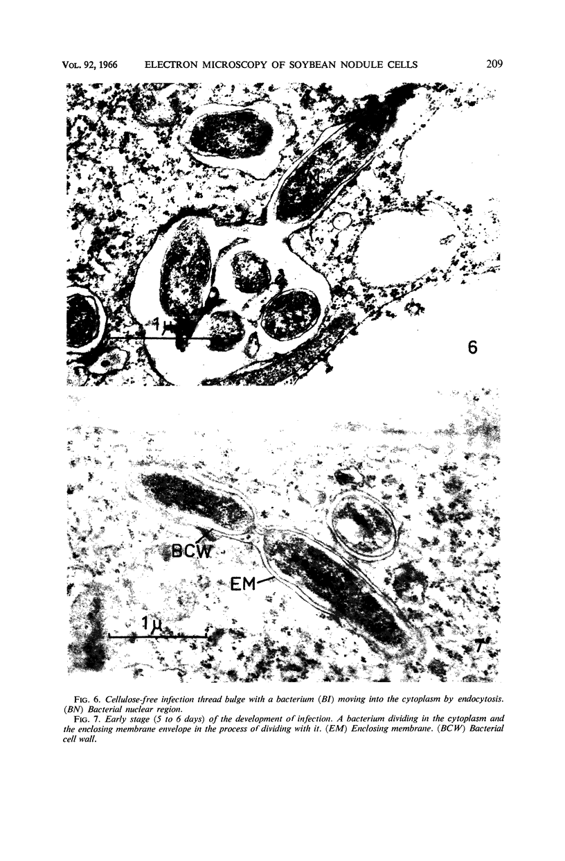
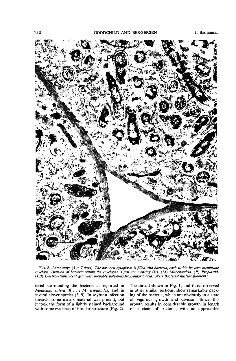
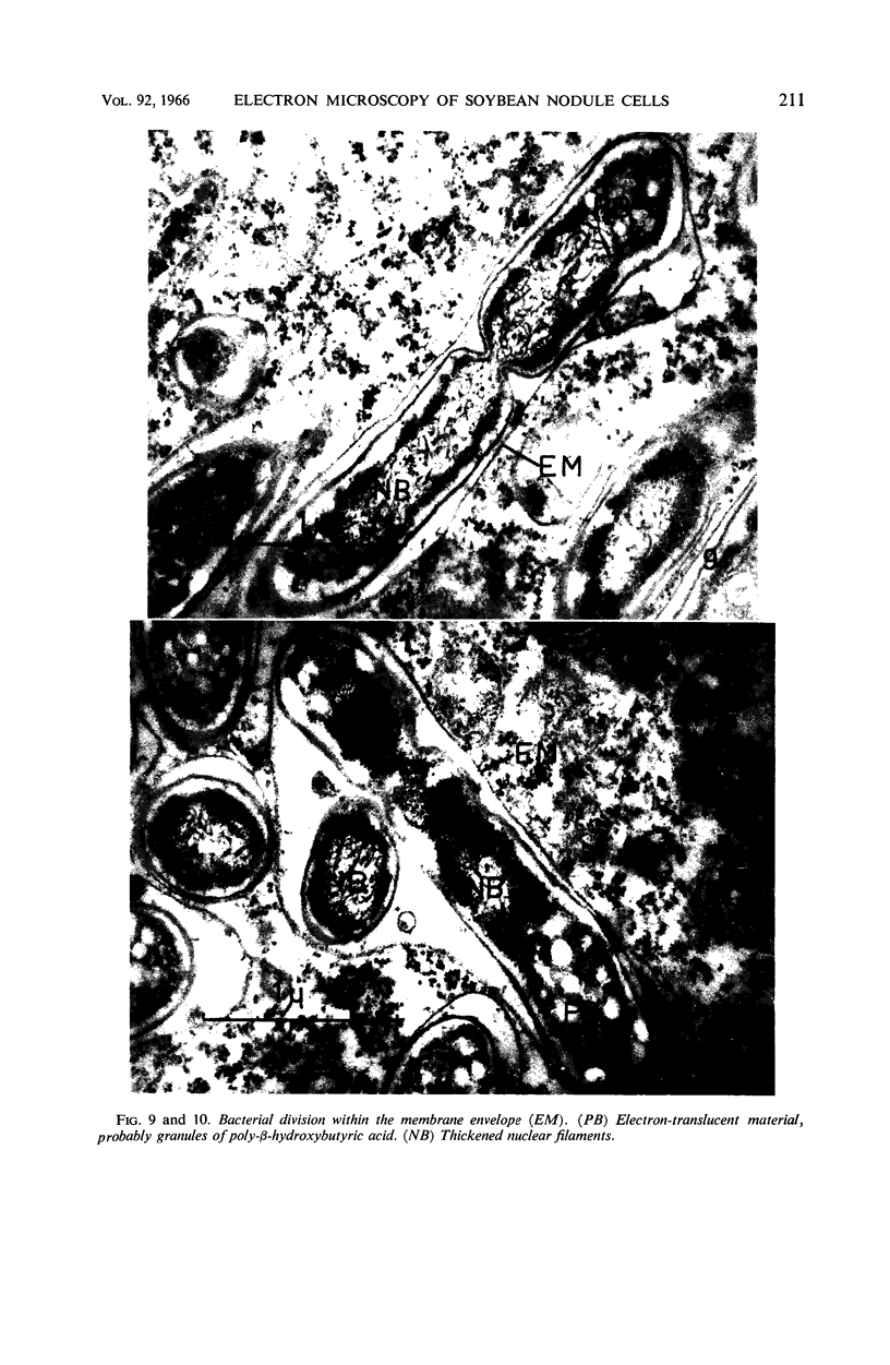
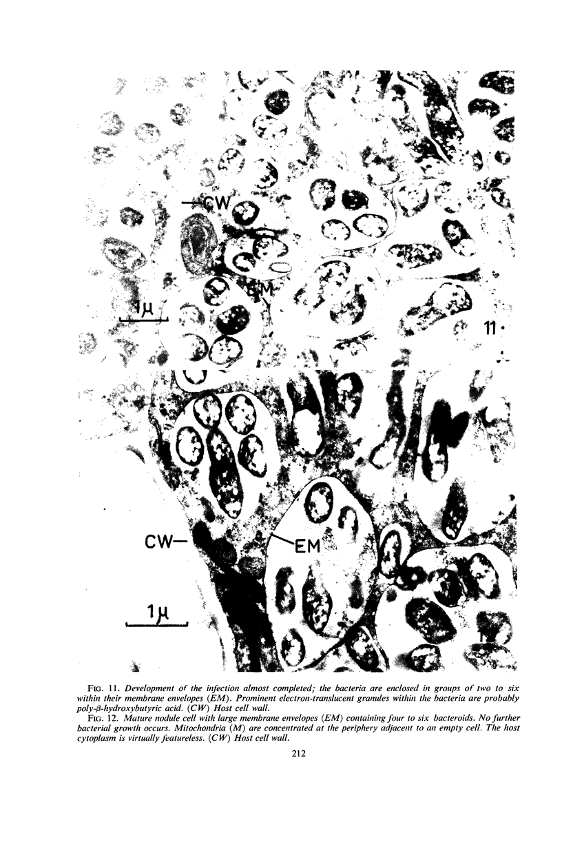
Images in this article
Selected References
These references are in PubMed. This may not be the complete list of references from this article.
- BERGERSEN F. J., BRIGGS M. J. Studies on the bacterial component of soybean root nodules: cytology and organization in the host tissue. J Gen Microbiol. 1958 Dec;19(3):482–490. doi: 10.1099/00221287-19-3-482. [DOI] [PubMed] [Google Scholar]
- BERGERSEN F. J. The bacterial component of soybean root nodules; changes in respiratory activity, cell dry weight and nucleic acid content with increasing nodule age. J Gen Microbiol. 1958 Oct;19(2):312–323. doi: 10.1099/00221287-19-2-312. [DOI] [PubMed] [Google Scholar]
- FOTHERGILL P. G., CHILD J. H. COMPARATIVE STUDIES OF THE MINERAL NUTRITION OF THREE SPECIES OF PHYTOPHTHORA. J Gen Microbiol. 1964 Jul;36:49–66. doi: 10.1099/00221287-36-1-49. [DOI] [PubMed] [Google Scholar]
- JORDAN D. C., GRINYER I., COULTER W. H. ELECTRON MICROSCOPY OF INFECTION THREADS AND BACTERIA IN YOUNG ROOT NODULES OF MEDICAGO SATIVA. J Bacteriol. 1963 Jul;86:125–137. doi: 10.1128/jb.86.1.125-137.1963. [DOI] [PMC free article] [PubMed] [Google Scholar]
- KARNOVSKY M. J. Simple methods for "staining with lead" at high pH in electron microscopy. J Biophys Biochem Cytol. 1961 Dec;11:729–732. doi: 10.1083/jcb.11.3.729. [DOI] [PMC free article] [PubMed] [Google Scholar]


