Abstract
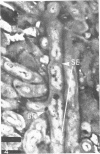
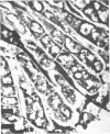
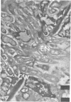
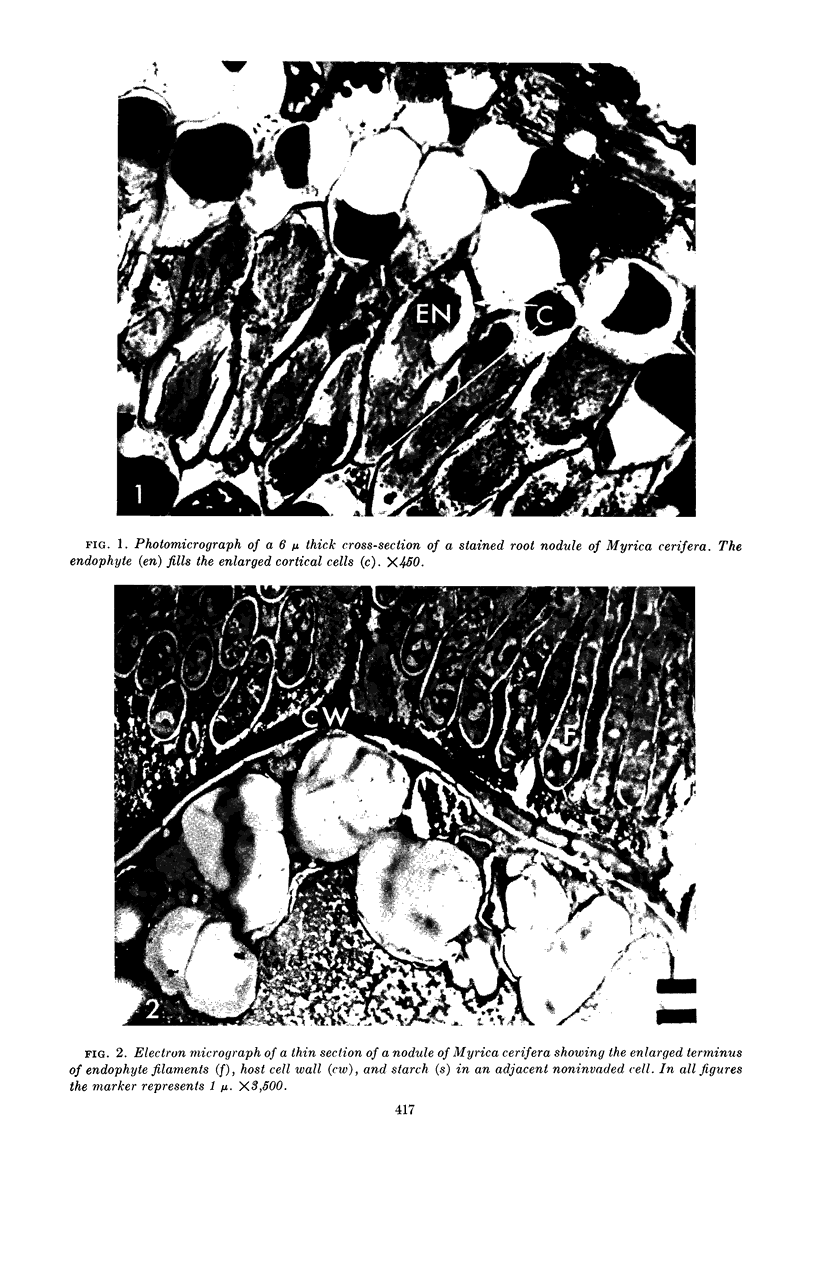
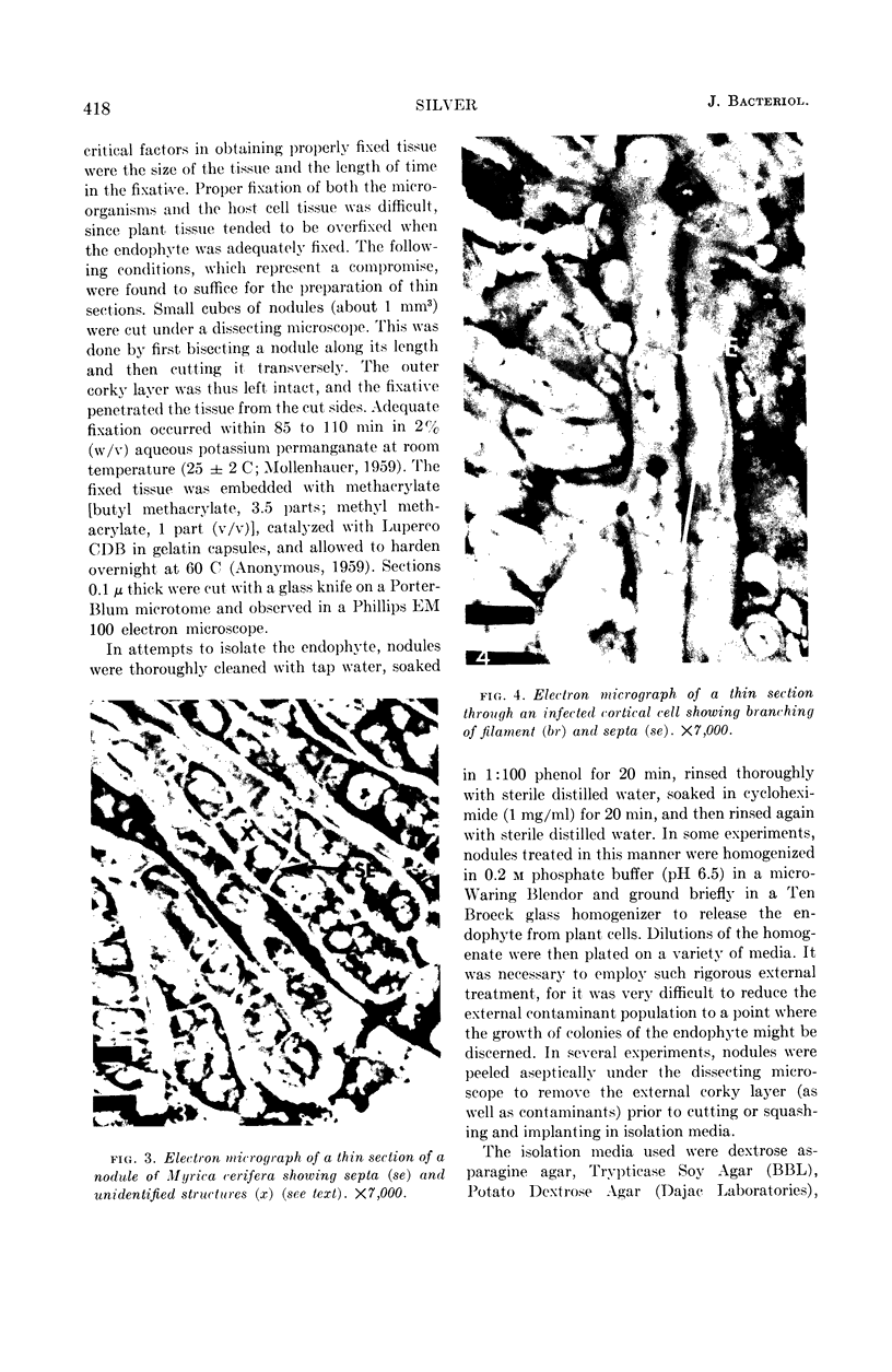
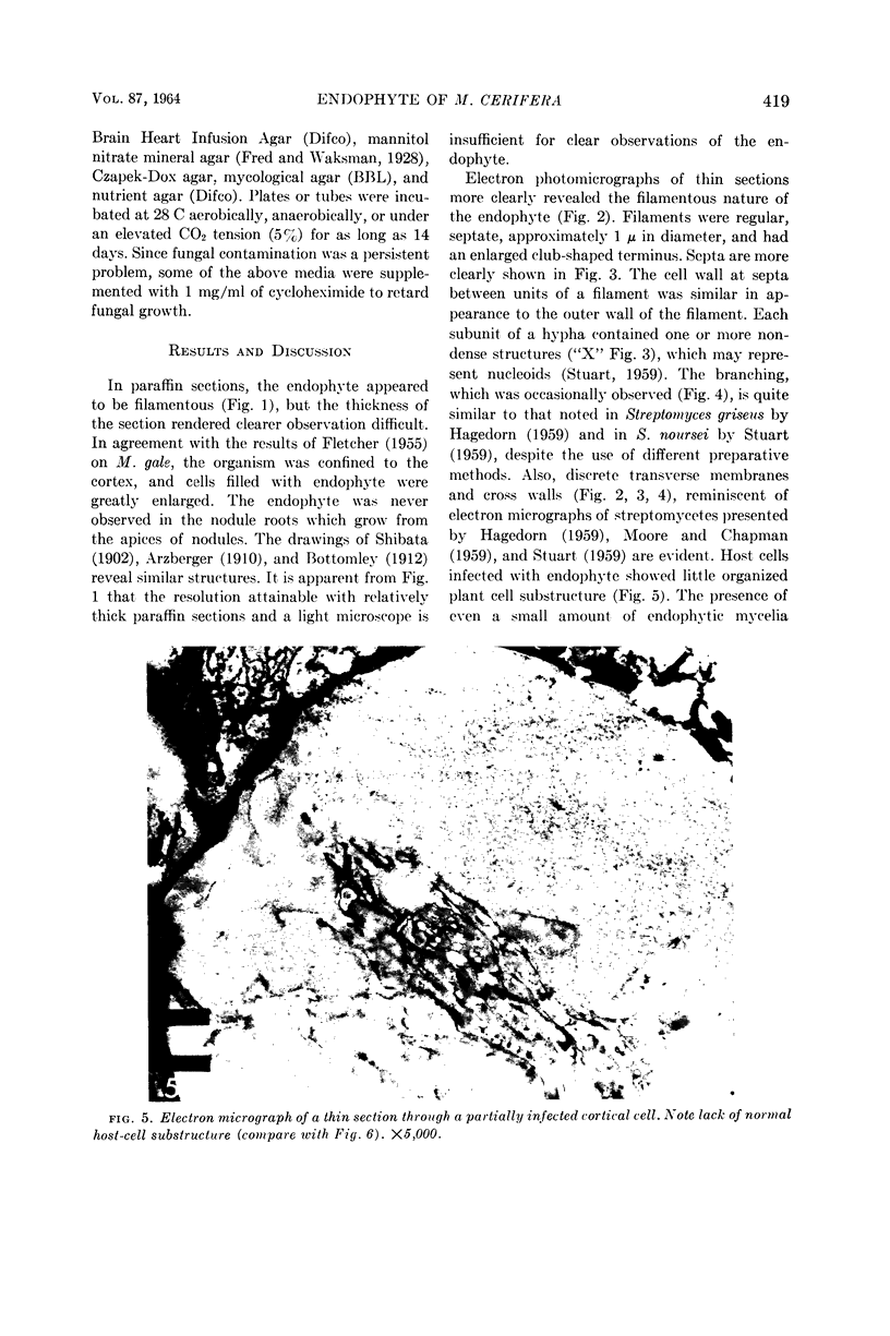
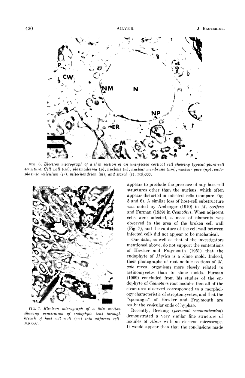
Silver, Warren S. (University of Florida, Gainesville). Root nodule symbiosis. I. Endophyte of Myrica cerifera. J. Bacteriol. 87:416–421. 1964.—Electron microscopy of 0.1-μ thick sections of root nodules, fixed with permanganate and embedded with methacrylate, showed that infected plant cells were filled with a mycelial endophyte. The endophyte was filamentous, 1 μ in diameter, septate, and had an enlarged, club-shaped terminus. Although structurally the endophyte strongly resembles an actinomycete, it was not isolated in pure culture on a variety of appropriate media.
Full text
PDF
Images in this article
Selected References
These references are in PubMed. This may not be the complete list of references from this article.
- HAWKER L. E., FRAYMOUTH J. A reinvestigation of the root-nodules of species of Elaeagnus, Hippophae, Alnus and Myrica, with special reference to the morphology and life histories of the causative organisms. J Gen Microbiol. 1951 May;5(2):369–386. doi: 10.1099/00221287-5-2-369. [DOI] [PubMed] [Google Scholar]
- MOORE R. T., CHAPMAN G. B. Observations of the fine structure and modes of growth of a streptomycete. J Bacteriol. 1959 Dec;78:878–885. doi: 10.1128/jb.78.6.878-885.1959. [DOI] [PMC free article] [PubMed] [Google Scholar]
- PETRAS E. [Electron microscopic studies on Streptomyces purpurascens Lindenbein]. Arch Mikrobiol. 1959;34:379–392. doi: 10.1007/BF00447099. [DOI] [PubMed] [Google Scholar]
- STUART D. C., Jr Fine structure of the nucleoid and internal membrane systems of Streptomyces. J Bacteriol. 1959 Aug;78:272–281. doi: 10.1128/jb.78.2.272-281.1959. [DOI] [PMC free article] [PubMed] [Google Scholar]


