Abstract
Epstein-Barr virus (EBV) is a human γ-herpesvirus that primarily infects B lymphocytes and epithelial cells. Entry of EBV into B cells requires the viral glycoproteins gp42, gH/gL and gB, while gp42 is not necessary for infection of epithelial cells. In EBV, gH and gL form two distinct complexes, a bipartite complex that contains only gH and gL, used for infection of epithelial cells, and a tripartite complex that additionally includes gp42, used for infection of B cells. The gH/gL complex is conserved within the herpesvirus family, but its exact role in entry and mechanism of fusion are not yet known. To understand more about the functionality of EBVgH/gL, we investigated the functional homology of gHs and gLs from human herpesvirus 8 (HHV8) and two primate (rhesus and marmoset) γ-herpesviruses in EBV-mediated virus-free cell fusion assay. Overall, gHs and gLs from the more homologous primate herpesviruses were better at complementing EBV gH and gL in fusion than HHV8 gH and gL. Interestingly, marmoset gH was able to complement fusion with epithelial cells, but not B cells. Further investigation of this led to the discovery that EBVgH is the binding partner of gp42 in the tripartite complex and the absence of fusion with B cells in the presence of marmoset gH/gL is due to its inability to bind gp42.
Keywords: Epstein-Barr virus, glycoprotein gH, glycoprotein gL, glycoprotein gp42, lymphocryptovirus, fusion, HHV8, cross-virus complementation
INTRODUCTION
Epstein-Barr virus (EBV) is a γ-herpesvirus, from lymphocryptovirus (LCV) subgroup, that readily infects B lymphocytes and epithelial cells (Kieff and Rickinson, 2001; Spear and Longnecker, 2003). The initiation of infection by EBV is driven by interaction of viral glycoproteins with cell surface receptors. The glycoproteins required for EBV entry differ between epithelial and B cells; gB, gH and gL are essential for infection of both epithelial and B cells, while gp42 is required only for B cell entry (Haan, Lee, and Longnecker, 2001; Li, Turk, and Hutt-Fletcher, 1995; Wang et al., 1998). Viral glycoprotein gp42 binds to B cell surface protein HLA class II, which triggers B cell fusion mediated by gB, gH and gL (Li et al., 1997b; Speck, Haan, and Longnecker, 2000). No EBV receptor for epithelial cells has been identified yet, but recent evidence suggests existence of an epithelial cell receptor for gH and the interaction of gH with its receptor has been proposed to serve as a trigger for epithelial cell fusion (Borza et al., 2004; Wu, Borza, and Hutt-Fletcher, 2005).
EBV glycoproteins gH and gL form a heteromeric complex which has been recently discovered to occur in a 1:1 ratio (Kirschner et al., 2006; Yaswen et al., 1993). The formation of a gH/gL complex is conserved throughout the herpesvirus family (Spear and Longnecker, 2003). gH is generally thought to be directly involved in the fusion process, while gL is thought to be important for proper processing and transport of gH to the cell surface (Pulford, Lowrey, and Morgan, 1994; Pulford, Lowrey, and Morgan, 1995; Yaswen et al., 1993). In addition to the bipartite gH/gL complex, EBV gH and gL also form a tripartite complex that includes gp42. These two complexes have a mutually exclusive ability to mediate infection of epithelial (gH/gL) and B cells (gH/gL/gp42), and are both present on EB virions (Wang et al., 1998).
EBV-related γ-herpesviruses, from Lymphocryptovirus genus, have been reported to naturally infect both Old and New World nonhuman primates (Ehlers et al., 2003; Wang, 2005). EBV and the Old World LCV genomes are highly homologous (i.e. homology between EBV and rhesus LCV is 75.6%) (Rivailler et al., 2002), and the repertoire of lytic and latent genes is virtually identical (Cho et al., 1999). The New World LCVs are more distant from EBV and Old World LCVs (i.e. homology between EBV and marmoset LCV is 47.3%) (Cho et al., 2001) but they are unified by their common biological properties (Wang et al., 2001). HHV8 is another γ-herpesvirus that infects humans but it belongs to the genus Rhadinovirus (Moore et al., 1996). The rhadinoviruses have also been reported to infect nonhuman primates (Damania and Desrosiers, 2001).
gH and gL play a central role in EBV entry and entry of other herpesviruses, but the exact role(s) of this complex in the fusion process is poorly understood. Interestingly, despite this functional conservation, a low sequence homology in gH and gL across and within subfamilies has been detected. Even though HHV8 and EBV are both members of γ-herpesvirus family, the homology between proteins of these two viruses is in general between 20 to 40% (Gupta et al., 2000; Lin et al., 1997; Lukac et al., 1998; Pertel, Spear, and Longnecker, 1998; Sun et al., 1998). Thus, HHV8 gH and gL have 26.5% and 24.2% identity and 48.3% and 53.8% similarity to EBV gH and gL, respectively (Russo et al., 1996). EBV gH and gL share a much higher degree of homology with gH and gL from nonhuman LCVs. For example, marmoset LCV gH and gL have 44.6% and 50.4% sequence homology with EBV, respectively, while the degree of conservation is even higher with rhesus LCV, 85.1% for gH and 81.6% for gL (Rivailler, Cho, and Wang, 2002; Rivailler et al., 2002). In this report we examined the ability of rhesus LCV (Rh), marmoset LCV (M) and HHV8 gH and gL to substitute for EBV gH and gL in fusion with epithelial and B cells. By performing this analysis, we have shown that gH/gL complexes are sensitive to cross-virus complementation.
RESULTS
Rhesus gH is functional in EBV-mediated fusion with epithelial and B cells
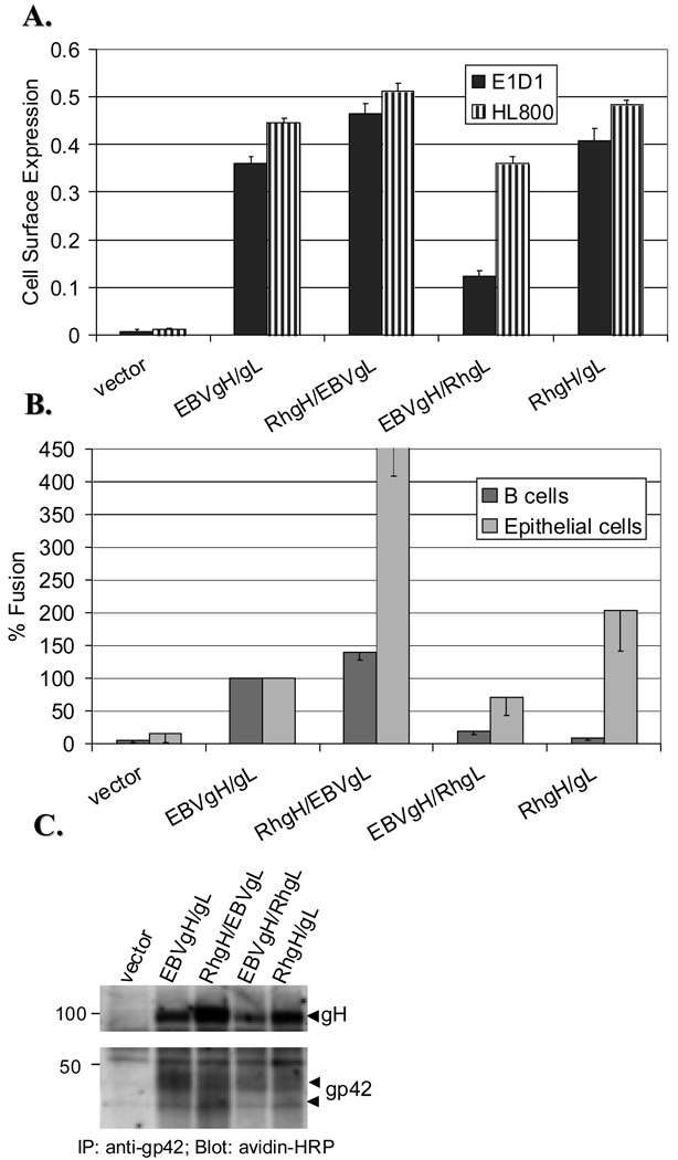
To investigate the functional homology between gH and gL from EBV and other related γ-herpesviruses, gH and gL from common marmoset LCV (M), rhesus monkey LCV (Rh) and human herpesvirus 8 (HHV8) were tested for their ability to substitute EBV gH and/or gL and mediate fusion with other EBV glycoproteins. For this purpose, Rh and M glycoproteins were cloned into a pCAGGS vector, previously used for expression of EBV and HHV8 glycoproteins (Haan, Lee, and Longnecker, 2001; Pertel, 2002). gH and gL from rhesus LCV were examined first since this virus is the most homologous to EBV. Rhesus LCV is capable of infecting human B cells in vitro (Moghaddam et al., 1998) but glycoproteins involved in entry of rhesus LCV have never been characterized. Prior to testing the heterologous Rh/EBV complexes in the fusion assay, we examined the cell surface expression of these complexes. Since no antibodies (Abs) against rhesus gH and gL were available we used two antibodies previously generated against EBVgH/gL complex, the E1D1 and the HL800 antibodies. The E1D1 recognizes the native gH/gL complex and blocks epithelial cell entry and fusion, while it has no effect on entry of EBV into B cells and B cell fusion (Li, Turk, and Hutt-Fletcher, 1995; McShane and Longnecker, 2004). The HL800 recognizes both EBV gH and gL (Omerovic, Lev, and Longnecker, 2005). Since there is a high degree of homology between EBV and Rh gH and gL, it was not surprising that both antibodies cross-reacted with rhesus glycoproteins and all of the complexes were detected at the cell surface (Fig. 1A). Interestingly, EBVgH/RhgL exhibited a greatly reduced binding to the E1D1 Ab. Since RhgH/gL bound this Ab as well as did EBVgH/gL this suggests that reduced binding was not due to the absence of residues recognized by the E1D1 antibody but rather a conformational change in the epitope due to a defect in gH/gL complex formation.
Figure 1. Rhesus gH is functional in EBV-mediated fusion with epithelial and B cells.
CHO-K1 cells were transiently transfected with EBV glycoproteins gp42 and gB and different combinations of gH and gL, as shown (Rh=rhesus). A. Postransfection, cells were transferred to 96 wells and assayed for gH/gL cell surface expression by CELISA with two different EBVgH/gL antibodies, E1D1 (black bars), a mouse monoclonal Ab, and HL800 (hatched bars), a rabbit polyclonal Ab. A representative experiment measured in triplicate is shown. B. Transfected CHO-K1 cells were overlaid with either Daudi B cells (black bars) or 293T kidney epithelial cells (gray bars) at 1:1 ratio and relative luciferase activity measured 24 hours later. Luciferase activity was normalized to wild-type levels, which was set to 100% for both cell types. Data are averages of three independent experiments with the standard deviations indicated by vertical lines. C. CHO-K1 cells were harvested 36 hours post transfection and the cell surface proteins labeled with biotin at 4°C. Biotinylated lysates were immunoprecipitated with the F-2-1 Ab (an anti-gp42 mouse monoclonal Ab) and probed with avidin-HRP in a Western blot. Proteins of interest are indicated with arrows. Positions of prestained protein markers (in kDa) are indicated.
We next examined the functionality of these complexes by testing their ability to mediate fusion with other EBV glycoproteins. For this purpose a cell-cell in vitro fusion assay was used (Haan, Lee, and Longnecker, 2001). All of the heterologous gH/gL complexes were functional in epithelial cell fusion (Fig. 1B). Interestingly, RhgH when paired with EBVgL mediated fusion with epithelial cells at greatly higher levels, as well as did RhgH/gL complex when compared to the EBVgH/gL. The complex of EBVgH and RhgL was still able to function in fusion with epithelial cells, but at somewhat lower levels. Since the conformation of this complex was altered according to the data with the E1D1 Ab and the epitope that E1D1 recognizes is important for epithelial cell fusion (Li, Turk, and Hutt-Fletcher, 1995; McShane and Longnecker, 2004), a decrease in fusion with epithelial cells was not surprising. However to our surprise the B cell fusion of EBVgH/RhgL complex was at only 20% of wild-type levels. In fact, the two complexes that contained RhgL (EBVgH/RhgL; RhgH/gL) had a very low, almost undetectable, fusion levels with B cells.
Since gp42 is required for EBV-mediated fusion with B cells and low levels of fusion with B cells could be due to a defect in gp42 association with gH/gL complex, we next examined whether these complexes were able to bind to gp42. In order to visualize gH and gL on the Western blot, the cell surface proteins of transfected cells were labeled with biotin and lysates immunoprecipitated for gp42. As shown in Fig. 1C, all of the gH/gL complexes immunoprecipitated with gp42. This suggested that the low levels of fusion seen when RhgL was paired with either Rh or EBV gH are not due to the inability of these complexes to bind to gp42 but rather some other defect such as an alteration in the overall structure of the complex or a failure of gp42 to trigger fusion. Interestingly, we constantly observed higher expression of RhgH in the Western blots, that was not dependent on the DNA preparation used, suggesting that the enhancement in fusion observed could be partially due to a higher cell surface expression.
Marmoset gH complements fusion with epithelial but not B cells
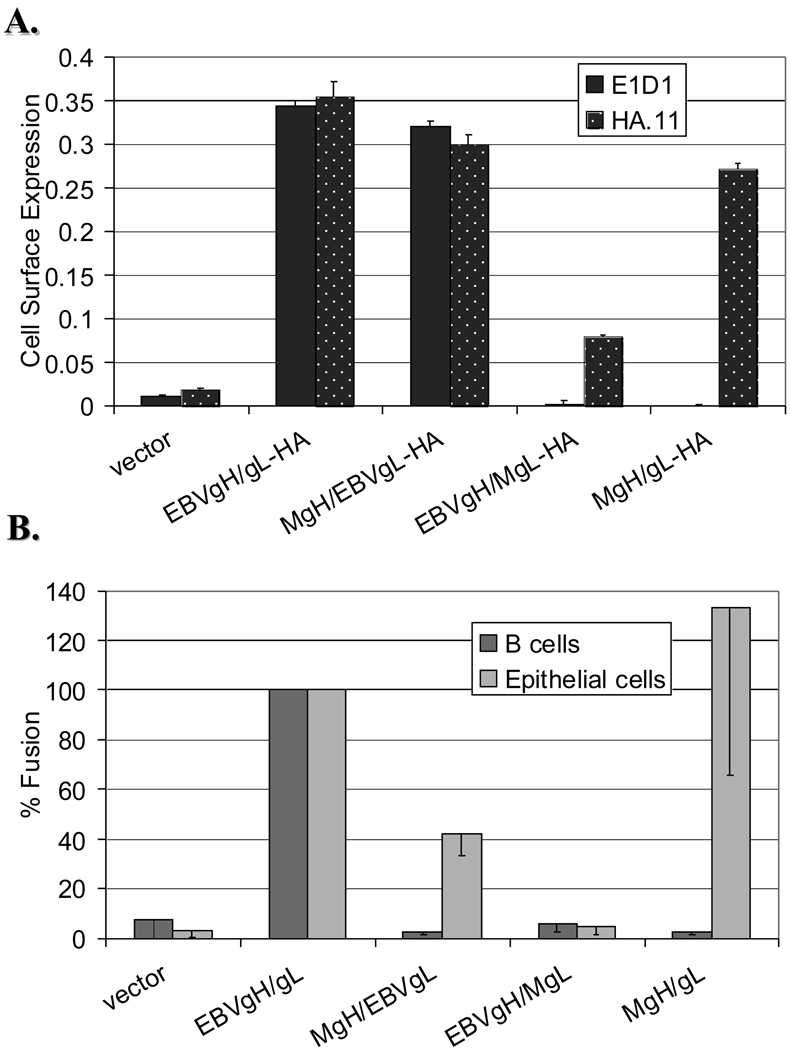
Similar to rhesus glycoproteins, the cell surface expression of heterologous complexes of marmoset (M) and EBV gH and gL was examined by CELISA (Fig. 2A). Only the complex of MgH and EBVgL was cross-reactive with the E1D1 Ab, showing it was expressed at the cell surface. The ability of MgH/EBVgL to bind the E1D1 Ab also suggested that this complex is in a similar conformation as the EBVgH/gL complex. In order to examine the cell surface expression of the rest of the complexes, that were not cross-reactive with the E1D1 Ab, we utilized HA-tagged constructs of EBV and M gL. The tagged constructs were used to more quantitatively measure the cell surface expression of given complexes. Marmoset and EBV gL were not expressed at the cell surface in the absence of gH, as measured by CELISA (data not shown). Thus, the levels of expression detected with an anti-HA Ab were directly related to the amount of gH/gL complexes present at the cell surface. As shown in Fig. 2A, the expression of MgH/gL and MgH/EBVgL complexes when analyzed with an anti-HA Ab was comparable to the EBVgH/gL cell surface expression. However, when MgL was substituted for EBVgL, only a low amount of gH/gL was detected at the cell surface. These results were similar to the data on EBVgH/RhgL suggesting that EBVgH/gL complex is sensitive to cross-species substitution in gL.
Figure 2. Marmoset gH complements fusion with epithelial but not B cells.
CHO-K1 cells transfected with EBVgp42, EBVgB and different combinations of marmoset and EBV gH and gL were used in CELISA (A) and in the fusion assay (B). The combinations of gH/gL tested are shown on the X-axes (M=marmoset). A. The cell surface expression of gH/gL measured by CELISA was performed using an anti-EBVgH/gL Ab, E1D1 (black bars), and an anti-HA Ab, HA.11 (dotted bars). Both MgL and EBVgL were tagged with an HA tag at the C terminus. B. As described in Fig. 1, CHO-K1 cells were overlaid with either Daudi B cells (dark bars) or kidney epithelial 293 cells (light bars) and luciferase activity measured 24 hours later. Luciferase activity was normalized to wild-type levels, which was set to 100% for both cell types. The fusion activity of HA-tagged complexes was comparable to the levels observed with the untagged constructs (data not shown). Data in B are averages of three independent experiments and a representative experiment measured in triplicate is shown in A.
Furthermore, we examined the heterologous complexes of EBV and marmoset gH and gL functionally in the fusion assay (Fig. 2B). As expected, the complex of EBVgH and MgL that was poorly expressed at the cell surface did not mediate fusion with either B cells or epithelial cells. Interestingly, when MgH was paired with either EBV or marmoset gL, fusion was observed with epithelial but not B cells. This raised a possibility that the MgH/EBVgL and MgH/gL complexes were unable to associate with gp42 and thus unable to mediate fusion with B cells. It is important to note that HA-tagged MgL and EBVgL constructs were confirmed to mediate fusion at levels comparable to untagged proteins (data not shown).
EBVgH is the binding partner of EBVgp42 and marmoset gH/gL is unable to bind to EBVgp42
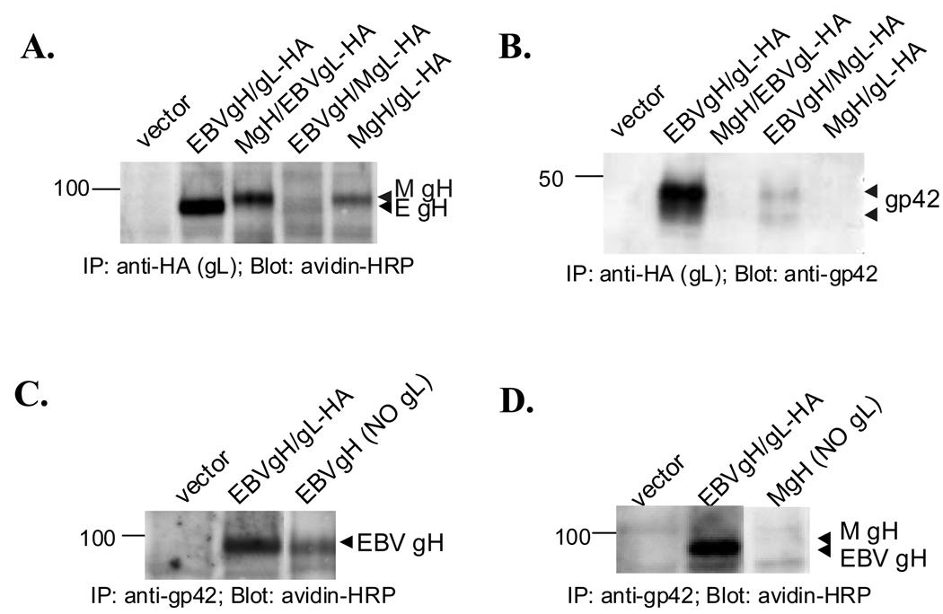
To explore the possibility that complexes containing MgH were unable to mediate fusion with B cells due to a failure to interact with gp42, we examined the association of gp42 and different combinations of gH/gL at the cell surface by co-immunoprecipitation. As shown in Fig. 3A, immunoprecipitation of gL with an-anti HA Ab co-immunoprecipitated gH. The levels of cell surface gH that co-immunoprecipitated correlated with the levels of given complexes expressed at the cells surface (Fig. 2A). Interestingly, neither the MgH/EBVgL nor MgH/gL complexes was able to bind to gp42 while the positive control of EBVgH/gL clearly co-immunoprecipitated gp42 (Fig. 3B). gp42 co-immunoprecipitated poorly with the EBVgH/MgL complex. In a reverse experiment, the MgH/EBVgL and MgH/gL complexes also did not co-immunoprecipitate with EBVgp42 (data not shown). Therefore, these data indicate the inability of MgH/EBVgL and MgH/gL complexes to mediate fusion with B cells is due to their failure to bind gp42.
Figure 3. EBVgH is the binding partner of EBVgp42 and marmoset gH/gL is unable to bind to EBVgp42.
CHO-K1 were transiently transfected with EBV gp42, gB and different combinations of marmoset and EBV gH and gL, and cell biotinylated at 4°C (M=marmoset). Cells were lysed, immunoprecipitated with either an anti-HA Ab (HA.11)(A & B) or an anti-gp42 Ab (F-2-1) (C & D) and SDS-PAGE performed. Avidin-HRP was used to detect biotinylated proteins (A, C & D) and an anti-gp42 rabbit polyclonal Ab (PB1114) (B) to detect gp42 associating with gH/gL. Both EBVgL and MgL were labeled with an HA tag at their C-termini. EBVgL was omitted from transfection in lane 3 in both C and D. The proteins of interest are indicated with arrows. Positions of prestained protein markers (in kDa) are indicated.
It is not clear yet how gp42 binds to gH/gL: whether it binds to both gH and gL or whether only one member of this complex is the binding partner of gp42. Since EBVgH/MgL was still able to bind gp42, it is suggestive that EBVgH is the binding partner of gp42. Thus, to further investigate the interaction between EBVgH/gL and gp42, we omitted either gH or gL from our transfections and immunoprecipitated for gp42. Fig. 3C shows that immunoprecipitation of gp42 resulted in co-immunoprecipitation of EBVgH in the absence of EBVgL. No EBVgL was coimmunoprecipitated with gp42 when expressed without EBVgH (data not shown). In contrast, neither MgH (Fig. 3D) nor MgL (data not shown) was immunoprecipitated with gp42. These data indicate that EBVgH is the binding partner of gp42 and suggest that the inability of MgH to bind EBVgp42 is due to absence of gp42 binding domain(s).
Heterologous complexes of EBV/HHV8 are not expressed at the cell surface and thus are nonfunctional in fusion
To further investigate difference in fusion function for gH and gL, we next studied gH and gL from HHV8 in EBV-mediated fusion. HHV8 gH and gL, along with HHV8gB, were shown previously to be required for HHV8-mediated fusion with B cells and epithelial cells (Pertel, 2002). Even though, EBV and HHV8 belong to the same subfamily of human herpesviruses, compared to the nonhuman primate LCVs, the sequence homology of EBV and HHV8 gH and gL is fairly low.
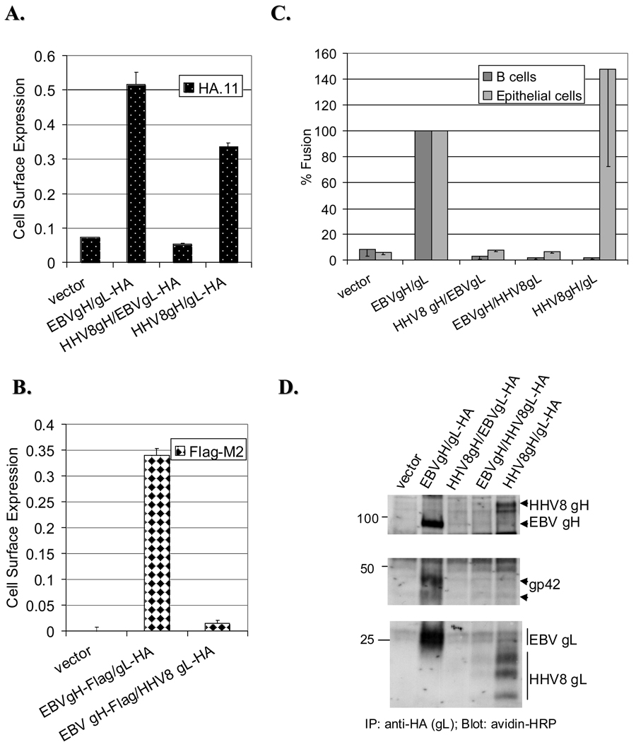
The cell surface expression of heterologous EBV/HHV8 complexes was first examined using the EBV-specific gH/gL Abs, but no cross-reactivity was observed with HHV8 gH and gL (data not shown). Also, the Ab that was previously used for HHV8 gH/gL did not give a sufficient signal in our system (data not shown) (Pertel, 2002). Thus, similar to above, in order to examine the cell surface expression we utilized HA-tagged EBV and HHV8 gL. As shown in Fig. 4A, the cell surface expression of HHV8gH/EBVgL complex was at background levels (vector), while the expression of EBVgH/gL and HHV8gH/gL complexes was at 7 and 4.5 fold above background, respectively. Since HHV8gL is expressed at the cell surface in the absence of gH (data not shown), the expression of EBVgH/HHV8gL complex was assessed by using a Flag-tagged EBVgH. The signal detected with an anti-Flag Ab was a measure of EBVgH/HHV8gL expression at the cell surface which was undetectable (Fig. 4Bl).
Figure 4. Heterologous complexes of EBV/HHV8 are not expressed at the cell surface and thus are nonfunctional in fusion.
As described above, CHO-K1 cells were transfected with EBV gp42, gB and different combinations of gH and gL, as indicated on the X-axes. A. The cell surface expression of different gH and gL complexes was assessed by CELISA as described before. Either an anti-HA (HA.11) (A) or anti-Flag (M2) antibody (B) was used. EBVgH construct with the Flag-tag was used to assess the cell surface expression of EBVgH/HHV8 gL complex since HHV8gL is expressed by itself at the cell surface (data not shown). No gp42 was included in transfection in B, since some EBVgH is expressed at the cell surface in the absence of EBVgL if gp42 is present (data not shown). Representative experiments performed in triplicate are shown. C. Posttransfection, CHO-K1 cells were overlaid with target cells and fusion assessed 24 hours later. Luciferase activity was normalized to wild-type levels, which was set to 100% for both cell types. Data are average of three independent experiments. D. Biotynylated lysates of transfected CHO-K1 cells were immunoprecipitated with an anti-HA Ab and Western probed with avidin-HRP. No association between HHV8 gH and/or gL with EBVgp42 was observed at the cell surface. The proteins of interest are indicated with arrows. Positions of prestained protein markers (in kDa) are indicated.
In our final series of experiments, HHV8 gH and gL were tested in fusion with EBV glycoproteins. Since none of the heterologous EBV/HHV8 complexes were expressed at the cell surface, it was not surprising that those complexes did not mediate fusion with either B cells or epithelial cells (Fig. 4C) and the complexes were also not able to associate with gp42 at the cell surface (Fig. 4D). The fusion levels of tagged constructs were comparable to the untagged proteins (data not shown). EBVgH-Flag was detected at the same size by Western blot as the biotinylated wild-type EBVgH (data not shown). Although HHV8gH/gL was unable to bind gp42 and thus unable to mediate fusion with B cells, when this complex was paired with EBVgB fusion with epithelial cells was observed. This indicated that the HHV8 gH and gL we used were functional. The HHV8 constructs we used in our study were the same ones previously used to described HHV8 cell-cell fusion (Pertel, 2002).
DISCUSSION
The main focus of this paper was to examine the functional conservation of gH and gL from human and nonhuman primate γ-herpesviruses in EBV-induced membrane fusion. Overall, gHs and gLs from more closely related primate herpesviruses were better at complementing EBV gH and gL in EBV-induced membrane fusion than HHV8 gH and gL and the most sensitive to cross-virus complementation were complexes in which EBVgL was substituted (Table 1). Interestingly, the results on heterologous complexes of EBV/HHV8 gH/gL contrast with earlier studies from Dr. Hutt-Fletcher examining the chaperone function of VZVgL in place of EBVgL (Li et al., 1997a). In Dr. Hutt-Fletcher’s study, it was shown that complexes of VZVgL and EBVgH are expressed at the cell surface of transfected cells (Li et al., 1997a). The heterologous complexes were not investigated for function in fusion, but it is interesting that gL from a more distantly related herpesvirus can function for the EBVgL chaperone function when the more closely related HHV8gL can not. One other difference of interest from earlier studies is our inability to detect gL bound to the cell surface which contrasts with earlier studies in which gL was detected at the cell surface (Li, Turk, and Hutt-Fletcher, 1995). This dissimilarity may be due to differences in cell lines used (CV-1 in the previous studies whereas we used CHO-K1 cells). In addition, a T7 expression system using vaccinia virus was used in the previous studies resulting in very high levels of expression of VZVgL. Whether the cell type used, level of expression, or if even vaccinia encoded proteins may influence gL expression on the cell surface will require further investigation. Interestingly, previous reports have indicated variation in whether or not gL is expressed at the cell surface in herpesvirus family members. VZVgL is also not expressed at the cell surface without gH; it is retained inside the cell (Duus and Grose, 1996). On the other hand, HHV8gL is detected at the cell surface even in the absence of gH (data not shown). Additionally, HSV-1gL is not expressed at the cell surface without gH, but unlike VZVgL, HSV-1gL is secreted into the culture medium (Dubin and Jiang, 1995). Therefore, even though the formation of gH/gL complex and its involvement in fusion is conserved across the herpesvirus family, virus specific differences exist in processing of gL and gH and the formation of the gH/gL complex.
Table 1.
Summary of Complementation Resultsa
| Glycoproteins | Cell Surface Expression |
Binding to gp42 |
B Cell Fusion |
Epithelial Cell Fusion |
|
|---|---|---|---|---|---|
| Rh gH | EBV gL | + | + | +b | +b |
| EBV gH | Rh gL | + | + | +/−−c | + |
| Rh gH | Rh gL | + | + | +/−−c | + |
| M gH | EBV gL | + | − | − | + |
| EBV gH | M gL | +/−−d | +/− | − | − |
| M gH | M gL | + | − | − | + |
| HHV8 gH | EBV gL | − | − | − | − |
| EBV gH | HHV8 gL | − | − | − | − |
| HHV8 gH | HHV8 gL | + | − | − | + |
Summary of complementation results on EBV-mediated fusion with rhesus (Rh), marmoset (M) and HHV8 gH and gL (column 3 and 4). Cell surface expression of these proteins when paired with either EBV gH or gL as well as the expression of virus specific gH/gL is shown in column 1. Both heterologous and homologous gH/gL complexes were also examined for their ability to bind to EBVgp42 (column 2).
Enhanced fusion, especially with epithelial cells.
Very low fusion, not higher than 30%, but still detectable over the background.
Expression lower or undetectable with the E1D1 Ab.
A very important finding from this study is identifying EBVgH as the sole binding partner of EBVgp42, similar to studies published while this work was under review (Wu and Hutt-Fletcher, 2007). To our surprise, EBVgH was also expressed at the cell surface in the absence of gL if gp42 was present, although at lower levels. Since MgH did not co-immunoprecipitate with gp42 and did not function in fusion with B cells, this finding suggested that MgH lacks domain(s) required for interaction with gp42 and that these domains are thus different from domains required for epithelial cell entry. Further work will need to be conducted in order to identify the exact domain(s) of EBVgH required for appropriate association with gp42.
Another particularly interesting result from the current study suggests that gL may be important for fusion. A dramatic reduction in B cell fusion was observed with RhgH/gL with fusion levels at only about 5% over background. Since RhgH/gL complex was expressed well at the cell surface, bound the E1D1 antibody, associated with gp42 and RhgH mediated fusion well when paired with EBVgL, these data in summary suggest that gL is somehow involved in fusion and substitution of EBVgL with RhgL was responsible for the reduction in fusion observed. It is important to note that rhesus LCV has been shown to infect human B cells in vitro (Moghaddam et al., 1998), thus low levels of fusion observed with RhgH/gL in our study could not be due an absence of a possible gH/gL receptor on B cells. EBVgL, as well as gLs of other herpesviruses, are thought to function as chaperones, essential for gH processing and transport to the cell surface. However, our data suggests that gL could also have a more direct role in fusion of at least B cells. This may also explain the fusion results obtained with EBVgH/RhgL complex. Even though this complex had lower cell surface expression in the Western blot, which could have resulted in a lower fusion, there was only a slight effect on epithelial cell fusion in contrast to the significant reduction in B cell fusion observed. Thus, it is possible that besides a defect in processing of EBVgH, RhgL is also defective in participating in fusion with B cells that is normally performed by EBVgL. A highly unlikely possibility is that RhgL interferes with EBVgH function and makes the complex nonfunctional, since RhgH/RhgL complex is also a very poor mediator of B cell fusion (Fig. 1B). Another possibility is that gH/gL complex functions differently than gp42/gH/gL complex in fusion, explaining the difference we observed with RhgL between epithelial and B cell fusion.
The work in this study was originally conducted to identify functional domains of EBVgH/gL based on sequence homology and complementation data. In this regard, the studies have been very successful. By comparing EBV and Rh gL protein sequences, excluding the signal sequences, there are differences at only 16 amino acids. Of these, 7 are conservative substitutions. Since the complex of EBVgH/RhgL has a reduced ability to bind to the E1D1 Ab, mutating these 9 amino acids could allow identification of residues important for a proper EBVgH/gL complex formation. Further studies will now focus on mutating individual amino acids within gL and the construction of chimeric gH proteins with the results of these studies providing an important basis to allow for the design and construction of the various chimeras.
The present study provides a detailed comparison of functionality of gH/gL complexes from different γ-herpesviruses in EBV-mediated fusion. Important clues have been obtained that will aid further understanding of the role and domain(s) necessary for proper function of EBVgH/gL in the fusion process. However, in order to completely understand the function of gH/gL complex in EBV entry, as well as other herpesviruses, it will be important to resolve the gH/gL X-ray crystal structure. Since gH/gL is essential for fusion, identification of functional domains will aid in the development of therapeutics to treat EBV infection and EBV-associated diseases.
MATERIALS AND METHODS
Cells and Antibodies
All cells were grown in medium containing 10% FetalPlex animal serum complex (Gemini Bio-Products) and 1% penicillin–streptomycin (BioWhittaker). Chinese hamster ovary cells (CHO-K1) kindly provided by Nanette Susmarski were grown in Ham’s F-12 medium (BioWhittaker). EBV-positive HLA class II- and CD21- expressing Daudi B lymphocytes were obtained from ATCC (Manassas, VA) and were grown in RPMI 1640 medium (BioWhittaker). To more easily monitor membrane fusion, the Daudi 29 cell line stably expressing T7 RNA polymerase was used (Silva et al., 2004). Human embryonic kidney 293T cells were passaged in DMEM medium (BioWhittaker). 293T cells stably expressing T7 RNA polymerase were utilized (Omerovic, Lev, and Longnecker, 2005). Cells were grown in 75-cm2 cell culture flasks (Corning), and adherent cells were detached by using either trypsin-Versene (BioWhittaker) or Versene (phosphate-buffer saline [PBS]-1mM EDTA).
Monoclonal antibodies E1D1 and F-2-1 were gifts from L. Hutt-Fletcher (Louisana State University Health Sciences Center, Shreveport) and recognize the EBVgH/gL complex and gp42, respectively (Balachandran, Oba, and Hutt-Fletcher, 1987; Strnad et al., 1982). A large-scale preparation of the E1D1 and the F-2-1 antibodies was made at the Northwestern University Monoclonal Antibody Facility. The HL800 Ab is a polyclonal antibody that recognizes EBV gH and gL as previously characterized (Omerovic, Lev, and Longnecker, 2005). HA.11 and Flag-M2 antibodies were purchased from Covance Research and Sigma, respectively. Anti-rabbit HRP-conjugated antibody was purchased from Cell Signaling.
Plasmid Constructs
All of the genes of interest were expressed in pCAGGS vector. Generation of plasmids expressing EBV glycoproteins have been discussed elsewhere (Haan, Lee, and Longnecker, 2001). Rhesus and marmoset LCV gH and gL were PCR subcloned from cosmids (rhesus: LV28, Cos9; marmoset: D6, A10) obtained from Dr. Fred Wang into pCAGGS vector. QuickChange Mutagenesis Kit was used to HA-tag both EBV and marmoset LCV gL at the C-terminus. Primers containing the HA-tag sequence flanked by either EBV or marmoset gL sequence at the 3’ end were used for this purpose. A similar method was employed in constructing Flag-tagged EBVgH, except the tag in this case was placed at the N-terminus after residue 23, following the signal sequence (Wu, Borza, and Hutt-Fletcher, 2005). HHV8 gH and gL, wild-type and HA-tagged, were obtained from a former colleague Dr. Peter Pertel. As for EBV and marmoset LCV gLs, HA-tag was placed at the C-terminus on HHV8gL.
Transfection
All of the transfections were performed by a standard protocol using Lipofectamine 2000 transfection reagent (Invitrogen). Twenty-four hours before transfection, CHO-K1 cells were seeded in 6-well plates and the next day were transiently transfected with 0.5 µg each of EBVgB and different combinations of gL and gH (EBV, marmoset, rhesus or HHV8), 2 µg of EBVgp42, and 0.8 µg of a luciferase-containing reporter plasmid with a T7 promoter (Haan, Lee, and Longnecker, 2001; Okuma et al., 1999). In samples where gp42 was omitted, 2µg of an empty pCAGGS vector was included to maintain the total amount of DNA constant. pCAGGS DNA was also used to compensate for missing gH or gL as well. For Western blot experiments, CHO-K1 cells were plated in T-25 cm2 cell culture flasks and one day later transfected with EBV gp42, gB and different combinations of gH and gL. In experiments examining the association between gH/gL and gp42, either gH or gL were omitted from transfection and an empty pCAGGS vector included instead to maintain the amount of DNA constant.
Fusion Assay
Effector CHO-K1 cells were transfected with plasmids encoding the glycoproteins and a luciferase reporter plasmid as stated above. Although the presence of gp42 can be inhibitory in epithelial cell fusion, fusion still occurs (Li, Turk, and Hutt-Fletcher, 1995; McShane and Longnecker, 2004). After 12 hours, CHO-K1 and 293 cells were washed with PBS and detached with Versene. All cells were counted with a Beckman Coulter Z1 particle counter, then the effector and the target cells were mixed in equal amounts (0.2 × 10^6 per sample) and plated into a 24-well plate in Ham’s F-12 medium (Haan, Lee, and Longnecker, 2001; McShane and Longnecker, 2004). Twenty-four hours later, the cells were washed with PBS and lysed, and luciferase was quantified by using the Promega Reporter Assay system. Relative luciferase activity was measured on a Perkin-Elmer Victor plate reader. Luciferase activity was normalized to wild-type levels, which was set to 100% for both B cells and epithelial cells.
Cell Enzyme-Linked Immunosorbent Assay (CELISA)
CHO-K1 cells used for the fusion assay as described above were also used to detect surface expression of the glycoproteins via CELISA as described before (McShane and Longnecker, 2004). Briefly, transfected CHO-K1 cells were transferred into 96-well plates and 24 hours later were incubated with different primary antibodies, depending on the experiment. The following primary antibodies were used: the mouse monoclonal E1D1 Ab, the rabbit polyclonal HL800 Ab, the mouse monoclonal HA.11 Ab and the mouse monoclonal Flag-M2 Ab. The cells were fixed and then incubated sequentially with the secondary biotin conjugated anti-mouse IgG or anti-rabbit IgG (Sigma) and tertiary antibodies. The plates were read as previously described (McShane and Longnecker, 2004).
Immunoprecipitation of Biotinylated Cells and Western Blotting
CHO-K1 cells were transfected as stated above. After 12 hours, the cells were washed with PBS and fresh Ham’s F-12 medium added. The cells were harvested twenty-four hours later and washed three times with ice-cold PBS. Following washes, cells were incubated with EZ-Link Sulfo-NHS-LC-Biotin (Pierce) by rotating for 20 min at 4°C. Biotin was inactivated by washing three times with ice-cold 100mM glycine-PBS. Cytoplasmic lysates were prepared by lysing the cells with 1% Triton-X-100 lysis buffer (Silva et al., 2004) and the insoluble material was removed by centrifugation at 4°C. Cleared lysates were immunoprecipitated overnight at 4°C with either the E1D1, HL800, F-2-1 or HA.11 Ab, depending on the experiment, and captured with protein G-Sepharose (Amersham). Samples were then washed three times in the lysis buffer, re-suspended in sodium dodecyl sulfate (SDS) sample buffer, heated at 95°C for 10 min, and pelleted by centrifugation. The supernatants were separated on Bio-Rad 12.5% or 10% Criterion SDS-PAGE gels, transferred to Immobilon-P membranes and blocked in Tris-buffered saline with Tween-20 (TBST) with 5% milk for 1 hour at room temperature (RT), or overnight at 4°C. The membranes were probed for 30 min at RT with a horseradish peroxidase (HRP)-conjugated-avidin (Bio-Rad) diluted at 1:2000 in blocking solution. For studies examining the association of gH/gL complex with gp42, membranes were incubated for 1 hour at RT with a rabbit polyclonal anti-gp42 antibody (PB1114) diluted at 1:2000 in blocking solution. Membranes were washed in TBST and an anti-rabbit HRP-conjugated secondary antibody (Cell Signaling) applied for 30 min at RT. Following five washes, blots were mixed in equal volumes of ECL solutions and exposed to hyperfilm (Amersham Biosciences).
ACKNOWLEDGEMENTS
We thank Dr. Lindsey Hutt-Fletcher for providing the E1D1 and the F-2-1 antibodies, Nanette Susmarski for cell line expertise, and the members of the Longnecker laboratory for help and support. We also thank Dr. Fred Wang for supplying us with cosmids containing primate gH and gL sequences and Lori Lev for cloning these into a mammalian expression vector. Finally, we thank Dr. Peter Pertel for untagged and HA-tagged HHV8 gH and gL constructs.
R. L. is supported by Public Health Service grants CA62234, CA73507, CA93444 and CA117794 from the National Cancer Institute and AID067048. This work is supported in part by a predoctoral fellowship from the American Heart Association, Midwest Affiliate (J.O.).
Footnotes
Publisher's Disclaimer: This is a PDF file of an unedited manuscript that has been accepted for publication. As a service to our customers we are providing this early version of the manuscript. The manuscript will undergo copyediting, typesetting, and review of the resulting proof before it is published in its final citable form. Please note that during the production process errors may be discovered which could affect the content, and all legal disclaimers that apply to the journal pertain.
REFERENCES
- Balachandran N, Oba DE, Hutt-Fletcher LM. Antigenic cross-reactions among herpes simplex virus types 1 and 2, Epstein-Barr virus, and cytomegalovirus. J Virol. 1987;61(4):1125–1135. doi: 10.1128/jvi.61.4.1125-1135.1987. [DOI] [PMC free article] [PubMed] [Google Scholar]
- Borza CM, Morgan AJ, Turk SM, Hutt-Fletcher LM. Use of gHgL for attachment of Epstein-Barr virus to epithelial cells compromises infection. J Virol. 2004;78(10):5007–5014. doi: 10.1128/JVI.78.10.5007-5014.2004. [DOI] [PMC free article] [PubMed] [Google Scholar]
- Cho Y, Ramer J, Rivailler P, Quink C, Garber RL, Beier DR, Wang F. An Epstein-Barr-related herpesvirus from marmoset lymphomas. Proc Natl Acad Sci U S A. 2001;98(3):1224–1229. doi: 10.1073/pnas.98.3.1224. [DOI] [PMC free article] [PubMed] [Google Scholar]
- Cho YG, Gordadze AV, Ling PD, Wang F. Evolution of two types of rhesus lymphocryptovirus similar to type 1 and type 2 Epstein-Barr virus. J Virol. 1999;73(11):9206–9212. doi: 10.1128/jvi.73.11.9206-9212.1999. [DOI] [PMC free article] [PubMed] [Google Scholar]
- Damania B, Desrosiers RC. Simian homologues of human herpesvirus 8. Philos Trans R Soc Lond B Biol Sci. 2001;356(1408):535–543. doi: 10.1098/rstb.2000.0782. [DOI] [PMC free article] [PubMed] [Google Scholar]
- Dubin G, Jiang H. Expression of herpes simplex virus type 1 glycoprotein L (gL) in transfected mammalian cells: evidence that gL is not independently anchored to cell membranes. J Virol. 1995;69(7):4564–4568. doi: 10.1128/jvi.69.7.4564-4568.1995. [DOI] [PMC free article] [PubMed] [Google Scholar]
- Duus KM, Grose C. Multiple regulatory effects of varicella-zoster virus (VZV) gL on trafficking patterns and fusogenic properties of VZV gH. J Virol. 1996;70(12):8961–8971. doi: 10.1128/jvi.70.12.8961-8971.1996. [DOI] [PMC free article] [PubMed] [Google Scholar]
- Ehlers B, Ochs A, Leendertz F, Goltz M, Boesch C, Matz-Rensing K. Novel simian homologues of Epstein-Barr virus. J Virol. 2003;77(19):10695–10699. doi: 10.1128/JVI.77.19.10695-10699.2003. [DOI] [PMC free article] [PubMed] [Google Scholar]
- Gupta AK, Ruvolo V, Patterson C, Swaminathan S. The human herpesvirus 8 homolog of Epstein-Barr virus SM protein (KS-SM) is a posttranscriptional activator of gene expression. J Virol. 2000;74(2):1038–1044. doi: 10.1128/jvi.74.2.1038-1044.2000. [DOI] [PMC free article] [PubMed] [Google Scholar]
- Haan KM, Lee SK, Longnecker R. Different functional domains in the cytoplasmic tail of glycoprotein B are involved in Epstein-Barr virus-induced membrane fusion. Virology. 2001;290(1):106–114. doi: 10.1006/viro.2001.1141. [DOI] [PubMed] [Google Scholar]
- Kieff E, Rickinson AB. Esptein-Barr virus. In: Knipe DM, Howley PM, editors. Fields Virology. 4th ed. Vol. 2. Philadelphia, P.A.: Lippincott Williams & Wilkins; 2001. pp. 2575–2627. [Google Scholar]
- Kirschner AN, Omerovic J, Popov B, Longnecker R, Jardetzky TS. Soluble Epstein-Barr virus glycoproteins gH, gL, and gp42 form a 1:1:1 stable complex that acts like soluble gp42 in B-cell fusion but not in epithelial cell fusion. J Virol. 2006;80(19):9444–9454. doi: 10.1128/JVI.00572-06. [DOI] [PMC free article] [PubMed] [Google Scholar]
- Li Q, Buranathai C, Grose C, Hutt-Fletcher LM. Chaperone functionscommon to nonhomologous Epstein-Barr virus gL and Varicella-Zoster virus gL proteins. J Virol. 1997a;71(2):1667–1670. doi: 10.1128/jvi.71.2.1667-1670.1997. [DOI] [PMC free article] [PubMed] [Google Scholar]
- Li Q, Spriggs MK, Kovats S, Turk SM, Comeau MR, Nepom B, Hutt-Fletcher LM. Epstein-Barr virus uses HLA class II as a cofactor for infection of B lymphocytes. J Virol. 1997b;71(6):4657–4662. doi: 10.1128/jvi.71.6.4657-4662.1997. [DOI] [PMC free article] [PubMed] [Google Scholar]
- Li Q, Turk SM, Hutt-Fletcher LM. The Epstein-Barr virus (EBV) BZLF2 gene product associates with the gH and gL homologs of EBV and carries an epitope critical to infection of B cells but not of epithelial cells. J Virol. 1995;69(7):3987–3994. doi: 10.1128/jvi.69.7.3987-3994.1995. [DOI] [PMC free article] [PubMed] [Google Scholar]
- Lin SF, Sun R, Heston L, Gradoville L, Shedd D, Haglund K, Rigsby M, Miller G. Identification, expression, and immunogenicity of Kaposi's sarcoma-associated herpesvirus-encoded small viral capsid antigen. J Virol. 1997;71(4):3069–3076. doi: 10.1128/jvi.71.4.3069-3076.1997. [DOI] [PMC free article] [PubMed] [Google Scholar]
- Lukac DM, Renne R, Kirshner JR, Ganem D. Reactivation of Kaposi's sarcoma-associated herpesvirus infection from latency by expression of the ORF 50 transactivator, a homolog of the EBV R protein. Virology. 1998;252(2):304–312. doi: 10.1006/viro.1998.9486. [DOI] [PubMed] [Google Scholar]
- McShane MP, Longnecker R. Cell-surface expression of a mutated Epstein-Barr virus glycoprotein B allows fusion independent of other viral proteins. Proc Natl Acad Sci U S A. 2004;101(50):17474–17479. doi: 10.1073/pnas.0404535101. [DOI] [PMC free article] [PubMed] [Google Scholar]
- Moghaddam A, Koch J, Annis B, Wang F. Infection of human B lymphocytes with lymphocryptoviruses related to Epstein-Barr virus. J Virol. 1998;72(4):3205–3212. doi: 10.1128/jvi.72.4.3205-3212.1998. [DOI] [PMC free article] [PubMed] [Google Scholar]
- Moore PS, Gao SJ, Dominguez G, Cesarman E, Lungu O, Knowles DM, Garber R, Pellett PE, McGeoch DJ, Chang Y. Primary characterization of a herpesvirus agent associated with Kaposi's sarcomae. J Virol. 1996;70(1):549–558. doi: 10.1128/jvi.70.1.549-558.1996. [DOI] [PMC free article] [PubMed] [Google Scholar]
- Okuma K, Nakamura M, Nakano S, Niho Y, Matsuura Y. Host range of human T-cell leukemia virus type I analyzed by a cell fusion-dependent reporter gene activation assay. Virology. 1999;254(2):235–244. doi: 10.1006/viro.1998.9530. [DOI] [PubMed] [Google Scholar]
- Omerovic J, Lev L, Longnecker R. The amino terminus of Epstein-Barr virus glycoprotein gH is important for fusion with epithelial and B cells. J Virol. 2005;79(19):12408–12415. doi: 10.1128/JVI.79.19.12408-12415.2005. [DOI] [PMC free article] [PubMed] [Google Scholar]
- Pertel PE. Human herpesvirus 8 glycoprotein B (gB), gH, and gL can mediate cell fusion. J Virol. 2002;76(9):4390–4400. doi: 10.1128/JVI.76.9.4390-4400.2002. [DOI] [PMC free article] [PubMed] [Google Scholar]
- Pertel PE, Spear PG, Longnecker R. Human herpesvirus-8 glycoprotein B interacts with Epstein-Barr virus (EBV) glycoprotein 110 but fails to complement the infectivity of EBV mutants. Virology. 1998;251(2):402–413. doi: 10.1006/viro.1998.9412. [DOI] [PubMed] [Google Scholar]
- Pulford D, Lowrey P, Morgan AJ. Expression of the Epstein-Barr virus envelope fusion glycoprotein gp85 gene by a recombinant baculovirus. J Gen Virol. 1994;75(Pt 11):3241–3248. doi: 10.1099/0022-1317-75-11-3241. [DOI] [PubMed] [Google Scholar]
- Pulford DJ, Lowrey P, Morgan AJ. Co-expression of the Epstein-Barr virus BXLF2 and BKRF2 genes with a recombinant baculovirus produces gp85 on the cell surface with antigenic similarity to the native protein. J Gen Virol. 1995;76(Pt 12):3145–3152. doi: 10.1099/0022-1317-76-12-3145. [DOI] [PubMed] [Google Scholar]
- Rivailler P, Cho YG, Wang F. Complete genomic sequence of an Epstein-Barr virus-related herpesvirus naturally infecting a new world primate: a defining point in the evolution of oncogenic lymphocryptoviruses. J Virol. 2002;76(23):12055–12068. doi: 10.1128/JVI.76.23.12055-12068.2002. [DOI] [PMC free article] [PubMed] [Google Scholar]
- Rivailler P, Jiang H, Cho YG, Quink C, Wang F. Complete nucleotide sequence of the rhesus lymphocryptovirus: genetic validation for an Epstein-Barr virus animal model. J Virol. 2002;76(1):421–426. doi: 10.1128/JVI.76.1.421-426.2002. [DOI] [PMC free article] [PubMed] [Google Scholar]
- Russo JJ, Bohenzky RA, Chien MC, Chen J, Yan M, Maddalena D, Parry JP, Peruzzi D, Edelman IS, Chang Y, Moore PS. Nucleotide sequence of the Kaposi sarcoma-associated herpesvirus (HHV8) Proc Natl Acad Sci U S A. 1996;93(25):14862–14867. doi: 10.1073/pnas.93.25.14862. [DOI] [PMC free article] [PubMed] [Google Scholar]
- Silva AL, Omerovic J, Jardetzky TS, Longnecker R. Mutational analyses of Epstein-Barr virus glycoprotein 42 reveal functional domains not involved in receptor binding but required for membrane fusion. J Virol. 2004;78(11):5946–5956. doi: 10.1128/JVI.78.11.5946-5956.2004. [DOI] [PMC free article] [PubMed] [Google Scholar]
- Spear PG, Longnecker R. Herpesvirus entry: an update. J Virol. 2003;77(19):10179–10185. doi: 10.1128/JVI.77.19.10179-10185.2003. [DOI] [PMC free article] [PubMed] [Google Scholar]
- Speck P, Haan KM, Longnecker R. Epstein-Barr virus entry into cells. Virology. 2000;277(1):1–5. doi: 10.1006/viro.2000.0624. [DOI] [PubMed] [Google Scholar]
- Strnad BC, Schuster T, Klein R, Hopkins RF, 3rd, Witmer T, Neubauer RH, Rabin H. Production and characterization of monoclonal antibodies against the Epstein-Barr virus membrane antigen. J Virol. 1982;41(1):258–264. doi: 10.1128/jvi.41.1.258-264.1982. [DOI] [PMC free article] [PubMed] [Google Scholar]
- Sun R, Lin SF, Gradoville L, Yuan Y, Zhu F, Miller G. A viral gene that activates lytic cycle expression of Kaposi's sarcoma-associated herpesvirus. Proc Natl Acad Sci U S A. 1998;95(18):10866–10871. doi: 10.1073/pnas.95.18.10866. [DOI] [PMC free article] [PubMed] [Google Scholar]
- Wang F. Epstein-Barr virus related lymphocryptoviruses of old and new world nonhuman primates. In: Robertson ES, editor. Epstein-Barr Virus. Caister Academic Press: Norfolk, England; 2005. pp. 691–709. [Google Scholar]
- Wang F, Rivailler P, Rao P, Cho Y. Simian homologues of Epstein-Barr virus. Philos Trans R Soc Lond B Biol Sci. 2001;356(1408):489–497. doi: 10.1098/rstb.2000.0776. [DOI] [PMC free article] [PubMed] [Google Scholar]
- Wang X, Kenyon WJ, Li Q, Mullberg J, Hutt-Fletcher LM. Epstein-Barr virus uses different complexes of glycoproteins gH and gL to infect B lymphocytes and epithelial cells. J Virol. 1998;72(7):5552–5558. doi: 10.1128/jvi.72.7.5552-5558.1998. [DOI] [PMC free article] [PubMed] [Google Scholar]
- Wu L, Borza CM, Hutt-Fletcher LM. Mutations of Epstein-Barr virus gH that are differentially able to support fusion with B cells or epithelial cells. J Virol. 2005;79(17):10923–10930. doi: 10.1128/JVI.79.17.10923-10930.2005. [DOI] [PMC free article] [PubMed] [Google Scholar]
- Wu L, Hutt-Fletcher LM. Point mutations in EBV gH that abrogate or differentially affect B cell and epithelial cell fusion. Virology. 2007 doi: 10.1016/j.virol.2007.01.025. [DOI] [PMC free article] [PubMed] [Google Scholar]
- Yaswen LR, Stephens EB, Davenport LC, Hutt-Fletcher LM. Epstein-Barr virus glycoprotein gp85 associates with the BKRF2 gene product and is incompletely processed as a recombinant protein. Virology. 1993;195(2):387–396. doi: 10.1006/viro.1993.1388. [DOI] [PubMed] [Google Scholar]


