Summary
During male meiosis, the X and Y chromosomes are transcriptionally silenced, a process termed meiotic sex chromosome inactivation (MSCI). Recent studies have shown that the sex chromosomes remain substantially transcriptionally repressed after meiosis in round spermatids, but the mechanisms involved in this later repression are poorly understood. Mice with deletions of the Y chromosome long arm (MSYq–) have increased spermatid expression of multicopy X and Y genes, and so represent a model for studying post-meiotic sex chromosome repression. Here, we show that the increase in sex chromosome transcription in spermatids from MSYq– mice affects not only multicopy but also single-copy XY genes, as well as an X-linked reporter gene. This increase in transcription is accompanied by specific changes in the sex chromosome histone code, including almost complete loss of H4K8Ac and reduction of H3K9me3 and CBX1. Together, these data show that an MSYq gene regulates sex chromosome gene expression as well as chromatin remodelling in spermatids.
Keywords: Spermatid, Sex chromosome, Post-meiotic sex chromatin, Chromatin marks
Introduction
Spermatogenesis describes a series of complex, developmental processes that are involved in the formation of mature haploid sperm from diploid spermatogonial stem cells. In mice, spermatogenesis occurs in the seminiferous epithelium of the testis and can be divided into three phases: mitotic division of spermatogonia, meiosis, and differentiation of round spermatids into sperm (spermiogenesis). Genes on the sex chromosomes play an essential role in spermatogenesis and, in mice, the Y chromosome encodes a limited number of functions that appear to be restricted to spermatogenesis and testis determination. Spermatogenesis-expressed genes are also over-represented on the X chromosome, with a 15-fold enrichment of spermatogonially expressed genes on the mouse X and a threefold increase in sex- or reproduction-related genes on the human X chromosome compared to the autosomes (Khil et al., 2004; Lercher et al., 2003; Saifi and Chandra, 1999; Wang et al., 2001). Furthermore, the mouse X chromosome is enriched in genes expressed after meiosis, with at least 273 genes (18% of 1555 protein-coding genes) being expressed predominantly or exclusively in spermatids (Mueller et al., 2008).
The transition from spermatogonium to mature sperm is accompanied by dramatic changes in X and Y gene transcription. During the spermatogonial divisions, the sex chromosomes are transcriptionally active, with almost all of the genes on the Y short arm (Yp) being transcribed. Although still transcriptionally active during the leptotene and zygotene stages of meiosis, the sex chromosomes are rapidly silenced at the zygotene-pachytene transition; a phenomenon termed meiotic sex chromosome inactivation (MSCI) (McKee and Handel, 1993). MSCI is a response to the largely unsynapsed state of the XY pair (Baarends et al., 2005; Turner et al., 2004; Turner et al., 2006; Turner et al., 2005) and is initiated when the tumour suppressor protein BRCA1 recruits the kinase ATR to the asynapsed axes of the X and Y chromosomes (Turner et al., 2004). ATR translocates from the axial elements to the chromatin loops, where it is hypothesised to phosphorylate the histone H2A variant, H2AX, at serine 139 to form γH2AX (Fernandez-Capetillo et al., 2003), instigating heterochromatinisation of the sex chromosomes. The XY bivalent is compartmentalised into a specialised chromatin domain known as the sex body (Solari, 1974; Turner, 2007) and undergoes further chromatin modifications, including histone H3 dimethylation, histone H3 and H4 deacetylation (Khalil et al., 2004), H2A ubiquitylation (Baarends et al., 1999), widespread replacement of the histones H3.1 and H3.2 with the histone variant H3.3 (van der Heijden et al., 2007) and incorporation of specific histone variants such as macroH2A.1 (Hoyer-Fender et al., 2000).
Although BRCA1, ATR and γH2AX dissociate from sex chromosomes before the first meiotic division (Mahadevaiah et al., 2001; Turner et al., 2004), the X and Y chromosomes remain substantially transcriptionally repressed after meiosis (Namekawa et al., 2006; Turner et al., 2006). Unlike MSCI, the maintenance of XY silencing in spermatids is incomplete, with genes showing low-level reactivation (Mueller et al., 2008). There is a correlation between X-linked gene copy number and escape from this post-meiotic repression, with the percentage of round spermatids expressing the gene increasing with increasing copy number, suggesting that gene amplification might compensate for the repressive effects initiated by MSCI. Cytologically, the spermatid X and Y form DAPI-dense heterochromatic domains known as post-meiotic sex chromatin (PMSC) (Namekawa et al., 2006), which are located next to the centromeric heterochromatin. PMSC are depleted in markers of transcription, including Cot1 and RNA polymerase II, and are enriched for several repressive chromatin marks present on the sex body, including histone H3 dimethylated at lysine 9 (H3K9me2) and CBX1 (chromobox protein homolog 1, previously known as HP1β) (Baarends et al., 2007; Greaves et al., 2006; Khalil et al., 2004; Namekawa et al., 2006; Turner et al., 2006). However, the X and Y chromosomes are continually remodelled during the transition between meiosis and spermiogenesis, and histone modifications associated with transcriptionally active chromatin [e.g. histone acetylation and histone H3 dimethylated on lysine 4 (H3K4me2)] are also enriched on PMSC in round spermatids (Baarends et al., 2007; Greaves et al., 2006; Khalil and Driscoll, 2006). These epigenetic changes could explain why XY spermatid silencing is less complete than MSCI, and might reflect a need to retain expression of some X- and Y-encoded genes that are essential for spermiogenesis.
Whereas the molecular events underlying MSCI are well characterised, those involved in controlling sex-linked gene expression in round spermatids are less clear. Mice lacking the ubiquitin-conjugating enzyme HR6B have changes to the epigenetic profile of the sex chromosomes in late meiotic and post-meiotic cells, including increased enrichment of the active chromatin mark H3K4me2 in diplotene spermatocytes and round spermatids (Baarends et al., 2007). This is accompanied by increased expression of several X-linked genes, suggesting that HR6B could be involved in spermatid XY repression, possibly by controlling the histone modifications associated with the sex chromosomes in late spermatocytes and round spermatids.
In mice, deletions of the Y chromosome long arm (MSYq) are associated with problems in sperm development and function. Whole testis microarray analysis has shown that mice with MSYq deletions (MSYq–) exhibit de-repression of several sex-linked genes in spermatids, including Ube1x, which is also de-repressed in Hr6b–/– spermatids (Baarends et al., 2007; Ellis et al., 2005). This suggests that genes encoded by the Y long arm are involved in spermatid sex chromosome repression. One MSYq candidate gene is Sly, a multicopy gene that has been shown to interact with histone-modifying enzymes, including the histone acetyltransferase KAT5 (Reynard et al., 2009). However, only a small percentage of spermatid-expressed sex-linked genes were reported to be de-repressed in the MSYq– models, and it is unclear whether this de-repression occurs at the transcriptional or post-transcriptional level. Ellis and colleagues noted that X-linked gene de-repression preferentially affected multiple-copy over single-copy genes, and suggested that there might be something particular about the chromosomal organisation of multicopy genes that is affected in the MSYq deletion mice (Ellis et al., 2005). However, a recent study has shown that in wild-type males, multicopy X genes exhibit spermatid-specific expression, whereas single-copy X genes are expressed abundantly before meiosis and at low levels in spermatids (Mueller et al., 2008). This would make potential increases in single-copy gene expression in spermatids from MSYq– mice more difficult to detect by microarray analysis because these increases would be masked by the abundant expression originating from earlier cell types.
Here, we show that MSYq deletions cause increased expression of sex-linked genes at the level of transcription. In addition, this affects not only multicopy but also single-copy genes and an X-linked transgene, demonstrating a global rather than a local defect in the maintenance of XY silencing. In addition, mice with MSYq deletions have specific abnormalities in the histone code of the round spermatid sex chromosome and centromeric heterochromatin.
Results
Upregulation of X- and Yp-linked genes in the MSYq deletion models is associated with increased nascent transcription
To determine whether the increased mRNA levels in MSYq deletion models is the result of increased transcription of the affected genes, we used RNA fluorescence in situ hybridisation (FISH) to monitor nascent transcription of genes in round spermatids, the cell type of interest. We analysed the expression of nine X, two Y and eight randomly selected autosomally located genes in XY males compared with two MSYq mutants, the first with a deletion of two thirds of the Y chromosome (2/3MSYq–) and a second with complete absence of MSYq (MSYq–). Round spermatid development occurs normally in the two deletion models, with the relative proportion of spermatids at each of the eight steps of round spermatid development unchanged relative to the XY control (Burgoyne et al., 1992; Conway et al., 1994) (L.N.R., unpublished observations).
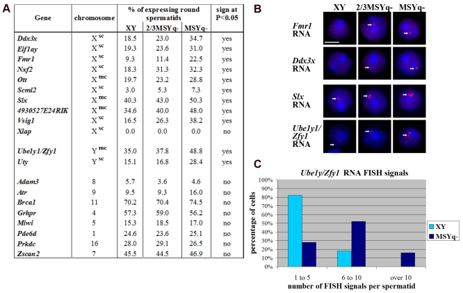
For all eleven sex-linked genes, RNA FISH signals were observed in a significantly (P<0.05) higher proportion of round spermatids from MSYq– mice than from XY control mice, with the expression increasing in proportion to the size of the deletion (Fig. 1A,B and supplementary material Table S1). This effect was independent of the copy-number, or location of the gene on the chromosome (supplementary material Fig. S1), and included three genes (Slx, 4930527E24RIK and Vsig1) previously reported to have increased testis mRNA levels in the MSYq– mice (Ellis et al., 2005), thus substantiating the link between the percentage of expressing spermatids and total mRNA levels. The most dramatic difference was seen for Vsig1, with the number of RNA FISH positive spermatids increasing from an average of 16.5% in XY mice to 38.2% in MSYq– mice. As well as an increase in the proportion of expressing round spermatids, there was also an increase in the number of RNA signals per spermatid for several of the sex-linked genes, as shown by using a probe that detects both Ube1y1 and Zfy1 transcripts (Fig. 1B,C and supplementary material Fig. S2A-C). The mean number of Ube1y1/Zfy1 signals per spermatid more than doubled, from 3.76 signals in the XY control (n=50) to 8.3 signals in MSYq– spermatids (n=50), with the maximum number of RNA signals observed in a single nucleus increasing from 10 to 18.
Fig. 1.
Increased transcription of sex-linked genes in round spermatids from mice with MSYq deletions. Gene-specific RNA FISH was performed on spermatogenic cells from mice with two-thirds (2/3MSYq–) or complete (MSYq–) deletion of the Y long arm, with wild-type XY mice as a control. (A) Table showing the average percentage of round spermatids expressing a given gene, on the basis of the presence of an RNA FISH signal. After angular transformation of the individual percentages, an ANOVA test was used to determine whether the difference between the three genotypes was significant at the P=0.05 level. sc, single copy gene; mc, multicopy gene. More details on gene copy number are given in supplementary material Fig. S1. (B) RNA FISH on XY (left panel), 2/3MSYq– (middle panel) and MSYq– (right panel) round spermatids using probes for Fmr1, Ddx3x, Slx and Ube1y1/Zfy1. DNA is stained with DAPI (blue) and the RNA FISH signal is in red. Scale bar: 5 μm. For Slx and Ube1y1/Zfy1, there is an increase in the number of RNA FISH signals per spermatid in the 2/3MSYq– and MSYq– deletion models compared with the XY control. (C) Quantification of the number of Ube1y1/Zfy1 RNA FISH signals per expressing round spermatid from XY and MSYq– mice.
For the eight autosomal control genes examined, there was no significant difference between the three genotypes with respect to the proportion of spermatids with RNA FISH signals or number of RNA signals per cell (Fig. 1A), implying that autosomal transcription remains unaffected in the MSYq deletion models and that the relative proportion of round spermatids at each step of development is unaffected. To further support this finding, dual RNA FISH was performed on round spermatids from the XY and MSYq– testes for an autosomal gene (Brca1 or Prdkc) together with either an X-linked (Vsig1) or Y-linked gene (Zfy1). The percentage of spermatids positive for a Brca1 or Prdkc RNA FISH signal did not differ between the two genotypes, indicating that the same populations of spermatids were analysed in the two models (supplementary material Fig. S3). Within the Brca1-positive population, the percentage of spermatids positive for Vsig1 (i.e. Brca1 Vsig1 double-positive spermatids) was more than doubled in the MSYq– testis (44.3%) compared with the XY control (21.5%). Additionally, the percentage of Prdkc-positive spermatids also positive for a Zfy1 RNA FISH signal was increased in the MSYq– testis relative to the XY control.
We also studied the expression of a tenth X-linked gene, Xiap, which unlike the other sex-linked genes studied shows no reactivation in XY spermatids (Mueller et al., 2008; Namekawa et al., 2006). Xiap was silent in spermatids from the 2/3MSYq– and MSYq– mice, suggesting that only genes normally transcribed in round spermatids are affected by deletions of MSYq.
To investigate whether the increase in transcription of sex-linked genes in MSYq mutants was restricted to spermiogenesis, we studied expression of the X-linked Fmr1, Nxf2 and Scml2 genes, and the Y-linked Uty gene during meiosis, when MSCI occurs. No expression of these genes was detected by RNA FISH in late pachytene spermatocytes from either XY or MSYq– testis (supplementary material Fig. S2D). In addition, Cot1 RNA FISH was performed on pachytene and diplotene cells from XY, 2/3MSYq– and MSYq– mice (n=55 spermatocytes per genotype). Cot1 DNA is enriched for repetitive sequences such as those found in introns and 3′ untranslated regions, and thus nascent transcripts hybridise to Cot1 probes. In XY spermatocytes, the γH2AX-positive sex-body chromatin domain is negative for Cot1 RNA (Namekawa et al., 2006; Turner et al., 2006), and Cot1 signals are also absent from the sex-body chromatin domain in 2/3MSYq– and MSYq– spermatocytes (supplementary material Fig. S2E), indicating that MSCI occurs normally in Yq deletion mice. Collectively, these findings indicate that the increased transcription of X- and Y-encoded genes in mice with MSYq deletions is restricted to the post-meiotic stages of germ cell development.
Taken together, these data show that the increased mRNA levels of X- and Y-linked genes in spermatids from mice with Y long arm deletions is associated with increased gene transcription. Furthermore, this transcriptional upregulation affects single as well as multicopy sex-linked genes that are distributed along the X and Yp chromosomes. We do not rule out the possibility that autosomal gene transcription is affected to some degree, but loss of MSYq clearly has a more prominent effect on sex chromosome gene expression.
Testis protein levels of upregulated sex-linked genes are increased in the MSYq deletion models
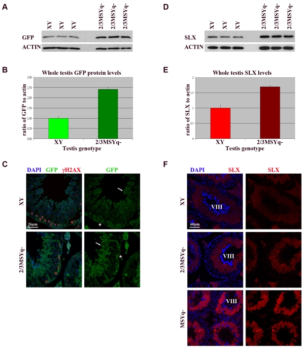
The RNA FISH data presented above show that there is an increase in transcription of genes located across the X chromosome, specifically in round spermatids from mice with large deletions of MSYq. To determine whether this effect was due to an intrinsic property of sex-linked testis-expressed genes, we examined the expression of an EGFP reporter transgene, which is inserted on the distal part of the mouse X chromosome (Hadjantonakis et al., 2001; Hadjantonakis et al., 1998). This transgene is transcribed in spermatogonia, inactivated in meiotic cells during MSCI and reactivated during spermiogenesis. As the increased transcription of genes in the MSYq deletion mice is thought to lead to a corresponding increase in their encoded proteins, we decided to examine the expression of the EGFP transgene at the protein level. Western blot analysis demonstrated that GFP levels are increased by more than twofold in the 2/3MSYq– testes compared to XY controls using both a whole testis (actin) or spermatid-specific (DPP9) (Dubois et al., 2009) protein as a loading control (Fig. 2A,B and supplementary material Fig. S4A,C). This indicates that the X-linked EGFP transgene is also affected by X de-repression in the MSYq deletion models. Immunofluorescence staining of testis sections from GFP-transgenic XY and 2/3MSYq– males showed that the pattern of GFP localisation is unaffected in the 2/3MSYq– testis; however, the level of GFP is increased in 2/3MSYq– mice, specifically in elongating spermatids (Fig. 2C, arrows), with spermatogonial GFP levels remaining unchanged (Fig. 2C, asterisks).
Fig. 2.
Increased testicular levels of GFP and SLX in mice with partial deletions of the Yq. (A) Western blot analysis of testicular GFP levels from XY and 2/3MSYq– mice carrying an X-linked reporter GFP transgene. Membranes were hybridised with an anti-GFP antibody (top panel), then washed and reprobed for actin (bottom panel) as a loading control. (B) Quantification of whole testis GFP levels from the western blot in A using ImageJ software. The level of GFP is represented as the ratio of GFP to actin and is given an arbitrary value of one in the XY testis. Error bars represent the s.e.m. The difference in testis GFP levels between the two genotypes was statistically significant (P<0.001, Student's t-test). (C) GFP immunostaining of testis sections from XY (top panel) and 2/3MSYq– (bottom panel) mice carrying an X-linked GFP transgene. The GFP protein is present in the cytoplasm of spermatogonia (asterisks), early meiotic cells, and elongating spermatids (arrows). There is an increase in GFP levels in the spermatid cytoplasm in the 2/3MSYq– testis compared to the XY control, but the spermatogonial levels of GFP are unchanged. (D) Whole testis western blot analysis of SLX levels in XY and 2/3MSYq– mice. Actin was used as a whole testis loading control. (E) Quantification of SLX levels in the testis of XY and 2/3MSYq– males relative to actin from the western blot in D using ImageJ software. Error bars represent the s.e.m. The difference in SLX levels between the two genotypes was found to be statistically significant (P<0.005, Student's t-test). (F) SLX immunostaining of spermatogenesis stage VIII seminiferous tubules from XY (top panel), 2/3MSYq– (middle panel) and MSYq– (bottom panel) mice. SLX is present in the cytoplasm of round and elongating spermatids from all three genotypes, although there is an increase in SLX levels in the two Yq deletion models compared with the XY control.
To further explore the effect of Yq deletions on sex-chromosome-encoded protein levels, the expression of spermatid-specific X-encoded SLX protein (Reynard et al., 2007) was examined in the 2/3MSYq– testis. Testis SLX levels are increased by over 50% in the 2/3MSYq– testis relative to the XY control (Fig. 2D,E and supplementary material Fig. S4B,D) Immunostaining of XY, 2/3MSYq–, and MSYq– testis sections revealed no change in the distribution and subcellular localisation of SLX, with staining restricted to the cytoplasm of round and early elongating spermatids (Fig. 2F and supplementary material Fig. S4E). However, staining of spermatids was much stronger in stage-matched seminiferous tubules from 2/3MSYq–, and MSYq– mice compared with the XY control. Thus, the increased amount of SLX in the testis of the MSYq deletion models does not lead to ectopic expression of SLX in other spermatogenic cell types, but results in elevated protein levels in the cells that normally express it.
Together, these RNA FISH and protein data show that many sex-linked genes expressed in round spermatids are upregulated in MSYq deletion models, resulting in a corresponding increase in their proteins, which potentially contribute to the phenotypes exhibited by these mice.
The epigenetic profile of the spermatid sex chromosomes is altered in mice with Yq deletions
The increase in sex-linked gene expression observed in the MSYq deletion models might be accompanied by changes in the epigenetic profile of the sex chromosomes. To explore this possibility, we examined the sex chromosome structure and epigenetic marks in round spermatids from XY, 2/3MSYq– and MSYq– mice.
Heterochromatic PMSC
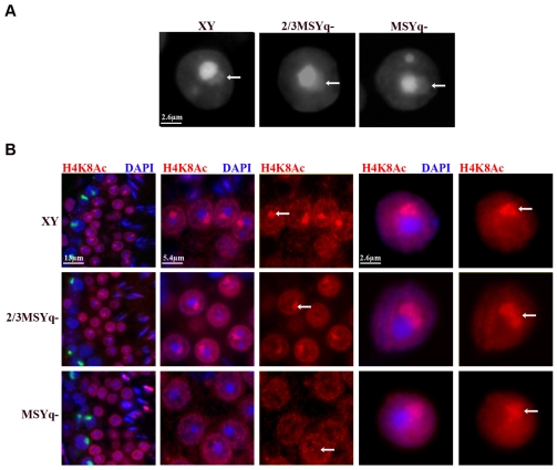
To determine whether the sex chromosomes in spermatids from MSYq deletion mice still form PMSC, spermatids from XY, 2/3MSYq– and MSYq– mice were stained with DAPI. PMSC was cytologically visible as a DAPI-dense structure located next to the chromocentre in 71.6% (514 out of 718) of XY round spermatids and 65.4% of 2/3MSYq– round spermatids (Fig. 3A, arrows). PMSC was discernable in 37.3% of round spermatids from MSYq– mice. Chromosome painting revealed that PMSC is only present in X-bearing round spermatids from MSYq– mice, and that the Y*x chromosome is not detected using DAPI (data not shown); this is not surprising in light of the small size of this chromosome. These results imply that the increased transcription of X-linked genes in the MSYq– spermatids is not associated with obvious X de-heterochromatinisation.
Fig. 3.
The epigenetic marks associated with PMSC are altered in MSYq– spermatids. (A) The DAPI-dense heterochromatic PMSC (arrows) still forms in round spermatids from the 2/3MSYq– and MSYq– mice. (B) The histone modification H48Ac is present on PMSC in round spermatids from XY (top row, arrows) and 2/3MSYq– (middle row, arrows) mice, but is lost from PMSC in the majority of round spermatids from MSYq– mice (bottom row, arrows). Far left: a stage I seminiferous tubule stained for H4K8Ac (red), DAPI (blue) and γH2AX (green). Left and right panel: higher magnification of round spermatids from a stage I tubule. H4K8Ac is present throughout the nucleus and is enriched on PMSC (arrows) but excluded from the chromocentre in spermatids from XY and 2/3MSYq– mice. H4K8Ac is still present in the nucleus of MSYq– spermatids but is not enriched on PMSC. Far right panel: H4K8Ac immunostaining of surface-spread round spermatids. H4K8Ac was enriched on PMSC in 36.6% of spermatids from XY mice but only 6.7% of MSYq– spermatids, where it is reduced compared to the XY control (see Table 1).
Histone H4 acetylation
In light of the interaction between the Yq-encoded SLY1 protein and the histone acetyltransferase KAT5 (Reynard et al., 2009), the most obvious candidate histone modification to be affected in 2/3MSYq– and MSYq– mice was H4 acetylation. Acetylation of H4K8 is associated with transcriptionally active regions of the genome in somatic tissue, and is enriched on the sex chromosomes in round spermatids (Greaves et al., 2006). Immunostaining of testis sections from XY and 2/3MSYq– mice revealed that H4K8Ac is present at low levels throughout the nuclei of round spermatids (Fig. 3B and supplementary material Fig. S5A), where it is excluded from the chromocentre, but enriched on PMSC in a stage-specific manner. By contrast, there was little or no enrichment of H4K8Ac on the PMSC in round spermatids from MSYq– mice (Fig. 3B and supplementary material Fig. S5A), although this modification was still present throughout the nucleus and was excluded from the chromocentre. Examination of surface-spread spermatids demonstrated that the percentage of round spermatids with H4K8Ac enrichment on the PMSC is significantly decreased (P<0.001, Chi-squared test) in the two MSYq deletion models in proportion to deletion size; from 36.6% in the XY control to 6.7% of round spermatids from MSYq– males (Table 1). Furthermore, the level of H4K8Ac is reduced on PMSC in those MSYq– spermatids that retain it, compared to the XY control (Fig. 3B; compare top and bottom rows). Analysis of testis sections indicated that there was no change in the levels or distribution of H4K8Ac in germ cells prior to the round spermatid stage (data not shown).
Table 1.
Summary of the epigenetic marks enriched on the PMSC and chromocentre in round spermatids from XY, 2/3MSYq– and MSYq– males
|
XY
|
2/3MSYq–
|
MSYq–
|
|||||
|---|---|---|---|---|---|---|---|
| Chromatin mark | Spermatid staining pattern | % | n/total | % | n/total | % | n/total |
| DAPI | PMSC | 71.6 | 514/718 | 65.4 | 399/610 | 37.3 | 265/710 |
| H4K8Ac | PMSC | 36.6 | 71/194 | 11.4 | 21/185 | 6.7 | 13/193 |
| H4K12Ac | Chromocentre enrichment | 16.6 | 51/308 | 7.9 | 24/305 | 0 | 0/303 |
| Uniform nuclear staining | 50.6 | 156/308 | 32.8 | 100/305 | 32.7 | 99/303 | |
| Chromocentre exclusion | 32.8 | 101/308 | 59.3 | 181/305 | 67.3 | 204/303 | |
| H3K9me2 | PMSC | 66.6 | 211/317 | 40.0 | 122/305 | 27.9 | 88/316 |
| Chromocentre | 57.1 | 181/317 | 43.3 | 132/305 | 39.6 | 125/316 | |
| Diffuse staining | 23.0 | 73/317 | 47.9 | 146/305 | 51.3 | 162/316 | |
| H3K9me3 | PMSC and chromocentre | 76.6 | 232/303 | 67.3 | 212/315 | 41.4 | 125/302 |
| Chromocentre only | 23.4 | 71/303 | 32.7 | 103/315 | 58.6 | 177/202 | |
| CBX1 | PMSC and chromocentre | 82.1 | 307/374 | 78.0 | 295/378 | 49.1 | 185/377 |
| Chromocentre only | 17.9 | 67/374 | 22.0 | 83/378 | 50.9 | 192/377 | |
For each genotype, testis material from three individuals was used to make a single cell suspension and analysed. The number (n) of round spermatids with enrichment of epigenetic marks on the PMSC is shown as a percentage of the total number of round spermatids counted (total). H4K8Ac is reduced on PMSC from mice with MSYq deletions. H4K12Ac is reduced from the chromocentre in MSYq mutants. Fewer spermatids have H3K9me2 on both the chromocentre and PMSC in MSYq mutants. The levels of H3K9me3 on PMSC is reduced in the MSYq mutants. The levels of CBX1 are reduced on PMSC in the MSYq mutants
Next, we examined the localisation of H4K12 acetylation on surface-spread round spermatids. In XY cells, three patterns of H4K12 localisation were observed: enrichment of H4K12Ac on the chromocentre (clustered centromeric heterochromatin), uniform nuclear staining and exclusion of H4K12Ac from the chromocentre (supplementary material Fig. S6A). These three patterns were also observed in 2/3MSYq– spermatids, although there was no enrichment of H412Ac on the chromocentre in MSYq– spermatids. Furthermore, the percentage of round spermatids with H4K12Ac excluded from the chromocentre was significantly increased in the two Yq deletion models (P<0.001, Chi-squared test), and was more than doubled in MSYq– mice compared with the XY control (Table 1).
Histone H3 methylation
The transcriptional upregulation and loss of H4K8Ac might reflect a global change in the conformation and activity of the PMSC in round spermatids. To investigate this further, we examined additional chromatin modifications associated with the PMSC. Methylation of H3K9 is a marker of transcriptionally silent chromatin.
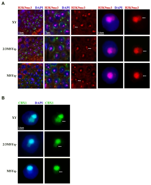
Trimethylation of H3K9 is enriched on PMSC and chromocentre or on the chromocentre only in round spermatids. On surface-spread testis cells, localisation of H3K9me3 to PMSC was seen in 76.6% of round spermatids from XY mice (Fig. 4A and Table 1), and 41.4% of round spermatids from MSYq– mice. In MSYq– mice, H3K9me3 was only observed on PMSC in X-bearing round spermatids (supplementary material Fig. S5C). Although there was no difference in the intensity of H3K9me3 chromocentre staining between the three genotypes, the extent of H3K9me3 enrichment on PMSC was reduced in MSYq– spermatids relative to that in normal males (Fig. 4A and supplementary material Fig. S5B).
Fig. 4.
Decreased H3K9me3 and CBX1 enrichment on PMSC in spermatids from MSYq– mice. (A) H3K9me3 is reduced on PMSC but not the chromocentre of MSYq– spermatids. Far left panel: a stage VIII seminiferous tubule stained for H3K9me3 (red), DAPI (blue) and the acrosomal protein DKKL1 (green). Left and right panel: higher magnification of round spermatids from a stage VIII tubule. H3K9me3 enrichment on the PMSC (arrows) is reduced in the MSYq– spermatids (bottom row) compared to XY (top row) and 2/3MSYq– (middle row) spermatids. Far right panel: H3K9me3 immunostaining of surface-spread round spermatids reveals that H3K9me3 enrichment on PMSC (arrows) is reduced in MSYq– spermatids relative to the XY control. (B) Antibody staining for the heterochromatin protein CBX1 (green) reveals that this protein is reduced on PMSC of MSYq– spermatids (bottom row, arrows) compared to XY (top row, arrows) and 2/3MSYq– spermatids (middle row, arrows).
On surface-spread testicular cells, three patterns of H3K9 dimethylation were identified in round spermatids from normal males: chromocentre and PMSC staining, PMSC staining only, or diffuse nuclear staining (Fig. S6B). These three staining patterns were also observed in round spermatids from 2/3MSYq– and MSYq– mice. There was a significant reduction in the number of round spermatids with H3K9me2 chromocentre staining, from 57.1% in XY spermatids to 43.3% and 39.6% in 2/3MSYq– and MSYq– spermatids, respectively (P<0.001, Chi-squared test). The level of H3K9me2 enrichment on PMSC in XY spermatids was comparable to that on the PMSC of 2/3MSYq– and MSYq– spermatids (supplementary material Fig. S6B), but the percentage of round spermatids with enrichment of H3K9me2 on the sex chromosome was decreased in the two Yq deletion models, in proportion to the extent of the deletion. No difference in the localisation or relative enrichment of H3K9 di- and trimethylation was observed in meiotic cells between the MSYq– testis and XY control (not shown).
Finally, we looked at the active histone modifications H3K36me3 and H3K4me3 in round spermatids from XY, 2/3MSYq– and MSYq– mice. There was no difference in the localisation of either histone mark between the three genotypes, with these modifications being excluded from both PMSC and chromocentre in round spermatids (supplementary material Fig. S7).
CBX1
The heterochromatin-associated protein CBX1 (also called M31 and HP1β) is recruited by H3K9 methylation (Lachner et al., 2001) and is considered a marker of inactive chromatin. In light of the changes in H3K9 tri- and dimethylation on the sex chromosome and chromocentre in MSYq– spermatids, respectively, we next analysed CBX1 in round spermatids. On surface-spread spermatogenic cells, CBX1 was present on the chromocentre of all round spermatids analysed from XY, 2/3MSYq– and MSYq– mice. In XY mice, CBX1 was enriched on the DAPI-dense X or Y PMSC in 82.1% of round spermatids (Fig. 4B and Table 1). In round spermatids from MSYq– mice, CBX1 localised to the sex chromosome domain in 49.1% of spermatids examined; chromosome painting confirmed that these were the X-bearing spermatids. As seen with H3K9me3, CBX1 staining was markedly reduced on the PMSC in MSYq– round spermatids compared to XY round spermatids, despite there being no difference in the intensity of chromocentre staining (Fig. 4B; compare top and bottom panels).
In conclusion, although the X chromosome still forms a DAPI-dense PMSC structure in round spermatids from 2/3MSYq– and MSYq– testes, the epigenetic marks associated with PMSC are altered compared to the XY control. These changes include reduced enrichment of H3K9me3 and CBX1, and, most strikingly, almost complete loss of H4K8Ac. There is also a significant reduction in the proportion of round spermatids with chromocentre H3K9me2 and H4K12Ac staining in MSYq– males compared with the XY control. A summary of these changes is given in Table 1.
Discussion
In this study, we have established that mice with deletions of the Y chromosome long arm have a global increase in transcription of sex chromosome genes specifically in round spermatids, leading to increased levels of their encoded proteins. Coupled with this, we report that loss of the mouse Y chromosome long arm affects histone modifications associated with the sex chromosomes and chromocentre in round spermatids, in particular exerting an effect on H4K8 acetylation and H3K9 methylation.
H3K9 methylation is a marker of transcriptionally inactive chromatin and so the finding that H3K9me3 is reduced on the PMSC in MSYq– spermatids is not unexpected given the increased transcription of sex-linked genes in these cells. By contrast, H4K8Ac is associated with active chromatin and has been hypothesised to play a role in transcription of the X chromosome in spermatids (Khalil et al., 2004). The reduction of H4K8Ac on PMSC in spermatids with MSYq deletions is therefore surprising, although H4K8Ac might be involved in replacement of histone variants such as H2A and macroH2A with H2A.Z (Greaves et al., 2006), which might be disturbed in these spermatids. Another unexpected finding is that there is loss of H3K9me2 and H4K12Ac histone modifications on the chromocentre in round spermatids from mice with Yq deletions. This suggests that the maintenance of epigenetic marks on the chromocentre and PMSC in round spermatids are linked, although it is not known whether the changes to the chromocentre histone modifications affects the transcriptional repression of centromeric repeats.
A similar phenotype to that described here occurs in mice deficient for the ubiquitin-conjugating enzyme HR6B (Baarends et al., 2007). HR6B appears to play a role in controlling histone modifications on the XY chromatin in late spermatocytes and round spermatids; Hr6b–/– round spermatids also have reduced levels of H3K9me2 on the centromeric heterochromatin. HR6B is involved in sex chromosome silencing during the transition from the meiotic and post-meiotic stages of spermatogenesis. By contrast, MSYq deletions only affect gene transcription and epigenetic markers associated with the sex chromosomes and centromeric heterochromatin in round spermatids.
The cause-effect relationship between the altered sex chromosome histone code and the increase in expression of sex-linked genes in MSYq– spermatids has not been explored. The increased expression in MSYq deletion mice might result from altered sex chromosome chromatin conformation caused by changes in their epigenetic profile, allowing increased access to the transcriptional machinery. For example, overexpression of the S. pombe Epe1 protein reduces the levels of H3K9me2 at heterochromatic domains, increasing transcription from these regions (Zofall and Gewal, 2006). Alternatively, increased transcription of the sex chromosomes might lead to the replacement of heterochromatin markers such as H3K9me3 with `active' modifications and proteins, although H3K4me3 and H3K36me3 remain excluded from the PMSC in 2/3MSYq– and MSYq– spermatids. The correlation between increased sex chromosome expression and changes to the sex chromosome epigenetic profile in mice with MSYq deletions implicates an MSYq-linked gene(s) in the establishment and maintenance of sex chromosome transcriptional repression during spermatid differentiation. This is the first gene(s) to have a role in sex chromosome repression that acts specifically in spermatids rather than during MSCI or during the meiotic to post-meiotic transition.
Several multicopy genes have been identified on the Yq that could potentially have a role in modulating the epigenetic marks and transcriptional activity of the sex chromosomes in round spermatids. These include the Ssty gene family, Asty, Orly and Sly, all of which are transcribed specifically in the testis and are absent in the MSYq– testis (Conway et al., 1994; Toure et al., 2005; Ellis et al., 2007). A good candidate gene is Sly, which encodes a protein (SLY1) that is present in the both the nucleus and cytoplasm of round and early elongating spermatids (Reynard et al., 2009). This protein is reduced in the 2/3MSYq– testis and absent in the MSYq– testis, and so the level of SLY1 in the Yq deletion models correlates with the severity of the spermatid sex chromosome de-repression.
SLY1 contains a COR1 domain, which is thought to facilitate chromatin binding, and this protein interacts with the acetyltransferase and chromatin remodelling protein KAT5 in round spermatids (Reynard et al., 2009). The transcription of Slx, the X-linked homologue of Sly, is increased in proportion to the decrease in Sly mRNA levels in the two MSYq deletion models, suggesting that there might be regulatory interactions between Sly and Slx in normal males (Ellis et al., 2005; Touré et al., 2005). It has been hypothesised that Sly and Slx might have evolved to reciprocally repress the expression of sex chromosome genes by their effect on sex chromatin (Ellis et al., 2005). However, Slx encodes a cytoplasmic protein present from step 2 of spermiogenesis (Reynard et al., 2007) and at present there is no evidence that SLX interacts with SLY1, KAT5 or other SLY1-interacting proteins (L.N.R., unpublished observations). In addition, reduction of H4K8Ac on the PMSC in MSYq– spermatids is seen in step 1 round spermatids, before Slx is transcribed and translated. Therefore, although Slx upregulation might contribute to the phenotypes exhibited by the MSYq deletion mice, it is not the primary cause of the increased expression and epigenetic changes to the sex chromosomes in round spermatids from these mice.
Another candidate gene is the Ssty family, which is present in at least 200 copies on the Yq and is composed of two distinct members, Ssty1 and Ssty2. Whereas both Ssty1 and Ssty2 are transcribed in round spermatids, only a subset of Ssty1 transcripts is thought to be translated, and no SSTY2 protein has been identified to date (Toure et al., 2004a). The subcellular localisation and function of the SSTY1 protein in spermatids is unknown, although the levels of this protein are increased by approximately twofold in the 2/3MSYq– testis (Toure et al., 2004b). The biological role of the Asty and Orly genes are also unknown, but might act as non-coding RNAs as these genes have no identifiable open reading frame (Touré et al., 2005; Ellis et al., 2007).
It seems counterintuitive that a Y-encoded gene would have evolved a role in repressing expression of sex-linked genes (presumably including itself) in round spermatids. However, the silencing of the mammalian sex chromosomes in the male germline by MSCI has been proposed to act as a genomic defence mechanism against sex-ratio distorters (SRDs; an allele or gene located on a sex chromosome that increases its own transmission to the next generation) by silencing these genes during and after meiosis (Ellis et al., 2005; Tao et al., 2007; Turner et al., 2006). This therefore maintains a 1:1 male to female sex ratio, the only evolutionary stable strategy (Fisher's principle) (Fisher, 1930; Hamilton, 1967). Several sex chromosome genes expressed during spermiogenesis are thought to have important functions in sperm maturation and fertility, and could evolve into SRDs. These SRDs reduce male reproductive fitness by reducing the number of functional sperm as well as by causing a deviation in the sex ratio; thus, there is strong selection for sex-linked or autosomal suppressors that ameliorate the deleterious effects of the distorter (Carvalho et al., 1997; Hamilton, 1967; Hurst, 1992; Montchamp-Moreau, 2006). However, new sex-ratio distorters might evolve that are unaffected by existing suppressor elements, allowing sex-ratio meiotic drive to continue ad infinitum. By evolving a function in sex chromosome silencing in spermatids, a Yq-linked suppressor element can suppress multiple sex-linked SRD elements at once rather than suppress only one.
In support of this hypothesis, a mild sex-ratio distortion in favour of females is present in the offspring of 2/3MSYq– mice, and this is hypothesised to be a consequence of a disruption in the equilibrium between an X-linked meiotic driver and a suppressor gene located on the Yq (Ellis et al., 2005). Unbalancing of the Drosophila Dox and Ste X-linked distorter genes from their suppressor results in a shortage of, or reduced fertilising ability of, Y-bearing sperm by an unknown mechanism and leads to a sex ratio in favour of females (Aravin et al., 2001; Tao et al., 2007a). Furthermore, gross overexpression of the X-linked distorter might be the cause of sterility in the MSYq– mice because unrepressed drivers (e.g. Stellate in D. melanogaster) can lead to sterility rather than drive.
In conclusion, the data we present in this paper provide evidence for the role of an MSYq encoded factor(s) in the regulation of histone modifications and transcription from the sex chromosomes in round spermatids. Analysis of MSYq– mice carrying transgenes or targeted mutations of the various MSYq-encoded genes will be invaluable in investigating which gene or genes are responsible.
Materials and Methods
Mice
All XY and 2/3MSYq– mice were produced at the MRC National Institute for Medical Research (London, UK) on a MF1 random bred background. XY mice carry an RIII strain of Y chromosome and are the appropriate controls for the 2/3MSYq– and MSYq– mice models. The 2/3MSYq– mice have an RIII strain Y chromosome with an interstitial deletion removing approximately two-thirds of the MSYq and were derived from a stock originating from the mice described by Conway and colleagues (Conway et al., 1994). MSYq– mice [XSxraY*X mice (Burgoyne et al., 1992) and XY*xSxra mice (Yamauchi et al., 2009)] lack the entire Y-specific (non-PAR) gene content of MSYq, with the only Y-specific material provided by the Y short-arm-derived factor Sxra. XSxraY*x males were produced by mating XY*X females (Burgoyne et al., 1998; Eicher et al., 1991) to XYSxra males (Cattanach, 1987; Cattanach et al., 1971). XY*xSxra mice were produced by Monika Ward (Institute for Biogenesis Research, John A. Burns School of Medicine, University of Hawaii, Honolulu, HI) using ICSI of sperm from XSxraY*x (Yamauchi et al., 2009). D4/XEGFP mice (Hadjantonakis et al., 2001; Hadjantonakis et al., 1998) were obtained from Andras Nagy (Samuel Lunenfeld Research Institute, Mount Sinai Hospital, Toronto, ON). Hemizygous EGFP transgenic XX females were bred with either XYRIII or 2/3MSYq– MF1 males to produce XY-GFP and 2/3MSYq-GFP mice, with non-transgenic littermates used as controls. Animal procedures were in accordance with the United Kingdom Animal Scientific Procedures Act 1986 and were subject to local ethical review.
RNA FISH
RNA fluorescence in situ hybridization (FISH) was performed on surface-spread spermatogenic cells from adult XY, 2/3MSYq– and MSYq– testes as described previously (Reynard et al., 2007) using the probes listed in supplementary material Table S2. For each gene, RNA FISH was replicated three times using three different mice per genotype, and the presence of an RNA FISH signal was examined in approximately 100 round spermatids per mouse (supplementary material Table S1). The percentage of expressing spermatids per mouse was converted into angles and a one way analysis of variance (ANOVA) test performed comparing the three genotypes. For dual RNA FISH, 200 round spermatids were counted per genotype for the presence of an RNA signal.
Western blot analysis
Testis protein extraction and western blotting was performed as described previously (Reynard et al., 2007) using the rabbit polyclonal anti-SLX antibody (Reynard et al., 2007) at 1:2500, anti-GFP antibody at 1:5000 (Invitrogen), rabbit anti-DPP9 antibody at 1:3000 (Abcam, ab42080) and mouse anti-actin antibody at 1:7000 (Sigma, A5441). After incubating with the appropriate secondary antibody (anti-mouse IgG, anti-rabbit IgG or anti-goat IgG) coupled to horseradish peroxidase (DAKO), the signal was revealed by chemiluminescence (SuperSignal, Pierce) and recorded on X-ray film.
Antibody staining and chromosome painting of surface spread spermatids
A single cell suspension was made from the testis of three mice per genotype and fixed for 5 minutes at room temperature (R/T) in 2.6 mM sucrose, 1.86% formaldehyde solution. The cell suspension was then centrifuged at 112 g for 5 minutes at R/T, the fixative removed and the cells resuspended in PBSA. Three drops of the cell suspension were added to Superfrost Plus slides (BHD) and allowed to air dry at R/T. The cells were permeabilised by adding 0.5% Triton X-100 to the slides for 10 minutes at R/T and then washed once in PBS for 5 minutes before being blocked in blocking buffer (PBS, 0.15% BSA, 0.1% Tween-20) for 1 hour at R/T. The slides were incubated overnight in the appropriate primary antibody diluted in blocking buffer at 37°C, washed in PBS three times, and incubated in secondary antibody diluted in PBS for 1 hour at 37°C. After washing the slides three times in PBS, the cells were mounted in Vectorshield mounting media with DAPI and observed. Chromosome painting was performed after antibody staining using FITC StarFISH mouse X or Y chromosome paint (Cambio, Cambridge, UK) as described previously (Reynard et al., 2007).
Preparation and immunostaining of testis section
After dissection, mouse testes were either fixed in 4% paraformaldehyde overnight at R/T, or pre-fixed in 4% paraformaldehyde for 4 hours at R/T before being fixed in dilute Bouin's solution (0.675% picric acid, 9.25% formaldehyde, 4.9% glacial acetic acid diluted in milliQ water) overnight at 4°C. Testes were then dehydrated, embedded, and cut into 3 μm sections as described previously (Reynard et al., 2007). The sections were mounted onto Superfrost slides, rehydrated and subjected to antigen retrieval by boiling in 0.01M sodium citrate solution pH 6.0. After boiling, the slides were rinsed in PBS before continuing the antibody staining protocol described above for surface-spread cells. A list of antibodies used in this study is given in supplementary material Table S3.
Supplementary Material
Supplementary material available online at http://jcs.biologists.org/cgi/content/full/122/22/4239/DC1
We thank Prim Singh (Division of Immunoepigenetics, Department of Immunology and Cell Biology, Research Center Borstel, Borstel, Germany) for the gift of an antibody against CBX1, and Monika Ward for providing XY*xSxra testis material. We are also grateful to Paul Burgoyne for mouse breeding, help with statistical analysis and the critical reading of this manuscript, Shantha Mahadevaiah for interesting discussions and Andrew Ojarikre for help with mouse breeding. This work was supported by the Medical Research Council. Deposited in PMC for release after 6 months.
References
- Aravin, A. A., Naumova, N. M., Tulin, A. V., Vagin, V. V., Rozovsky, Y. M. and Gvozdev, V. A. (2001). Double-stranded RNA-mediated silencing of the genomic tandem repeats and transposable elements in the D. melanogaster germline. Curr. Biol. 11, 1017-1027. [DOI] [PubMed] [Google Scholar]
- Baarends, W. M., Hoogerbrugge, J. W., Roest, H. P., Oooms, M., Vreeburg, J., Hoeijmakers, J. H. J. and Grootegoed, J. A. (1999). Histone ubiquitination and chromatin remodeling in mouse spermatogenesis. Dev. Biol. 207, 322-333. [DOI] [PubMed] [Google Scholar]
- Baarends, W. M., Wassenaar, E., van der Laan, R., Hoogerbrugge, J., Sleddens-Linkels, E., Hoeijmakers, J. H., de Boer, P. and Grootegoed, J. A. (2005). Silencing of unpaired chromatin and histone H2A ubiquitination in mammalian meiosis. Mol. Cell. Biol. 25, 1041-1053. [DOI] [PMC free article] [PubMed] [Google Scholar]
- Baarends, W. M., Wassenaar, E., Hoogerbrugge, J. W., Schoenmakers, S., Sun, Z. W. and Grootegoed, J. A. (2007). Increased phosphorylation and dimethylation of XY body histones in the Hr6b-knockout mouse is associated with de-repression of the X chromosome. J. Cell Sci. 120, 1841-1851. [DOI] [PubMed] [Google Scholar]
- Burgoyne, P. S., Mahadevaiah, S. K., Sutcliffe, M. J. and Palmer, S. J. (1992). Fertility in mice requires X-Y pairing and a Y-chromosomal “spermiogenesis” gene mapping to the long arm. Cell 71, 391-398. [DOI] [PubMed] [Google Scholar]
- Burgoyne, P. S., Mahadevaiah, S. K., Perry, J., Palmer, S. J. and Ashworth, A. (1998). The Y* rearrangement in mice: new insights into a perplexing PAR. Cytogenet. Cell Genet. 80, 37-40. [DOI] [PubMed] [Google Scholar]
- Carvalho, A. B., Vaz, S. C. and Klaczko, L. B. (1997). Polymorphism for Y-linked suppressors of sex-ratio in two natural populations of Drosophila mediopunctata. Genetics 146, 891-902. [DOI] [PMC free article] [PubMed] [Google Scholar]
- Cattanach, B. M. (1987). Sex-reversed mice and sex determination. Ann. N. Y. Acad. Sci. 513, 27-29. [DOI] [PubMed] [Google Scholar]
- Cattanach, B. M., Pollard, C. E. and Hawkes, S. G. (1971). Sex reversed mice: XX and XO males. Cytogenetics 10, 318-337. [DOI] [PubMed] [Google Scholar]
- Conway, S. J., Mahadevaiah, S. K., Darling, S. M., Capel, B., Rattigan, Á. M. and Burgoyne, P. S. (1994). Y353/B: a candidate multiple-copy spermiogenesis gene on the mouse Y chromosome. Mamm. Genome 5, 203-210. [DOI] [PubMed] [Google Scholar]
- Dubois, V., Van Ginneke, C., De Cock, H., Lambeir, A. M., Van der Veken, P., Augustyns, K., Chen, X., Scharpe, S. and De Meester, I. (2009). Enzyme activity and immunohistochemical localization of dipeptidyl peptidase 8 and 9 in male reproductive tissues. J. Histochem. Cytochem. 57, 531-541. [DOI] [PMC free article] [PubMed] [Google Scholar]
- Eicher, E. M., Hale, D. W., Hunt, P. A., Lee, B. K., Tucker, P. K., King, T. R., Eppig, J. T. and Washburn, L. L. (1991). The mouse Y* chromosome involves a complex rearrangement, including interstitial positioning of the pseudoautosomal region. Cytogenet. Cell Genet 57, 221-230. [DOI] [PubMed] [Google Scholar]
- Ellis, P. J. I., Clemente, E. J., Ball, P., Toure, A., Ferguson, L., Turner, J. M. A., Loveland, K. L., Affara, N. A. and Burgoyne, P. S. (2005). Deletions on mouse Yq lead to up-regulation of multiple X- and Y-linked transcripts in spermatids. Hum. Mol. Genet. 14, 2705-2715. [DOI] [PubMed] [Google Scholar]
- Ellis, P. J., Ferguson, L., Clemente, E. J. and Affara, N. A. (2007). Bidirectional transcription of a novel chimeric gene mapping to mouse chromosome Yq. BMC Evol. Biol. 7, 171. [DOI] [PMC free article] [PubMed] [Google Scholar]
- Fernandez-Capetillo, O., Mahadevaiah, S. K., Celeste, A., Romanienko, P. J., Camerini-Otero, R. D., Bonner, W. M., Manova, K., Burgoyne, P. and Nussenzweig, A. (2003). H2AX is required for chromatin remodeling and inactivation of sex chromosomes in male mouse meiosis. Dev. Cell 4, 497-508. [DOI] [PubMed] [Google Scholar]
- Fisher, R. A. (1930). The Genetical Theory of Natural Selection (ed. H. Bennett). Oxford: Clarendon Press.
- Greaves, I. K., Rangasamy, D., Devoy, M., Marshall Graves, J. A. and Tremethick, D. J. (2006). The X and Y chromosomes assemble into H2A.Z, containing facultative heterochromatin, following meiosis. Mol. Cell. Biol. 26, 5394-5405. [DOI] [PMC free article] [PubMed] [Google Scholar]
- Hadjantonakis, A. K., Gertsenstein, M., Ikawa, M., Okabe, M. and Nagy, A. (1998). Non-invasive sexing of preimplantation stage mammalian embryos. Nature Genetics 19, 220-222. [DOI] [PubMed] [Google Scholar]
- Hadjantonakis, A.-K., Cox, L. L., Tam, P. P. L. and Nagy, A. (2001). An X-linked GFP transgene reveals unexpected paternal X-chromosome activity in trophoblast giant cells of the mouse placenta. Genesis 29, 133-140. [DOI] [PubMed] [Google Scholar]
- Hamilton, W. D. (1967). Extraordinary sex ratios. A sex-ratio theory for sex linkage and inbreeding has new implications in cytogenetics and entomology. Science 156, 477-488. [DOI] [PubMed] [Google Scholar]
- Hoyer-Fender, S., Costanzi, C. and Pehrson, J. R. (2000). Histone macroH2A1.2 is concentrated in the XY-body by the early pachytene stage of spermatogenesis. Exp. Cell Res. 258, 254-260. [DOI] [PubMed] [Google Scholar]
- Hurst, L. D. (1992). Is Stellate a relict meiotic driver? Genetics 130, 229-230. [DOI] [PMC free article] [PubMed] [Google Scholar]
- Khalil, A. M., Boyar, F. Z. and Driscoll, D. J. (2004). Dynamic histone modifications mark sex chromosome inactivation and reactivation during mammalian spermatogenesis. Proc. Natl. Acad. Sci. USA 101, 16583-16587. [DOI] [PMC free article] [PubMed] [Google Scholar]
- Khalil, A. M. and Driscoll, D. J. (2006). Histone H3 lysine 4 dimethylation is enriched on the inactive sex chromosomes in male meiosis but absent on the inactive X in female somatic cells. Cytogenet. Genome Res. 112, 11-15. [DOI] [PubMed] [Google Scholar]
- Khil, P. P., Smirnova, N. A., Romanienko, P. J. and Camerini-Otero, R. D. (2004). The mouse X chromosome is enriched for sex-biased genes not subject to selection by meiotic sex chromosome inactivation. Nat. Genet. 36, 642-646. [DOI] [PubMed] [Google Scholar]
- Lachner, M., O'Carroll, D., Rea, S., Mechtler, K. and Jenuwein, T. (2001). Methylation of histone H3 lysine 9 creates a binding site for HP1 proteins. Nature 410, 116-120. [DOI] [PubMed] [Google Scholar]
- Lercher, M. J., Urrutia, A. O. and Hurst, L. D. (2003). Evidence that the human X chromosome is enriched for male-specific but not female-specific genes. Mol. Biol. Evol. 20, 1113-1116. [DOI] [PubMed] [Google Scholar]
- Mahadevaiah, S. K., Turner, J. M. A., Baudat, F., Rogakou, E. P., de Boer, P., Blanco-Rodriguez, J., Jasin, M., Keeney, S., Bonner, W. M. and Burgoyne, P. S. (2001). Recombinational DNA double-strand breaks in mice precede synapsis. Nat. Genet. 27, 271-276. [DOI] [PubMed] [Google Scholar]
- McKee, B. D. and Handel, M. A. (1993). Sex chromosomes, recombination, and chromatin conformation. Chromosoma (Berlin) 102, 71-80. [DOI] [PubMed] [Google Scholar]
- Montchamp-Moreau, C. (2006). Sex-ratio meiotic drive in Drosophila simulans: cellular mechanism, candidate genes and evolution. Biochem. Soc. Trans. 34, 562-565. [DOI] [PubMed] [Google Scholar]
- Mueller, J. L., Mahadevaiah, S. K., Park, P. J., Warburton, P. E., Page, D. C. and Turner, J. M. (2008). The mouse X chromosome is enriched for multicopy testis genes showing postmeiotic expression. Nat. Genet. 40, 794-799. [DOI] [PMC free article] [PubMed] [Google Scholar]
- Namekawa, S. H., Park, P. J., Zhang, L. F., Shima, J. E., McCarrey, J. R., Griswold, M. D. and Lee, J. T. (2006). Postmeiotic sex chromatin in the male germline of mice. Curr. Biol. 16, 660-667. [DOI] [PubMed] [Google Scholar]
- Reynard, L. N., Turner, J. M., Cocquet, J., Mahadevaiah, S. K., Toure, A., Hoog, C. and Burgoyne, P. S. (2007). Expression analysis of the mouse multi-copy X-linked gene Xlr-related, meiosis-regulated (Xmr), reveals that Xmr encodes a spermatid-expressed cytoplasmic protein, SLX/XMR. Biol. Reprod. 77, 329-335. [DOI] [PubMed] [Google Scholar]
- Reynard, L. N., Cocquet, J. and Burgoyne, P. S. (2009). The mouse multi-copy gene Sycp3-like Y-linked (Sly) encodes an abundant spermatid protein that interacts with a histone acetyltransferase and an acrosomal protein. Biol. Reprod. 81, 250-257. [DOI] [PMC free article] [PubMed] [Google Scholar]
- Saifi, G. M. and Chandra, H. S. (1999). An apparent excess of sex- and reproduction-related genes on the human X chromosome. Proc. Biol. Sci. 266, 203-209. [DOI] [PMC free article] [PubMed] [Google Scholar]
- Solari, A. J. (1974). The behaviour of the XY pair in mammals. Int. Rev. Cytol. 38, 273-317. [DOI] [PubMed] [Google Scholar]
- Tao, Y., Araripe, L., Kingan, S. B., Ke, Y., Xiao, H. and Hartl, D. L. (2007). A sex-ratio meiotic drive system in Drosophila simulans. II: an X-linked distorter. PLoS Biol. 5, e293. [DOI] [PMC free article] [PubMed] [Google Scholar]
- Touré, A., Grigoriev, V., Mahadevaiah, S. K., Rattigan, A., Ojarikre, O. A. and Burgoyne, P. S. (2004a). A protein encoded by a member of the multicopy Ssty gene family located on the long arm of the mouse Y chromosome is expressed during sperm development. Genomics 83, 140-147. [DOI] [PubMed] [Google Scholar]
- Touré, A., Szot, M., Mahadevaiah, S. K., Rattigan, A., Ojarikre, O. A. and Burgoyne, P. S. (2004b). A new deletion of the mouse Y chromosome long arm associated with loss of Ssty expression, abnormal sperm development and sterility. Genetics 166, 901-912. [DOI] [PMC free article] [PubMed] [Google Scholar]
- Touré, A., Clemente, E. J., Ellis, P., Mahadevaiah, S. K., Ojarikre, O. A., Ball, P. A. F., Reynard, L., Loveland, K. L., Burgoyne, P. S. and Affara, N. A. (2005). Identification of novel Y chromosome encoded transcripts by testis transcriptome analysis of mice with deletions of the Y chromosome long arm. Genome Biol. 6, R102. [DOI] [PMC free article] [PubMed] [Google Scholar]
- Turner, J. M. (2007). Meiotic sex chromosome inactivation. Development 134, 1823-1831. [DOI] [PubMed] [Google Scholar]
- Turner, J. M., Aprelikova, O., Xu, X., Wang, R., Kim, S., Chandramouli, G. V., Barrett, J. C., Burgoyne, P. S. and Deng, C. X. (2004). BRCA1, histone H2AX phosphorylation, and male meiotic sex chromosome inactivation. Curr. Biol. 14, 2135-2142. [DOI] [PubMed] [Google Scholar]
- Turner, J. M., Mahadevaiah, S. K., Ellis, P. J., Mitchell, M. J. and Burgoyne, P. S. (2006). Pachytene asynapsis drives meiotic sex chromosome inactivation and leads to substantial postmeiotic repression in spermatids. Dev. Cell 10, 521-529. [DOI] [PubMed] [Google Scholar]
- Turner, J. M., Mahadevaiah, S. K., Fernandez-Capetillo, O., Nussenzweig, A., Xu, X., Deng, C. X. and Burgoyne, P. S. (2005). Silencing of unsynapsed meiotic chromosomes in the mouse. Nat. Genet. 37, 41-47. [DOI] [PubMed] [Google Scholar]
- van der Heijden, G. W., Derijck, A. A., Posfai, E., Giele, M., Pelczar, P., Ramos, L., Wansink, D. G., van der Vlag, J., Peters, A. H. and de Boer, P. (2007). Chromosome-wide nucleosome replacement and H3.3 incorporation during mammalian meiotic sex chromosome inactivation. Nat. Genet. 39, 251-258. [DOI] [PubMed] [Google Scholar]
- Wang, P. J., McCarrey, J. R., Yang, F. and Page, D. C. (2001). An abundance of X-linked genes expressed in spermatogonia. Nat. Genet. 27, 422-426. [DOI] [PubMed] [Google Scholar]
- Yamauchi, Y., Riel, J. M., Wong, S. J., Ojarikre, O. A., Burgoyne, P. S. and Ward, M. A. (2009). Live offspring from mice lacking the Y chromosome long arm gene complement. Biol. Reprod. 81, 353-361. [DOI] [PMC free article] [PubMed] [Google Scholar]
- Zofall, M. and Grewal, S. (2006). Swi6/HP1 recruits a JmjC domain protein to facilitate transcription of heterochromatic repeats. Mol. Cell. 22, 681-692. [DOI] [PubMed] [Google Scholar]
Associated Data
This section collects any data citations, data availability statements, or supplementary materials included in this article.


