Abstract
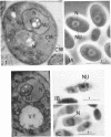
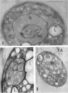
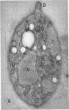
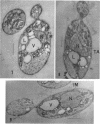
Thyagarajan, T. R. (Dartmouth Medical School, Hanover, N. H.), S. F. Conti, and H. B. Naylor. Electron microscopy of Rhodotorula glutinis. J. Bacteriol. 83:381–394. 1962.—The structure and manner of nuclear division in Rhodotorula glutinis was studied by electron microscopy of ultrathin sections. Parallel studies with the light microscope, employing conventional staining techniques and phase-contrast microscope observations on nuclei in living cells, were carried out.
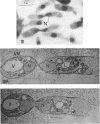
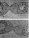
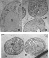
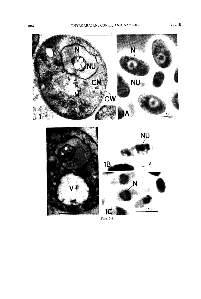
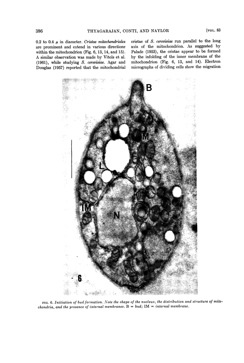
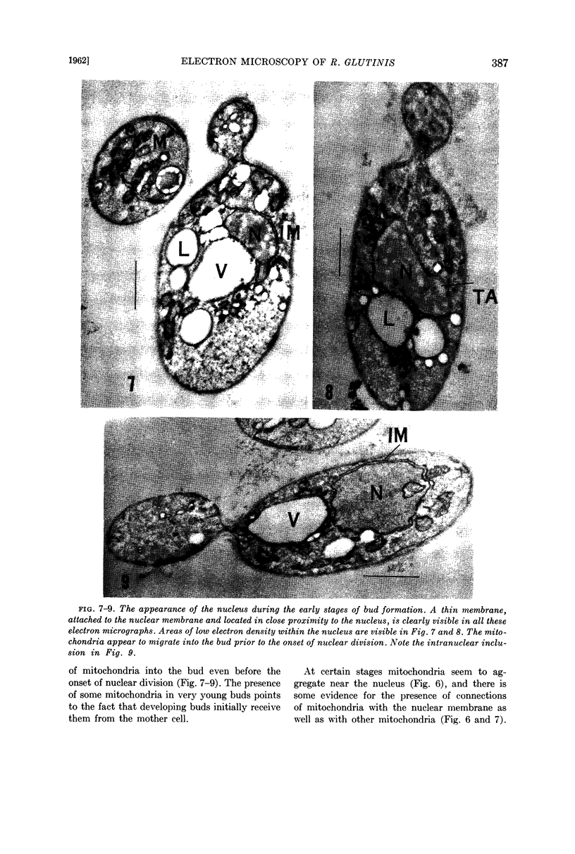
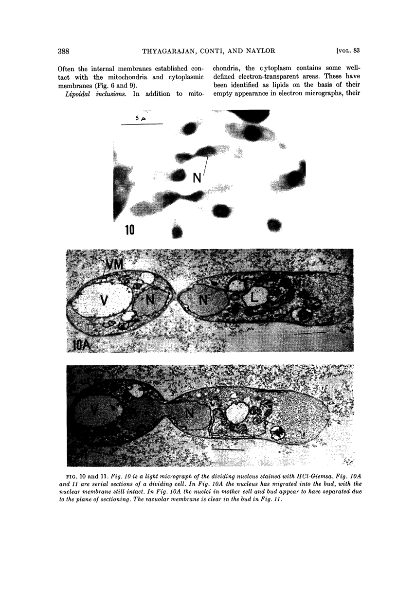
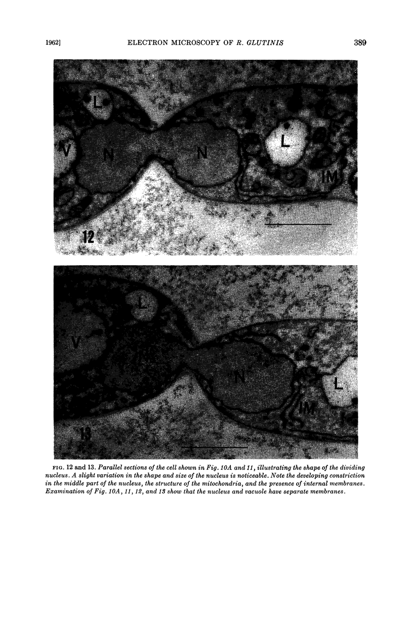
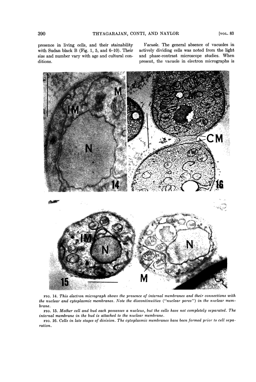
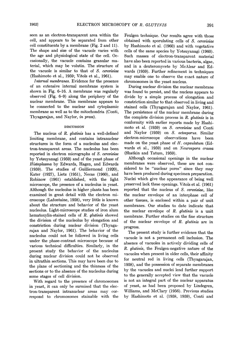
The nucleus is spherical to oval and is bounded by a nuclear membrane. Intranuclear structures, identified as nucleoli, and electron-transparent areas were observed. The nuclear membrane persists throughout the various stages of cell division. Observations of the nucleus with the electron microscope revealed that nuclear division occurs by a process of elongation and constriction similar to that seen in both living and stained cells.
The fine structure of mitochondria and other components of the yeast cell and their behavior during cell division are described. The absence of vacuoles in actively dividing cells of Rhodotorula glutinis lends further support to the view that the vacuole is not an integral part of the nucleus. The results with the electron microscope generally support and considerably extend those obtained with living and stained cells.
Full text
PDF
Images in this article
Selected References
These references are in PubMed. This may not be the complete list of references from this article.
- AGAR H. D., DOUGLAS H. C. Studies on the cytological structure of yeast: electron microscopy of thin sections. J Bacteriol. 1957 Mar;73(3):365–375. doi: 10.1128/jb.73.3.365-375.1957. [DOI] [PMC free article] [PubMed] [Google Scholar]
- BARTHOLOMEW J. W., LEVIN R. The structure of Saccharomyces carlsbergensis and S. cerevisiae as determined by ultra-thin sectioning methods and electron microscopy. J Gen Microbiol. 1955 Jun;12(3):473–477. doi: 10.1099/00221287-12-3-473. [DOI] [PubMed] [Google Scholar]
- BRIEGER E. M., GLAUERT A. M., ALLEN J. M. ytoplasmic structure in Mycobacterium leprae. Exp Cell Res. 1959 Oct;18:418–421. doi: 10.1016/0014-4827(59)90032-1. [DOI] [PubMed] [Google Scholar]
- CONTI S. F., NAYLOR H. B. Electron microscopy of ultrathin sections of Schizosaccharomyces octosporus. I. Cell division. J Bacteriol. 1959 Dec;78:868–877. doi: 10.1128/jb.78.6.868-877.1959. [DOI] [PMC free article] [PubMed] [Google Scholar]
- CONTI S. F., NAYLOR H. B. Electron microscopy of ultrathin sections of Schizosaccharomyces octosporus. II. Morphological and cytological changes preceding ascospore formation. J Bacteriol. 1960 Mar;79:331–340. doi: 10.1128/jb.79.3.331-340.1960. [DOI] [PMC free article] [PubMed] [Google Scholar]
- CONTI S. F., NAYLOR H. B. Electron microscopy of ultrathin sections of Schizosaccharomyces octosporus. III. Ascosporogenesis, ascospore structure, and germination. J Bacteriol. 1960 Mar;79:417–425. doi: 10.1128/jb.79.3.417-425.1960. [DOI] [PMC free article] [PubMed] [Google Scholar]
- DEMPSEY E. W. Variations in the structure of mitochondria. J Biophys Biochem Cytol. 1956 Jul 25;2(4 Suppl):305–312. doi: 10.1083/jcb.2.4.305. [DOI] [PMC free article] [PubMed] [Google Scholar]
- EDWARDS M. R., HAZEN E. L., EDWARDS G. A. The fine structure of the yeast-like cells of Histoplasma in culture. J Gen Microbiol. 1959 Jun;20(3):496–503. doi: 10.1099/00221287-20-3-496. [DOI] [PubMed] [Google Scholar]
- GLAUERT A. M., HOPWOOD D. A. The fine structure of Streptomyces coelicolor. I. The cytoplasmic membrane system. J Biophys Biochem Cytol. 1960 Jun;7:479–488. doi: 10.1083/jcb.7.3.479. [DOI] [PMC free article] [PubMed] [Google Scholar]
- HASHIMOTO T., CONTI S. F., NAYLOR H. B. Fine structure of microorganisms. III. Electron microscopy of resting and germinating ascospores of Saccharomyces cerevisiae. J Bacteriol. 1958 Oct;76(4):406–416. doi: 10.1128/jb.76.4.406-416.1958. [DOI] [PMC free article] [PubMed] [Google Scholar]
- HASHIMOTO T., CONTI S. F., NAYLOR H. B. Studies of the fine structure of microorganisms. IV. Observations on budding Saccharomyces cerevisiae by light and electron microscopy. J Bacteriol. 1959 Mar;77(3):344–354. doi: 10.1128/jb.77.3.344-354.1959. [DOI] [PMC free article] [PubMed] [Google Scholar]
- HASHIMOTO T., GERHARDT P., CONTI S. F., NAYLOR H. B. Studies on the fine structure of microorganisms. V. Morphogenesis of nuclear and membrane structures during ascospore formation in yeast. J Biophys Biochem Cytol. 1960 Apr;7:305–310. doi: 10.1083/jcb.7.2.305. [DOI] [PMC free article] [PubMed] [Google Scholar]
- LAFONTAINE J. G. Structure and mode of formation of the nucleolus in meristematic cells of Vicia faba and Allium cepa. J Biophys Biochem Cytol. 1958 Nov 25;4(6):777–784. doi: 10.1083/jcb.4.6.777. [DOI] [PMC free article] [PubMed] [Google Scholar]
- LINDEGREN C. C., WILLIAMS M. A., MCCLARY D. O. The distribution of chromatin in budding yeast cells. Antonie Van Leeuwenhoek. 1956;22(1):1–20. doi: 10.1007/BF02538308. [DOI] [PubMed] [Google Scholar]
- MOSES M. J. Studies on nuclei using correlated cytochemical, light, and electron microscope techniques. J Biophys Biochem Cytol. 1956 Jul 25;2(4 Suppl):397–406. doi: 10.1083/jcb.2.4.397. [DOI] [PMC free article] [PubMed] [Google Scholar]
- MUNDKUR B. Electron microscopical studies of frozen-dried yeast. I. Localization of polysaccharides. Exp Cell Res. 1960 Jun;20:28–42. doi: 10.1016/0014-4827(60)90219-6. [DOI] [PubMed] [Google Scholar]
- MUNDKUR B. Submicroscopic morphology of frozen-dried yeast. Exp Cell Res. 1960 Oct;21:201–205. doi: 10.1016/0014-4827(60)90361-x. [DOI] [PubMed] [Google Scholar]
- McALEAR J. H., EDWARDS G. A. [Continuity of plasma membrane and nuclear membrane]. Exp Cell Res. 1959 Mar;16(3):689–692. doi: 10.1016/0014-4827(59)90139-9. [DOI] [PubMed] [Google Scholar]
- PALADE G. E. An electron microscope study of the mitochondrial structure. J Histochem Cytochem. 1953 Jul;1(4):188–211. doi: 10.1177/1.4.188. [DOI] [PubMed] [Google Scholar]
- ROBINOW C. F. Mitosis in the yeast Lipomyces lipofer. J Biophys Biochem Cytol. 1961 Apr;9:879–892. doi: 10.1083/jcb.9.4.879. [DOI] [PMC free article] [PubMed] [Google Scholar]
- SATIR P. G., PEACHEY L. D. Thin sections. II. A simple method for reducing compression artifacts. J Biophys Biochem Cytol. 1958 May 25;4(3):345–348. doi: 10.1083/jcb.4.3.345. [DOI] [PMC free article] [PubMed] [Google Scholar]
- SHATKIN A. J., TATUM E. L. Electron microscopy of Neurospora crassa mycelia. J Biophys Biochem Cytol. 1959 Dec;6:423–426. doi: 10.1083/jcb.6.3.423. [DOI] [PMC free article] [PubMed] [Google Scholar]
- SJOSTRAND F. S. Electron microscopy of mitochondria and cytoplasmic double membranes. Nature. 1953 Jan 3;171(4340):30–32. doi: 10.1038/171030a0. [DOI] [PubMed] [Google Scholar]
- TOKUYASU K., YAMADA E. Fine structure of Bacillus subtilis. I. Fixation. J Biophys Biochem Cytol. 1959 Jan 25;5(1):123–128. doi: 10.1083/jcb.5.1.123. [DOI] [PMC free article] [PubMed] [Google Scholar]
- VITOLS E., NORTH R. J., LINNANE A. W. Studies on the oxidative metabolism of Saccharomyces cerevisiae. I. Observations on the fine structure of the yeast cell. J Biophys Biochem Cytol. 1961 Mar;9:689–699. doi: 10.1083/jcb.9.3.689. [DOI] [PMC free article] [PubMed] [Google Scholar]
- YOTSUYANAGI Y. Etude au microscope électronique des coupes ultra-fines de la levure. C R Hebd Seances Acad Sci. 1959 Jan 12;248(2):274–277. [PubMed] [Google Scholar]
- YOTSUYANAGI Y. [Electron-microscopic demonstration of chromosomes in yeasts by means of specific staining]. C R Hebd Seances Acad Sci. 1960 Feb 22;250:1522–1524. [PubMed] [Google Scholar]


