

Abstract
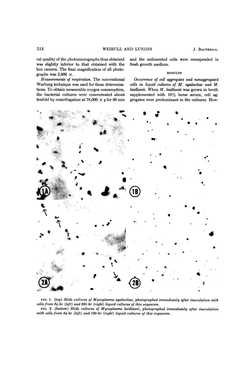
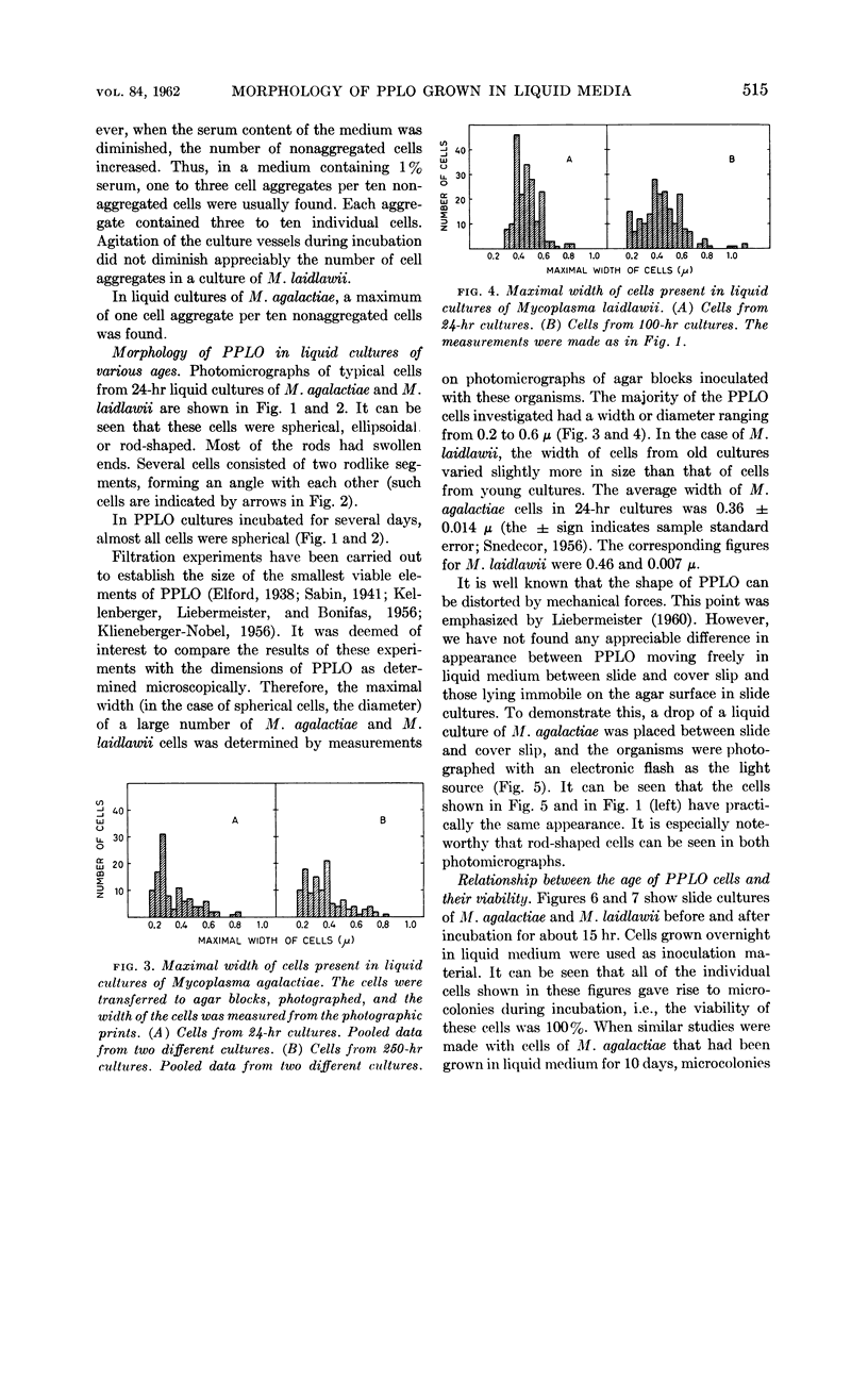
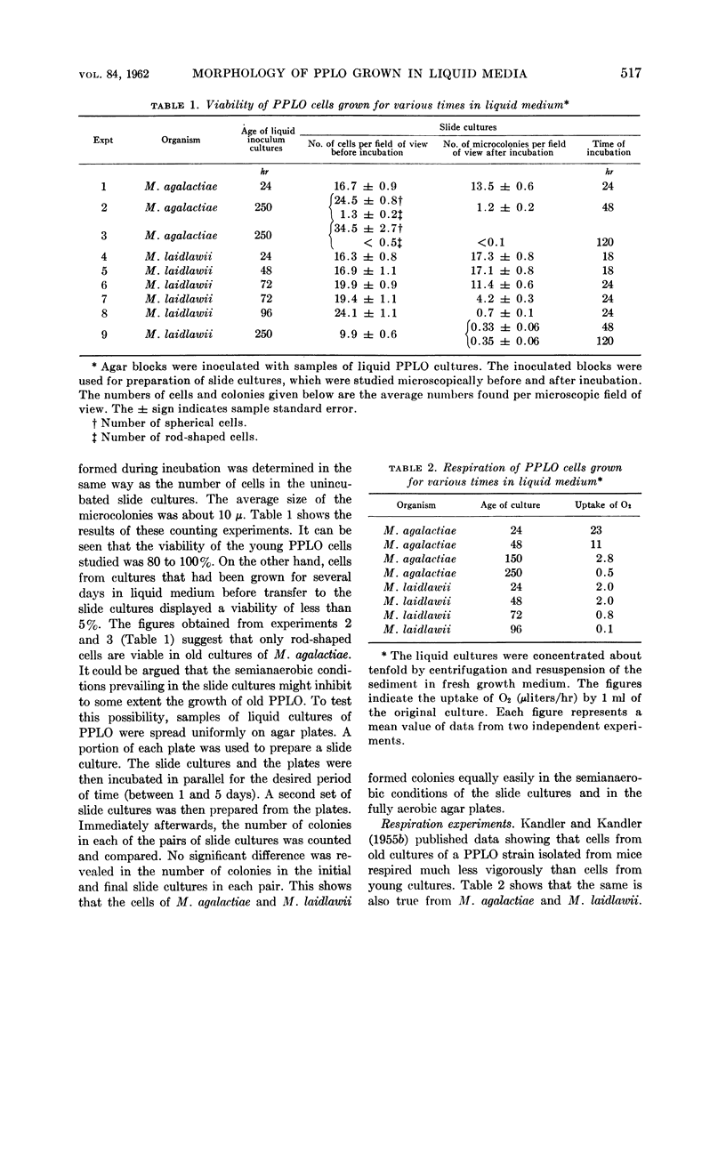
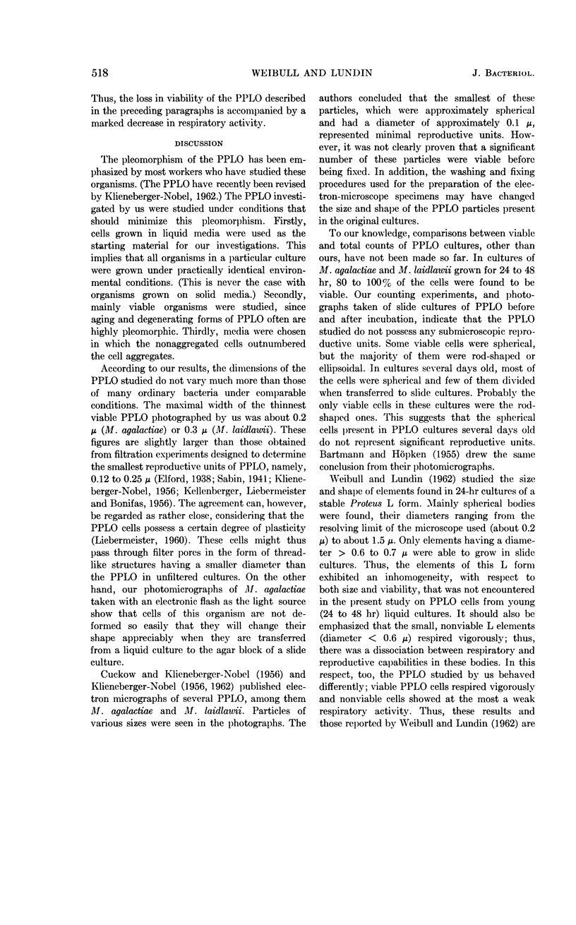
Weibull, C. (Central Bacteriological Laboratory of Stockholm City, Stockholm, Sweden), and Britt-Marie Lundin. Size and shape of pleuropneumonia-like organisms grown in liquid media. J. Bacteriol. 84:513–519. 1962.—Samples of liquid cultures containing mainly nonaggregated cells of Mycoplasma agalactiae or M. laidlawii were transferred to agar blocks containing the same medium as the liquid cultures. By use of a phase-contrast microscope, photomicrographs were made of the slide cultures immediately after they had been prepared, and the dimensions of a large number of pleuropneumonia-like organisms (PPLO) were measured. These measurements indicated that, in young cultures (incubated for 24 to 48 hr), the size of the cells did not vary much more than that of ordinary bacteria; 95% of the cells had a width of 0.2 to 0.6 μ. The growth of individual PPLO was followed during incubation of the slide cultures. It was found that 80 to 100% of the cells present in liquid overnight cultures divided and gave rise to microcolonies within a few hours. Rod-shaped, ellipsoidal, and spherical cells were seen in these cultures. Liquid cultures incubated for several days contained mainly spherical cells. Fewer than 5% of the cells in these cultures showed any indication of growth during incubation in slide cultures for 5 days. Photomicrographs of cells of M. agalactiae moving freely in liquid medium were taken with an electronic flash as the light source. The photographs thus obtained directly demonstrated the existence of rod-shaped cells.
Full text
PDF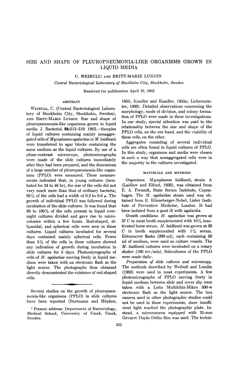
Images in this article
Selected References
These references are in PubMed. This may not be the complete list of references from this article.
- BARTMANN K., HOPKEN W. Phasenkontrastmikroskopische Beobachtungen zur Morphologie der peripneumonieähnlichen Organismen (PPLO). Zentralbl Bakteriol Orig. 1955 Aug;163(5-6):319–332. [PubMed] [Google Scholar]
- CUCKOW F. W., KLIENEBERGER-NOBEL E. Further studies of organisms of the pleuropneumonia group by electron microscopy. J Gen Microbiol. 1955 Aug;13(1):149–154. doi: 10.1099/00221287-13-1-149. [DOI] [PubMed] [Google Scholar]
- KANDLER G., KANDLER O. Untersuchungen über die Morphologie und die Vermehrung der pleuropneumonie-ähnlichen Organismen und der L-Phase der Bakterien. I. Licht mikroskopische Untersuchungen. Arch Mikrobiol. 1954;21(2):178–201. [PubMed] [Google Scholar]
- LIEBERMEISTER K. Morphology of the PPLO and L forms of Proteus. Ann N Y Acad Sci. 1960 Jan 15;79:326–343. doi: 10.1111/j.1749-6632.1960.tb42694.x. [DOI] [PubMed] [Google Scholar]
- Sabin A. B. THE FILTRABLE MICROORGANISMS OF THE PLEUROPNEUMONIA GROUP. Bacteriol Rev. 1941 Mar;5(1):1–67. doi: 10.1128/br.5.1.1-67.1941. [DOI] [PMC free article] [PubMed] [Google Scholar]
- WEIBULL C., LUNDIN B. M. Growth of elements of various sizes found in cultures of a stable Proteus L form. J Gen Microbiol. 1962 Feb;27:241–248. doi: 10.1099/00221287-27-2-241. [DOI] [PubMed] [Google Scholar]