Abstract
Objective:
To present the characteristics and create awareness of symptomatic carpal bossing and discuss potential etiologies and the role of conservative management through the presentation of an athlete with traumatic onset of symptomatic carpal bossing.
Clinical features:
This case report outlines the presentation and conservative management of an elite eighteen year old hockey player with symptomatic carpal bossing after a traumatic on ice collision. Carpal bossing is a bony, dorsal prominence in the quadrangular joint of the wrist that is inconsistently symptomatic.
Intervention and outcome:
A conservative treatment plan consisting of education, reassurance, avoidance of aggravation, and soft tissue therapy allowed return to play in two weeks without restrictions or need for surgical consultation.
Conclusion:
With inconsistent recurrence rates and surgical complications, the role of conservative management for symptomatic carpal bossing deserves further exploration. The conservative practitioner should be aware of the signs and symptoms of symptomatic carpal bossing to institute suitable treatment.
Keywords: boss, carpal, athlete, hockey, trauma
Abstract
Objectif :
Présenter les caractéristiques et faire de la sensibilisation concernant le carpe bossu symptomatique et discuter des étiologies potentielles et du rôle du traitement conservateur par la présentation d’un athlète avec une apparition traumatique du carpe bossu symptomatique.
Caractéristiques cliniques :
Ce rapport de cas indique la présentation et le traitement conservateur d’un joueur de hockey d’élite de 18 ans présentant un carpe bossu symptomatique à la suite d’une collision traumatique sur la glace. Le carpe bossu est une protubérance osseuse dorsale de l’articulation quadrangulaire du poignet qui est symptomatique de façon non constante.
Intervention et résultats :
Un plan de traitement conservateur de sensibilisation, de rassurance, d’évitement de l’aggravation et de traitement des tissus mous a permis un retour au jeu après deux semaines sans restrictions ou besoin de consultation chirurgicale.
Conclusion :
Des taux de récidive non constants et des complications chirurgicales font en sorte que le rôle du traitement conservateur pour le carpe bossu symptomatique mérite qu’on s’y attarde davantage. Le praticien conservateur doit savoir reconnaître les signes et symptômes du carpe bossu symptomatique afin de prodiguer le traitement approprié.
Introduction
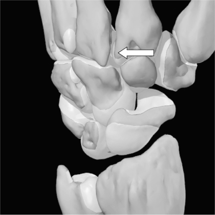
The carpal boss is a bony and inconsistently symptomatic prominence appearing between the base of the second and third metacarpals, the trapezoid, and capitate on the dorsal wrist (figure 1).1 This joint, termed the quadrangular joint of the wrist, is the only area in which carpal bossing appears.2 Carpal boss etiology is a subject of contention with speculative reports on trauma, degeneration, instability, an accessory ossicle and most recently partial or complete osseous coalition appearing in the literature.2–6 The undefined etiology makes the incidence difficult to ascertain. The pathophysiology of carpal bossing has never been elucidated with only speculative reports on why this bony prominence occurs.
Figure 1.
The dorsal wrist with the white arrow illustrating the quadrangular joint. (Image courtesy of and copyright to Primal Images ltd)
Literature on the treatment of the symptomatic carpal boss is surgical.1,4,7–13 Conservative management is attempted prior to surgical care, however the descriptions of outcome measures, course of conservative therapy and treatment regimen are poor. The indications and contraindications for surgery are similarly inadequate. Further confounding the topic are reports in prospective and retrospective longitudinal studies stating an inconsistent recurrence rate between 0 and 50% for simple resection, and a lack of sufficient follow up for recent advances in arthrodesis.1,4,7,8,11–14 This questions the appropriateness of surgical management while leaving the conservative practitioner without definitive management criteria or prognosis. In an athlete, surgery results in missed playing time and a rehabilitation protocol that can take weeks to months.
This report will highlight a case of symptomatic carpal bossing in an elite eighteen year old hockey player and examine the relevant diagnosis, anatomy, etiology, pathophysiology, and the role of conservative management for symptomatic carpal bossing.
Case presentation
An eighteen year old male provincial level hockey player was acutely injured in game, and presented to the team chiropractor on the bench. The player, a defenceman, was hit into the boards by an opposing player with the dorsal aspect of his right wrist first contacting the boards while flexed. The subsequent addition of his mass and that of the opposing player further compressed the wrist between the boards and the players own body.
The player, right hand dominant, was otherwise healthy based upon a preseason assessment with no previous injury to the wrist. Upon equipment removal edema was noted on the dorsal aspect of the right hand and wrist. The primary complaint was poorly localized to the dorsal aspect of the right wrist over the second and third carpometacarpal joints, and the length of the second and third metacarpals. The player rated the intensity of pain as 8 out of 10. Active range of motion of the right wrist revealed limitations in flexion by fifty percent compared to the left due to moderate pain on the dorsal aspect of the right wrist in the area of the primary complaint. Mild pain was produced in the same area at end range of extension, ulnar and radial deviation. Active supination and pronation were full and pain free. Passive range of motion of the right wrist produced identical findings as active range of motion testing. Resisted extension of the right wrist produced moderate pain in the area of the primary complaint and was graded four out of five compared to the left wrist. Resisted flexion, ulnar deviation, radial deviation, supination and pronation were graded five out of five with mild pain. Grip strength was graded as four out of five when compared to the left side. Resisted metacarpophalangeal joint extension produced mild pain in the area of primary complaint. All other active, passive and resisted range of motion testing of the digits was within normal limits.
A neurological evaluation including sensory, motor and deep tendon reflexes of the upper limb was performed and deemed within normal limits.
Palpation revealed tenderness over the length of the right second and third metacarpals, the capitate and trapezoid. Anterior/posterior shear testing of the second and third metacarpals caused moderate poorly localized pain. Axial compression of the second and third carpometacarpal joints reproduced the patient’s dorsal wrist pain. A 128Hz tuning fork was felt as painful at the base of the second and third metacarpals, the trapezoid and capitate compared to vibration in the same areas on the left wrist.
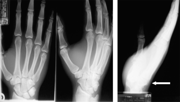
The player was given ice and was not cleared to return to play. Suspecting fracture, the player was instructed to attend a facility for radiographic imaging. The following day the player had not gone for imaging and presented with the same physical findings but only mild swelling allowing palpation of a bony projection in the area of the quadrangular joint. The patient had radiographic imaging of the right wrist seen in figure 2.
Figure 2.
x-ray imaging series with white arrow showing a dorsal projection on the lateral view at the area of the quadrangular joint diagnosed as an os styloideum.
The patient was diagnosed by a chiropractic radiologist with an acute os styloideum of the right wrist and was held out of competition for two days. A treatment plan consisting of reassurance and education on symptomatic carpal bossing to reduce fears of a more ominous wrist injury and Active Release Technique (ART®) soft tissue therapy of the extensor carpi radialis longus and brevis, extensor digitorum and extensor retinaculum was instituted twice weekly for two weeks. The player returned to practice without contact for the first two practices post injury. One week from the injury the player returned to full play with intensity rated at 0 out of 10. The player was only symptomatic with full active or passive wrist flexion combined with direct pressure over the bony prominence. The player continued play for the final two months of the hockey season without re-aggravation.
Discussion
Athletes sustain a variety of muskuloskeletal injuries. Hand and wrist injuries account for 3–9% of all athletic injuries.15–16 A ten year longitudinal study of injuries sustained at the Olympic Training Centre in the United States revealed 8.7% of 8311 injuries involved the wrist and hand with 64% of the diagnoses representing sprains and contusions.15 In collision sports, the incidence of hand and wrist injuries accounts for 15% of all injuries.15
The incidence of the symptomatic carpal boss is not well defined. There is a paucity of reports of symptomatic carpal bossing in athletes where a literature search produced one case report in a swimmer.3 A dorsal ossicle in the quadrangular joint was first described by Saltzmann in 1725.18 In 1931 Fiolle described a case of a bony wrist exostosis producing a functional disturbance providing the name carpe bossu.19 Bassoe and Bassoe were the first to describe the incidence of bossing based on an x-ray study of 450 hands in 1955 suggesting the carpal boss as an accessory ossicle present in 1–4% of the population.20 Because the etiology has been challenged, so too has the accuracy of the reported incidence. In a cadaveric anatomic study of the second through fifth carpometacarpal joints, Nakamura found carpal coalitions only in the area of the second and third carpometacarpal joints in 14/80 or 18% of dissected wrists.6 Alemohammed found similar results, uncovering a partial osseous coalition in all cadavers with clinical signs of a bony prominence representing 19% of their sample.2 These two studies suggest the true incidence of carpal bossing to be much greater than previously thought, however because the studies were cadaveric it is not possible to conclude the incidence of symptomatic carpal bossing.
The clinical presentation of symptomatic carpal bossing is variable. Data from surgical publications describing patient characteristics taken as continuous data suggests a mean age of 32.25 with a range of 11–75.1,4,7,8,11,12 Presentation is most often in the dominant arm with varied reports on male to female distribution.1,4,7,8,11,12 A review suggests patients may present secondary to direct trauma, advanced age and recurrent strain.20 The literature demonstrates antecedent trauma in just 23–27% of cases, however a traumatic incident was present in this case.1,4,7,8 Advancing age does not seem likely as the aforementioned mean age in the literature is 32 years. Repetitive strain, specifically forced wrist extension, is thought to aggravate bossing symptoms due to a tenosynovitis of the extensor carpi radialis longus and brevis at their insertions on the dorsal aspect of the base of the second and third metacarpals. There is variable presentation of ganglions seen in 0–30% of subjects.1,4,7,8 The pain presentation is also variable, but improves with rest.20
Diagnosis of carpal bossing is made on clinical and imaging findings after ruling out various differential diagnoses. Ganglion cysts are the most common cause of dorsal wrist protuberances.21 Differential diagnosis between a ganglion and carpal bossing is based on location and palpation. Between 60 and 70% of all ganglions occur over the scapholunate ligament.21 Carpal bossing is always over the quadrangular joint. Carpal bossing has a hard consistency, while ganglions are mucin filled creating a cystic consistency that can be transilluminated.20 Dorsal ganglia have been suggested to spontaneously resolve in 58% of cases.22 Because the carpal boss is a bony prominence, there are no reports of spontaneous remission. Intraosseous ganglions can cause a similar pain presentation as the carpal boss.23 Additional differential diagnoses include benign bony lesions such as aneurysmal bone cysts, unicameral bone cysts, enchondromas, osteochondromas and osteoid osteomas as well as malignant tumors and locally invasive tumors such as osteosarcomas, and giant cell tumors.20
Physical diagnosis begins with direct observation and palpation for a dorsal bony prominence accentuated with wrist flexion, using the opposite wrist for contrast. The clinician must keep in mind that when present 11–21% of bossing is seen bilateral.2,6 Swelling and a dorsal ache are observed inconsistently.20 Fusi describe the malalignment test where the examiner distracts the second and third metacarpal with concomitant supination and pronation while the metacarpalphalangeal joints are held in flexion.8 The authors claim this distorts the anatomic relationship of the quadrangular joint. When explored, the second and third carpometacarpal joints have one degree of freedom (flexion and extension), while the fourth and fifth have three.24 Studies indicate only 1–3 degrees of flexion occur through the quadrangular joint, while 15 and 30 occur through the fourth and fifth carpometacarpal joints respectively indicating this area has very little motion.24 This calls into question if distorting the anatomy at this joint can occur at all via the malalignment test. Lorea found the malalignment test positive in all 32 patients with a surgically confirmed carpal boss but only when utilizing the third digit.11 Clarke described the stress test with palmar directed pressure on the second and third metacarpalphalangeal joints while stabilizing the metacarpals from the ventral side meant to aggravate degenerative arthritis. However degeneration has not been confirmed in carpal bossing. Anatomic studies only found degeneration in the metacarpal articulations 3 to 5, with none in the quadrangular joint.6 Neither the malalignment or stress tests were utilized in this case example.
Though an acute os styloideum appeared on the routine wrist imaging series seen in Figure A, radiographic imaging of the carpal boss is difficult due to superimposition of structures on a standard wrist series.18 The carpe bossu view was originally described as a lateral view of the wrist with the hand flexed and supinated thirty to forty degrees.18 This projection mimicks the 30–40 degree dorsoradial projection of the boss.18 The normal axis of the metacarpocapitate articular surface is oblique by twenty to thirty degrees. By adding twenty to thirty degrees of ulnar deviation the articulation of the boss if present will be exposed.4 Hultgren found that four of sixteen patients with a surgically confirmed boss had preoperative radiographs that were inconclusive.7 A negative radiograph cannot rule out carpal bossing. When bossing is present on radiographic imaging, there is no sclerosis or reduction of joint space typically noted indicating that a degenerative process may not be the etiology of the carpal boss.1 Apple noted increased local uptake in the quadrangular joint utilizing bone scintigraphy in a case report on carpal bossing.24 In a larger sample, Clarke found a positive bone scintigraphy in 12 out of 18 carpal bosses confirmed surgically.12 Magnetic Resonance Imaging has been shown to pick up the bony anomaly however the high cost and long wait times call into question if this imaging modality is practical.26
Carpal boss etiology is undefined. Hypotheses of trauma, minor stress, instability, ganglion, accessory ossicle, ligamentous microrupture, chronic periositis, ununited fracture, degeneration, partial bony coalition and combinations of the above inundate the reader with theories based on scant evidence.1–20 Commonly proposed etiological explanations for carpal bossing involve an os styloideum (accessory ossicle) causing aberrant biomechanics and a resultant degeneration, and a recently suggested etiology of a partial incomplete bony coalition.
An accessory ossicle, or os styloideum, in the quadrangular joint was identified in 1894 by Thompson. When present, this ossicle is suggested to be completely isolated in 2% of cases, but more commonly fused to the proximal third metacarpal (94%), the capitate (3.5%) or trapezoid (0.5%).20 The relationship to the carpal boss was not drawn until 1955 by Bassoe and Bassoe who suggested that the os styloideum represented the symptomatic carpal boss.17 This abnormal joint configuration could predispose an individual to the development of a highly localized degenerative arthritis making this area more susceptible to the effects of repetitive trauma. Normal ossification of the capitate occurs in infancy, and the trapezoid by 7 to 8 years.27 The metacarpals have ossification centres in the distal epiphysis for metacarpals 2 through 5, however a second epiphyseal center in the proximal second and fifth metacarpal has been observed in up to 6% of the population.28 The second metacarpal represents 33% of the pseudoepiphyses, however this fuses with the diaphyses by age fifteen to twenty.28 These explanations for the etiology of the accessory ossicle are deficient and require further exploration. The presence of an os styloideum is rare, however was diagnosed on plain film radiograph in this case.
The major axes of stress in the normal wrist are the scapholunate and quadrangular joints.29,30 The middle longitudinal arch is said to act as a fulcrum for the opposition of the first ray with the fourth and fifth rays.30 Because the third metacarpocapitate joint receives great stress, degenerative joint disease may result from an inability of this abnormal joint configuration to withstand ordinary daily stresses. The evidence for degeneration is murky. Anatomically, dissection studies only show degeneration in the fourth and fifth carpometacarpal joints.2,6 The age range of carpal boss does not coincide with degenerative changes revealing bossing in individuals as young as eleven years old, and a mean age of 32.1,4,7,8,11,12 The radiographic appearance of carpal bossing shows no sclerosis of the joint margin or narrowing of the cartilage space.1 The second and third carpometacarpal joints are extremely stable with one degree of freedom questioning if this stable joint is prone to degeneration at all.24 In a study of 3156 residents of Tecumseh Michigan, there was no evidence of degeneration via x-ray in any carpometacarpal joints other than the first ray.31
The first suggestion of a partial osseous coalition occurred in a 1995 surgical study which identified a bony anomaly in the quadrangular joint with an occurrence of 63%, most frequently a separate ossicle that was fused in part or completely to the metacarpals. Frequently the fused ossicle bridged both the respective metacarpal and carpal bones.8 This possible etiology was further explored by Nakamura and again by Alemohammad.2,6 Both studies found partial osseous coalition only in the area of the quadrangular joint, present in all cadavers with clinical signs of a carpal bossing.2,6 The partial coalition was always on the dorsal aspect of the wrist. When present none of the five normal dorsal ligaments that cross the dorsal quadrangular joint were present.6 It was hypothesized that carpal bossing is actually a congenital partial carpal coalition. A symptomatic carpal boss may result from a fracture or breakdown of the coalition similar to those seen in tarsal coalitions.2 Though limitations in these studies as they pertain to this case exist; the in vitro nature of the study, the lack of strong objective criteria to establish clinically the dorsal protuberance and a sample not representative of a symptomatic bossing population, these studies do provide evidence to support a partial coalition as an etiological explanation. Coalitions of carpal bones are a failure of demarcation of joint spaces during embryogenesis caused by incomplete cavitation of a common embryologic carpal precursor during the fourth to eighth week intrauterine.32–34 Coalitions can be cartilaginous or bony.33 A partial coalition or synchondrosis occurs when there is some degree of joint space formation, often resembling a pseudoarthrosis.32 Partial coalitions are more likely to cause symptoms than complete coalitions due to stress loading activities, while complete coalitions are symptomatic only after fracture.32 The absence of adequate intaarticular cartilage between the incomplete coalition and normal carpus is thought to cause intolerance of forceful activity and is seen on magnetic resonance imaging as bone marrow edema.34 The most commonly cited carpal coalition is the lunotriquetral coalition.33 Minaar found thirty six wrists with carpal coalitions, classifying them in four types (table 1). Type 1 (Proximal pseudarthrosis) and Type 2 (Proximal osseous bridge with distal notch) are reported as potentially symptomatic, while Type 3 (complete fusion) and Type 4 (fusion with other carpal anomalies) are suggested to be asymptomatic.33 The incidence of carpal coalition has been cited as 0.1–0.7% of the population with a familial tendency.32–34 No studies on carpal bossing have explored the role of genetics. Carpal coalition has been cited as bilateral in up to 61.5%, while the anatomic carpal boss studies cite a bilateral occurrence of 11–21%.2,6 The etiological explanation of a partial or complete congenital osseous coalition is the most promising explanation available. It is possible that the os styloideum may represent a partial or complete osseous coalition seen on radiographic imaging.
Table 1.
Minnaar Classification of lunotriquetral coalitions
| Type 1 | Proximal Pseudarthrosis |
| Type 2 | Proximal osseous bridge with distal notch |
| Type 3 | Complete Fusion |
| Type 4 | Fusion with Other Carpal Anomalies |
No studies with adequate size on the efficacy of conservative management of carpal bossing exist. The treatment literature available is surgical and offers scant accounts of failed conservative therapy as an indication for surgery, however the conservative care attempted is variable and poorly defined.1,4,7,8,11,12 Clarke utilized corticosteroid injections in a sample of 13 surgical candidates with no relief in 11 though the authors noted difficulty with the injection due to the distorted anatomy present.12 Curtiss suggested immobilizing the hand and wrist for three to four weeks after initial diagnosis.35 In a recent study Lorea included a conservative treatment regimen of splinting at night and during heavy manual activities and corticosteroid injections. This was responsive in 16 patients, with only 7 of the remaining 16 opting for surgery.11 A twenty year review by Fusi suggested four to six weeks of rest, minimally restrictive reinforced elastic wrist brace, and the use of oral non-steroidal anti-inflammatory agents with no report on success.8 The literature suggests that surgery is reserved only for those whose symptoms are chronic and interfere with either occupational or recreational activities, and not just for cosmetic deformities or only occasional discomfort (table 2).1,8 The only conservative study with follow up was performed on 34 patients with 43 lesions treated with an explanation of bossing and advice on avoidance of provocation. Only 11 patients were followed from 1 to 7 years. All reported the boss remained stable in size, with disappearance of pain seen in 3.1 The athlete presented in this case report was treated with a conservative approach based upon the recommendation of conservative management prior to surgical consultation. A treatment regimen of soft tissue therapy, education and avoidance of provocation produced superb results as qualified by the athlete’s speedy return to play and decrease on numerical rating scale.
Table 2.
Artz and Posch Indications and Contraindications for Surgery
| Surigcal indications |
|
| Surgical contra-indications |
|
Surgical management of symptomatic carpal bossing has evolved greatly since it was first described by Carter in 1941 who suggested prompt recurrence advocating against surgery.36 The surgical technique of wedge excision to the level of normal cartilage and cancellous bone followed by reconstitution of the dorsal capsular structures predominates.4 The recurrence rate has a reported range of 0 to 50% in the literature, with great variation based on the definition of successful outcome.1,4,7,8,11,12 Citteur explored the cause of recurrence suggesting the excision of the dorsal ligamentous structures causing continued instability in the joint.37 However when a partial osseous coalition is present, there is complete absence of these ligaments questioning the validity of this study.6 Vermeulen suggested the depth of wedge excision caused resultant instability showing a 55% excision resulting in instability, yet a clinical wedge excision does not exceed 33%.38 Lorea explored the use of resection utilizing radial bone grafting and staple arthrodesis resulting in no complications, however the sample was only seven and the follow up only seventeen months.11 Clarke looked at excision compared to fusion surgery, finding a fifty percent good and fifty percent poor outcome in each group where the definition of successful outcome was complete resolution of symptoms and patient satisfaction.12 Because surgical complications and recurrence abound, an extended conservative management trial is advisable saving surgery for the most symptomatic patients with functional and occupational disturbances.
Conclusion
The role of conservative management for the symptomatic carpal boss deserves further exploration. Currently, there is an undefined best surgical procedure and unsatisfactory surgical outcome. In an athlete a surgical procedure would require four to six weeks without play in addition to the consult and procedure, significantly impacting their season. With a conservative approach of reassurance, education, a graded return to play and therapy, the player in this case was able to continue play one week after the original injury. It must be noted that there are limitations of basing a conclusion on one subject. Further research contrasting conservative and surgical care is required to compare the outcomes of these management styles.
References
- 1.Artz T, Posch JL. The carpometacarpal boss. J Bone Jt Surg. 1973;55-A(4):747–752. [PubMed] [Google Scholar]
- 2.Alemohammad AM, et al. Incidence of carpal boss and osseous coalition: an anatomic study. J Hand Surg. 2009;34A:1–6. doi: 10.1016/j.jhsa.2008.08.025. [DOI] [PubMed] [Google Scholar]
- 3.Maquirriain J, Ghisi JP. Acute os styloideum in an elite athlete. Skeletal Radiology. 2006;35:394–396. doi: 10.1007/s00256-005-0027-7. [DOI] [PubMed] [Google Scholar]
- 4.Cuono CB, Watson HK. The carpal boss: Surgical treatment and etiological considerations. Plastic & Reconstructive Surgery. 1979;63(1):88–93. [PubMed] [Google Scholar]
- 5.Geutjens G. Carpal bossing with capitate-trapezoid fusion: a case report. Acta Orthopaedica Scandinavia. 1994;65(1):97–98. doi: 10.3109/17453679408993728. [DOI] [PubMed] [Google Scholar]
- 6.Nakamura K, et al. The ligament and skeletal anatomy of the second through fifth carpometacarpal joints and adjacent structures. J Hand Surg. 2001;26A(6):1016–1028. doi: 10.1053/jhsu.2001.26329. [DOI] [PubMed] [Google Scholar]
- 7.Hultgren T, Lugnegard H. Carpal boss. Acta Orthopaedica. 1986;57:547–550. doi: 10.3109/17453678609014791. [DOI] [PubMed] [Google Scholar]
- 8.Fusi S, et al. The carpal boss – a 20-year review of operative management. J Hand Surg. 1995;20B(3):405–408. doi: 10.1016/s0266-7681(05)80104-4. [DOI] [PubMed] [Google Scholar]
- 9.Williams MR, Fullilove SM. A carpal boss leading to extensor tendon ruptures – a case report. J Hand Surg (European Volume) 2008;33:223. doi: 10.1177/1753193408087111. [DOI] [PubMed] [Google Scholar]
- 10.Tielliu IFJ, Van Wellen PAJ. Carpal boss caused by an accessory capitate – case report. Acta Orthopaedica Belgica. 1998;64:107–108. [PubMed] [Google Scholar]
- 11.Lorea P, et al. The preliminary results of treatment of symptomatic carpal boss by wedge joint resection, radial bone grafting and arthrodesis with a shape memory staple. J Hand Surg (European Volume) 2008;33E(2):174–178. doi: 10.1177/1753193408087068. [DOI] [PubMed] [Google Scholar]
- 12.Clarke AM, et al. The symptomatic carpal boss is simple excision enough. J Hand Surg (British and European Volume) 1999;24B(5):591–595. doi: 10.1054/jhsb.1999.0238. [DOI] [PubMed] [Google Scholar]
- 13.Hazlett JW. The third metacarpal boss. Int Ortho. 1992;16:369–371. doi: 10.1007/BF00189621. [DOI] [PubMed] [Google Scholar]
- 14.Lenoble E, Foucher G. Anaales de Chirurgie de la Main et due Membre Superieur. 1992;11:46–50. doi: 10.1016/s0753-9053(05)80052-3. [DOI] [PubMed] [Google Scholar]
- 15.Rettig AC. Athletic injuries of the wrist and hand. Am J Sports Med. 2003;31:1038–1049. doi: 10.1177/03635465030310060801. [DOI] [PubMed] [Google Scholar]
- 16.Rettig AC. Athletic injuries of the wrist and hand Part 2: overuse injuries of the wrist and traumatic injuries to the hand. Am J Sports Med. 2004;32:262–273. doi: 10.1177/0363546503261422. [DOI] [PubMed] [Google Scholar]
- 17.Bassoe E, Bassoe HH. The styloid bone and carpe Bossu Disease. Am J Roentgenology, Radium Therapy and Nuclear Medicine. 1955;74:886–888. [PubMed] [Google Scholar]
- 18.Conway WF, et al. The carpal boss: an overview of radiographic evaluation. Radiology. 1985;156(1):29–31. doi: 10.1148/radiology.156.1.3923555. [DOI] [PubMed] [Google Scholar]
- 19.Fiolle J. Le ‘Carpe Bossu’ Bull et Mem Soc Nat’l de Chir. 1931;57:1587–1690. [Google Scholar]
- 20.Park MJ, et al. The carpal boss: review of diagnosis and treatment. J Hand Surg. 2008;33A:446–449. doi: 10.1016/j.jhsa.2007.11.029. [DOI] [PubMed] [Google Scholar]
- 21.Goldsmith S, Yang SS. Magnetic resonance imaging in the diagnosis of occult dorsal wrist ganglions. J Hand Surg (European Volume) 2008;33:595–599. doi: 10.1177/1753193408092041. [DOI] [PubMed] [Google Scholar]
- 22.Dias JJ, et al. The natural history of untreated dorsal wrist ganglia and patient reported outcome 6 years after intervention. J Hand Surg (European Volume. 2007;32E(5):502–508. doi: 10.1016/J.JHSE.2007.05.007. [DOI] [PubMed] [Google Scholar]
- 23.Magee TM, et al. Intraosseous ganglia of the wrist. Radiology. 1995;195:517–520. doi: 10.1148/radiology.195.2.7724776. [DOI] [PubMed] [Google Scholar]
- 24.El-Shennawy M, et al. Three-dimensional kinematic analysis of the second through fifth carpometacarpal joints. J Hand Surg. 2001;26A(6):1031–1035. doi: 10.1053/jhsu.2001.28761. [DOI] [PubMed] [Google Scholar]
- 25.Apple JS, et al. Parinful os styloideum: bone scintigraphy in Carpe Bossu Disease. Am J Roentgenometry. 1984;142:181–182. doi: 10.2214/ajr.142.1.181. [DOI] [PubMed] [Google Scholar]
- 26.Zanetti M, et al. Role of MR imaging in chronic wrist pain. Eur Radiology. 2007;17:927–938. doi: 10.1007/s00330-006-0365-4. [DOI] [PubMed] [Google Scholar]
- 27.Srivastav A, et al. A study of wrist ossification for age estimation in pediatric group in central Rajasthan. JIAFM. 2004;26(4):132–135. [Google Scholar]
- 28.Schmidt HM, Lanz U. Surgical anatomy of the hand. Thieme; 2004. [Google Scholar]
- 29.Tang JB. General concepts of wrist biomechanics and a view from other species. J of Hand Surg (European Volume) 2008;33:519–525. doi: 10.1177/1753193408090108. [DOI] [PubMed] [Google Scholar]
- 30.Marzke MW, Marzke RF. The third metacarpal styloid process in humans: origin and functions. Am J Phys Anthro. 1987;73:415–431. doi: 10.1002/ajpa.1330730403. [DOI] [PubMed] [Google Scholar]
- 31.Butler WJ. Prevalence of radiologically defined osteoarthritis in the finger and wrist joints of adult residents of Tecumseh, Michigan 1962–65. J Clin Epi. 1988;41(5):467–473. doi: 10.1016/0895-4356(88)90048-0. [DOI] [PubMed] [Google Scholar]
- 32.Ganos DL. Symptomatic congenital coalition of the pisiform and hamate. J Hand Surg. 1991;16A(4):646–650. doi: 10.1016/0363-5023(91)90188-h. [DOI] [PubMed] [Google Scholar]
- 33.Delaney TJ. Carpal coalitions. J Hand Surg. 1992;17A:28–31. doi: 10.1016/0363-5023(92)90108-2. [DOI] [PubMed] [Google Scholar]
- 34.Haliloglu N, Sahin G. Symptomatic carpal coalition with degenerative changes: report of two cases. Eur J Radiology Extra. 2007;63:11–15. [Google Scholar]
- 35.Curtiss PH. The hunchback carpal bone. J Bone Jt Surg. 1961;43A(3):392–394. [PubMed] [Google Scholar]
- 36.Carter RM. Carpal boss: commonly overlooked deformity of the carpus. J Bone Jt Surg. 1941;23:935–940. [Google Scholar]
- 37.Citteur JME, et al. Carpal boss: destabilization of the third carpometacarpal joint after a wedge excision. J Hand Surg. 1998;23B(1):76–78. doi: 10.1016/s0266-7681(98)80225-8. [DOI] [PubMed] [Google Scholar]
- 38.Vermeulen GM, et al. Carpal boss: effect of wedge excision depth on third carpometacarpal joint stability. J Hand Surg. 2009;34A:7–13. doi: 10.1016/j.jhsa.2008.09.010. [DOI] [PubMed] [Google Scholar]


