Abstract
Object The recently developed vascular space occupancy (VASO) fMRI technique is gaining popularity as it facilitates the measurement of cerebral blood volume (CBV) changes concomitant with brain activation, without the use of contrast agents. Thus far, VASO fMRI has only been used in conjunction with a GE-EPI (gradient-echo echo planar imaging) sequence, which is proceeded by an inversion recovery (IR) experiment to selectively null the blood signal. The use of GE-EPI has potential disadvantages: (a) the non-zero TE may lead to BOLD contamination and (b) images suffer from the EPI-typical inhomogeneity artefacts.
Materials and methods Here, we propose the use of VASO based on an IR-HASTE (inversion recovery half-Fourier acquisition single-shot turbo spin echo) sequence.
Results Results from a visual stimulation study (n = 8) show a 43% higher functional contrast-to-noise (CNR) of HASTE compared to EPI, with a strongly increased count of active voxels at the same significance threshold. Sensitivity to inflow effects was investigated and found to be similar for both methods.
Conclusion As HASTE VASO yields essentially artefact-free images, it appears to be the method of choice for measuring relative CBV changes with VASO.
Keywords: VASO fMRI, FSE, RARE, HASTE, CBV, BOLD undershoot
Introduction
The vascular space occupancy (VASO) technique [1] has proven valuable for measuring relative changes in cerebral blood volume (CBV) upon brain activation. The original method was implemented with a standard echo-planar imaging (EPI) sequence [1], preceded by a non-selective adiabatic inversion pulse. The inversion time (TI) is chosen such that the blood magnetization is passing the zero-point at the moment of excitation. CBV increases as a result of increased brain activation, thereby leading to a negative signal change in VASO as a larger fraction of the tissue volume is taken up by blood.
Employment of EPI-based acquisitions may result in T2* blurring and geometric distortions, and therefore a potential reduction in spatial specificity. Furthermore, the echo time (TE) should be as short as possible (ideally zero) to avoid any BOLD-like signal contributions, but the duration of the EPI readout often requires a TE above 10 ms even at standard spatial resolution and matrix size. Shorter TEs can only achieved by use of a very high acquisition bandwidth or accelerated parallel acquisition techniques, thereby incurring SNR penalties, or segmented acquisitions at the cost of temporal resolution. If the TE is too long this can lead to contamination of the VASO signal by the positive, and thus opposite, BOLD signal change, especially at the more and more routinely used higher field strengths. The TE dependence of the VASO signal has been observed in multi-echo EPI measurements [2]. Such measurements have been used for extrapolating the VASO signal to zero TE, and predicting extravascular parenchymal BOLD effects and oxygen extraction fractions [2]; however, acquisitions at a single short TE would generally be favourable. One way to achieve this is the use of a spiral-out sampling scheme which allows minimal TE, but will still suffer from some measure of T2* contamination. With the inherently low signal levels in VASO images, due to the inversion recovery (IR) preparation and therefore considerable reduction of the tissue signal, the method has a much reduced sensitivity compared to BOLD fMRI. Improvement of the functional sensitivity of VASO is hence desirable.
The impact of blood flow effects has been reported for EPI-based VASO [3]. The VASO method relies on the assumption of steady-state conditions, while in reality the inflow of fresh spins from outside the inversion volume effectively makes the TI too short. The result is a larger negative signal change than would be observed in the steady state. It was demonstrated at high spatial resolution that typical short-TR VASO measurements therefore show a considerably larger effect than long-TR measurements.
In this paper, we investigate the application of the HASTE (half-Fourier acquisition single-shot turbo spin echo) sequence to VASO fMRI. HASTE [4] is a half-Fourier, single-shot RARE/FSE sequence [5,6]. It consists of a train of RF refocusing pulses which, following excitation, repeatedly form a spin echo half-way between the refocusing pulses. These echoes are acquired with different phase encoding gradients to cover just more than half of k-space. The unrecorded lines are generated by Hermitian conjugation of the acquired lines. The resulting complex image undergoes Margosian phase correction, using a low resolution phase map that is extracted from the fully-covered central portion of k-space. Being a spin echo sequence, HASTE has the advantage that it yields pure T2 contrast. As tissue T2 is much longer than T2*, and at short TE the BOLD changes related to T2 are considerably less than those related to T2*, a short TE readout using HASTE should suppress BOLD contributions almost entirely. At the same time, such T2-weighted images are essentially free of susceptibility artefacts. Results from an activation study using a visual stimulation paradigm are compared to conventional EPI-based VASO. Differences in the sensitivity to inflow effects between the two methods are also investigated.
Methods
Data acquisition
The following were the scan parameters common to all acquisitions: matrix size 64 × 64, FoV = 224 mm, voxel size 3.5 × 3.5× 5mm3, TI = 710 ms, TR = 2 s, 1slice, non-selective adiabatic inversion pulse applied with the body coil to achieve the largest possible inversion volume, and fat saturation. For the HASTE VASO measurements, an IR-HASTE sequence was used with the following features: ascending phase encoding with 36 echoes (i.e., 56% partial-Fourier, with the eight central lines used for phase correction) starting on the undersampled side of k-space; flip angle was 180° and a readout bandwidth of 700 Hz was used. This yields an inter-echo spacing of 3.5 ms, an actual TE of 14 ms for the central line of k-space, and a total echo train length of 115 ms. Conventional VASO-EPI acquisitions were made with an IR-GE-EPI sequence, using 75% partial-Fourier to shorten TE to 11 ms, and bandwidth 2,700 Hz. T2*-weighted GE-EPI BOLD scans (TE = 40 ms) were made for comparison of the activation time courses, and to extract regions of interest for subsequent analysis. Additional VASO measurements were made at TR = 5 s (TI = 1,054 ms) and TR = 8 s (TI = 1,116 ms) to assess inflow effects.
All data were acquired on a 3T Siemens Magnetom Trio system (Siemens Medical Solutions, Erlangen, Germany) using the product 8-channel head coil.
Stimulus material and subjects
Visual stimuli consisting of 30 s rest followed by 21 s of 8 Hz inverting black and white checkerboards were shown using Presentation software (Neurobehavioral Systems, Inc., USA). Eight subjects were scanned for the main experiment at TR = 2 s, five at TR = 5 s and three at TR = 8 s. Each run lasted 6 min, with the exception of the GE-EPI measurement which lasted 3 min. All subjects had normal or corrected-to-normal vision, and gave written consent according to local regulations.
Data processing and analysis
Linear trend removal and high-pass filtering of all time courses was done in Brainvoyager2000 (Brain Innovation, The Netherlands). No data smoothing was applied as part of the pre-processing. Motion correction was deliberately not used to avoid the introduction of a possible bias due to the strong signal change the stimulus induces in large portion of the slice; however, using the SPM2 realignment algorithm (http://www.fil.ion.ucl.ac.uk/spm) it was verified that the data were not affected by excessive motion (average maximum detected motion below 10% of the voxel size). For unbiased functional analysis, a ROI mask was extracted from the activation in GE BOLD scan at P < 0.0001. For the purpose of the ROI analysis, distortion in the EPI data was assumed to be negligible, thus permitting the activation mask from the BOLD data to be applied to both EPI and HASTE VASO data. Amplitude signal changes, and mean and maximum t-scores of the HASTE and EPI VASO activation time courses were noted. For all subjects, temporal SNR (tSNR) was determined from a manually selected non-activated gray matter region in the preprocessed data (40 pixels, tSNR calculated first pixel-wise, then averaged). This was used for calculation of functional CNR, and estimation of the relative contribution of thermal and physiological noise. For the investigation of inflow effects, signal changes in HASTE and EPI VASO were extracted separately by t tests (i.e., without the use of a mask).
Results
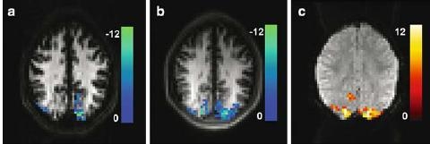
Typical EPI and HASTE VASO images with activation overlays are shown in Fig. 1. As can be seen from the images, the use of a pure spin echo sequence results in a higher intrinsic signal level in grey matter for HASTE than for EPI: at TR = 2 s, the mean gray matter intensities (n = 8) are 336 ± 30 and 202 ± 19 for HASTE and EPI, respectively, for the same receiver gain and scaling parameters in the reconstruction. With 4.38 ± 0.54 and 4.13 ± 0.58 units the average temporal noise in the non-activated gray matter ROI is nearly identical in HASTE and EPI. Purely on the basis of bandwidth considerations, i.e., taking the square root ratio of the effective ADC time which is spent acquiring the MR signal, intrinsic thermal noise in HASTE is expected to be a factor of 1.70 lower than thermal noise in EPI. Under the assumptions that non-thermal noise is of physiological origin and scales proportionately with signal strength [7], and that the experimentally determined temporal noise reflects the contributions of thermal (σth) and physiological (σph) noise as √(gs2th + σ2ph), then the relative contribution of the two noise sources can be estimated for each method. This yields 66% physiological to 34% thermal noise for HASTE, and a corresponding ratio of 41–59% for EPI.
Fig. 1.

Typical EPI (a) and HASTE (b) VASO images with activation overlays (t scores, P < 0.0004). Activation regions detected in the HASTE data are considerably larger, suggesting superior functional sensitivity.As a result, the activation pattern of HASTE more closely resembles that of the BOLD measurements (c). The use of pure spin echoes results in nearly factor 1.7 higher grey matter intensity in the HASTE images as compared to the EPI images
Defining the BOLD activation (P < 0.0001) as a neutral ROI for comparison, the VASO signal changes are 3.42±1.34 for EPI and 5.18±1.96 for HASTE, i.e., considerably larger for the spin-echo sequence (errorbars = SD across subjects). Functional CNR was calculated as CNR = tSNR · ΔS/S. Given the observed noise levels, this yields a 43% higher CNR of the HASTE method. Calculation of mean t-scores in the same ROI yielded a subject average for 3.40 ± 1.16 for EPI and 4.78 ± 1.56 for HASTE. The ratio of 1.41 is in excellent agreement with the direct CNR measurements; this is not unexpected as t-scores and CNR are equivalent measures in a blocked design.
When analysed separately, this increased sensitivity resulted in amore than doubled count of active voxels in HASTE (61 voxels) compared to EPI (26 voxels) at the same significance threshold (P < 0.0004), averaged over all subjects. Visual assessment of the activation maps indicates that as a consequence the HASTE VASO activation covers a similar volume to the BOLD activation (Fig. 1).
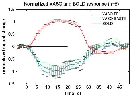
Normalized stimulus response curves of the two VASO measurements, as well the BOLD response, are shown in Fig. 2.
Fig. 2.

Averaged stimulus response (n = 8) of the EPI and HASTE VASO activation (P < 0.0004). Note the decline of the CBV well before the end of the stimulus; this behaviour is much less pronounced in the BOLD response. The VASO curves show a nearly identical signal timecourse, with a return to baseline that essentially coincides with that of the BOLD (error bars = SEM). The stimulation period is indicated by the solid line on the time axis. The baseline was taken as the average of the 5 s before stimulus onset
Measurements at longer TR were performed with both methods for the investigation of their sensitivity to inflow effects, and did not reveal a significant difference. The results from the present data are shown in Table 1. Within the limits of experimental error, mainly due to the dominant inter-subject differences, both methods appear to be equally affected.
Table 1.
VASO signal changes measured with EPI and HASTE at different TR
| TR (s) | TI (ms) | ΔSEPI (a.u.) | ΔSHASTE (a.u.) |
|---|---|---|---|
| 2* | 713 | 5.60±1.06 | 6.27±1.50 |
| 5** | 1054 | 5.20±1.26 | 4.66±0.89 |
| 8*** | 1116 | 4.12±1.08 | 3.67±0.33 |
Within measurement errors (SD over subjects) CBV signals equally change with TR for both methods. This suggests that both are equally affected by CBF increases upon activation.
Activation was thresholded at *P < 0.0004, **P < 0.0017 and ***P < 0.01 to account for the reduced number of measurements available with increasing TR and to keep activation volume approximately equal for each case. All significantly activated voxels were taken into account for the calculation of signal change
Discussion
In the choice of sequence parameters care was taken that the HASTE echo train is sufficiently short so as not to lead to T2 blurring along the phase-encode dimension: this might potentially lead to ‘smearing’ of high white matter signal into the low-intensity gray matter areas if the acquisition window were too long. The FWHM of the PSF due to T2 decay is given byΔf = 1/(π · T2). Thus for typical T2 values of approximately 80 ms [8] and the short readout train of approximately 115 ms, a linewidth in excess of one pixel cannot be expected. T2 blurring may pose a problem for larger matrix sizes or higher image resolution that would require a longer echo train, but here parallel imaging techniques can be employed to shorten the readout appropriately. Particularly for longer echo trains, the use a ‘centre-out’ phase encoding scheme might be beneficial as it typically permits use of shorter TE than a linearly ascending scheme.
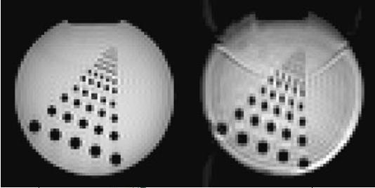
The possibility of blurring in HASTE due to partial Fourier sampling and imperfect Margosian phase correction was investigated by performing scans on a resolution phantom. The results (Fig. 3) showed very sharp images for HASTE and thus blurring could be excluded from causing an ‘artificial’ reconstruction related SNR increase in HASTE.
Fig. 3.

Phantom images acquired with HASTE (left) and EPI (right) using the same parameters as for the functional experiment, but without inversion pulse
BOLD signal changes are expected to contribute considerably less to the T2-weighted HASTE VASO signal than to EPI; with the short TE of 14 ms the HASTE sequence is almost entirely insensitive BOLD related signal changes which would peak at the TE of approximately 80 ms. As is evident from the multi-echo data shown by Lu et al. [2], some degree of BOLD contamination can be expected for the typical gray matter T2* of 40 ms and the TE of 11 ms used here for EPI.
The estimation of noise ratios did not distinguish between the different sources of physiological fluctuations, and possible interdependencies between them, such as a cross-correlation of noise in T2*(t) and signal intensity S0(t). This has been proposed in a more elaborate noise model [9]. For the very short TEs used in this study, it is reasonable to assume that the contribution of T2* noise to the total physiological noise (including cross-terms) can be neglected.
Considering the relative noise contributions to EPI and HASTE VASO, it appears that for the voxel size used here EPI is in the thermal noise dominated regime, whereas HASTE is physiological noise dominated (66%). If physiological noise is the major contributor to HASTE, then the relative sensitivity penalty for increasing the spatial resolution would thus be much lower than for EPI. This suggests HASTE as the more suitable method for high resolution VASO measurements. VASO scans at higher spatial resolution than currently used for BOLD would appear attractive because of the stronger microvascular weighting in VASO [1].
Despite its advantage of permitting CBV measurements without contrast agents, the fact that VASO is a single slice experiment is clearly disadvantageous in a typical routine clinical or cognitive fMRI setup. A multi-slice VASO acquisition scheme using global inversion cycling (MAGIC) [10] was successfully demonstrated at 1.5 T, taking advantage of the relatively low BOLD contribution and the fast EPI readout. As an alternative to multi-slice acquisition the SE-based approach proposed here could be extended to 3D acquisitions by using a GRASE (gradient and spin echo) scheme. This would appear attractive on the basis of SNR and image artefact considerations: reasonable slab acquisition times could be achieved by the use of parallel imaging techniques. That GRASE offers potential benefits has recently been shown by Gunther et al. [11] for perfusion imaging. For VASO, a phase encoding scheme by which T2 and T2* effects are separated into two different directions [12] should permit acquisitions with minimal BOLD contamination and high image quality.
In conclusion, the application of HASTE to VASO fMRI was investigated. EPI VASO time courses were faithfully reproduced using IR-HASTE measurements. HASTE VASO showed a 43% higher sensitivity than EPI. This results in a large increase in number of active pixels at the same significance threshold. As a consequence VASO activation maps cover a similar volume to their BOLD counterparts, rather than showing activation in only a subset of the BOLD activated pixels. This is physiologically reasonable, as BOLD activation is a result of increased CBF, which must have a corresponding increase in CBV associated with it. The data also provide further evidence of increased microvascular weighting of VASO compared to BOLD. With the additional advantage of yielding images free of susceptibility artefacts, the present results suggest that a HASTE acquisition scheme is significantly superior to EPI and should therefore be the method of choice for measuring VASO signal changes.
Acknowledgment
The work presented in this paper was supported by STW grant NGT.6154. The authors thank Markus Barth for useful discussions, and two anonymous reviewers for helpful comments.
References
- 1.Lu H, Golay X, Pekar JJ, van Zijl PCM (2003) Functional magnetic resonance Imaging based on changes in vascular space occupancy. Magn Reson Med 50(2):263–274 [DOI] [PubMed]
- 2.Lu H, van Zijl PC (2005) Experimental measurement of extravascular parenchymal BOLD effects and tissue oxygen extraction fractions using multi-echo VASO fMRI at 1.5 and 3.0 T. Magn Reson Med 53(4):808–816 [DOI] [PubMed]
- 3.Donahue MJ, Lu H, Jones CK, Edden RA, Pekar JJ, van Zijl PC (2006) Theoretical and experimental investigation of the VASO contrast mechanism. Magn Reson Med 56(6):1261–1273 [DOI] [PubMed]
- 4.Kiefer B, Grässner J, Hausmann R (1994) Image acquisition in a second with half-Fourier-acquisition single-shot turbo spin echo. supplement to JMRI volume 4(P), abstract#436; p. 86
- 5.Hennig J, Nauerth A, Friedburg H (1986) RARE imaging: a fast imaging method for clinical MR. Magn Reson Med 3(6):823–833 [DOI] [PubMed]
- 6.Norris DG, Bornert P, Reese T, Leibfritz D (1992) On the application of ultra-fast RARE experiments. Magn Reson Med 27(1):142–164 [DOI] [PubMed]
- 7.Kruger G, Glover GH (2001) Physiological noise in oxygenation-sensitive magnetic resonance imaging. Magn Reson Med 46(4):631–637 [DOI] [PubMed]
- 8.Wansapura JP, Holland SK, Dunn RS, Ball WS Jr (1999) NMR relaxation times in the human brain at 3.0 tesla. J Magn Reson Imaging 9(4):531–538 [DOI] [PubMed]
- 9.Wu G, Li SJ (2005) Theoretical noise model for oxygenation-sensitive magnetic resonance imaging. Magn Reson Med 53(5):1046–1054 [DOI] [PubMed]
- 10.Lu H, van Zijl PC, Hendrikse J, Golay X (2004) Multiple acquisitions with global inversion cycling (MAGIC): a multislice technique for vascular-space-occupancy dependent fMRI. Magn Reson Med 51(1):9–15 [DOI] [PubMed]
- 11.Gunther M, Oshio K, Feinberg DA. (2005) Single-shot 3D imaging techniques improve arterial spin labeling perfusion measurements. Magn Reson Med 54(2):491–498 [DOI] [PubMed]
- 12.Mugler JP 3rd (1999) Improved three-dimensional GRASE imaging with the SORT phase-encoding strategy. J Magn Reson Imaging 9(4):604–612 [DOI] [PubMed]


