Abstract
Botulinum neurotoxin (BoNT) heavy chain (Hc) facilitates receptor-mediated endocytosis into neuronal cells and transport of the light chain (Lc) protease to the cytosol where neurotransmission is inhibited as a result of SNARE protein cleavage. Here we show that the role of BoNT Hc in cell intoxication can be replaced by commercial lipid-based and polycationic polymer DNA transfection reagents. BoNT “transduction” by these reagents permits efficient intoxication of neuronal cells as well as some non-neuronal cell lines normally refractory to BoNT. Surprisingly, the reagents facilitate delivery of recombinant BoNT Lc protease to the cytosol of both neuronal and non-neuronal cells in the absence of BoNT Hc, and with sensitivities approaching that of BoNT holotoxin. Transduction of BoNT, as with natural intoxication, is inhibited by bafilomycin A1, methylamine and ammonium chloride indicating that both pathways require endosome acidification. DNA transfection reagents facilitate intoxication by holotoxins, or isolated Lc proteases, of all three BoNT serotypes tested (A, B, E). These results suggest that lipid and cationic polymer transfection reagents facilitate cytosolic delivery of BoNT holotoxins and isolated Lc proteases by an endosomal uptake pathway.
Keywords: Botulinum toxin, transfection reagent, protein transduction, bafilomycins, intoxication
1. Introduction
Botulinum neurotoxins (BoNTs) are extremely potent protein toxins and cause flaccid paralysis by entering motor neurons and inhibiting vesicular neurotransmitter release. There are seven BoNT serotypes, A–G, each cleaving SNARE (soluble NSF attachment receptor) proteins SNAP-25, synaptobrevin, and/or syntaxin at different sites to inhibit neurotransmitter exocytosis (Binz et al., 1994; Blasi et al., 1993; Schiavo et al., 1992; Schiavo et al., 1993a; Schiavo et al., 1993b). Each BoNT contains a heavy chain (Hc) and light chain (Lc) tethered by a single disulfide bond (DasGupta and Sugiyama, 1972). The Hc includes a receptor binding domain and a translocation domain while the Lc contains zinc metalloprotease activity (Hoch et al., 1985; Lacy et al., 1998; Montecucco and Schiavo, 1993). During the intoxication process, botulinum toxin binds with high specificity to receptors on the nerve termini at the neuromuscular junction (Kozaki et al., 1989; Lacy et al., 1998; Lalli et al., 1999). These receptors for BoNT include both a ganglioside and a cell surface protein (Kitamura et al., 1980; Montecucco, 1986; Nishiki et al., 1996). The synaptic vesicle protein 2 (SV2) (Dong et al., 2008; Mahrhold et al., 2006) is the receptor for BoNT/A while synaptotagmins I and II are the protein receptors for serotypes B and G respectively (Dong et al., 2007; Nishiki et al., 1994; Nishiki et al., 1996; Rummel et al., 2004). Following receptor mediated endocytosis (Binz and Rummel, 2009; Humeau et al., 2000; Simpson, 1980, 1981, 2000; Verderio et al., 2007) and endosome formation (Fisher and Montal, 2006; Keller et al., 2004; Montecucco et al., 1994), the Lc is translocated across vesicle membrane into the cytosol through a protein channel formed by the amino terminus of the Hc (Blaustein et al., 1987; Blocker et al., 2003; Cai et al., 2006b; Fisher and Montal, 2006; Hoch et al., 1985; Humeau et al., 2000; Koriazova and Montal, 2003; Schmid et al., 1993; Shone et al., 1987; Simpson, 1980, 1981). The translocation of the Lc to the cytosol occurs following a conformational change that is induced by acidification of the endosome (Cai et al., 2006b; Hoch et al., 1985). Drugs that inhibit endosome acidification, such as bafilomycin A1, are antagonists of BoNT intoxication (Deshpande et al., 1997; Simpson, 1983; Simpson et al., 1994). Once in the cytosol, the different BoNT Lcs cleave and inactivate their SNARE protein substrates leading to intoxication.
Botulinum toxin proteases are useful reagents in studies on cellular transport and secretion because of their ability to specifically cleave SNARE proteins. Because of the potential of BoNT as a weapon in bioterrorism, major research efforts are also underway to develop both antitoxins and antidotes for botulism. Botulinum toxin research, though, has been hampered by the fact that the toxins naturally enter only those cells which express the surface receptors recognized by the BoNT serotype under study. These receptors are generally present in significant amounts on primary neuronal cells but are often poorly expressed or even lacking in neuronal cell lines. Neuroblastoma cell lines are available that can become intoxicated but often require high doses of toxin to produce evidence of intoxication if it occurs at all (Ibanez et al., 2004; Purkiss et al., 2001). Primary neuronal cells are sensitive to most BoNT serotypes although these are difficult and costly to prepare and to maintain (Keller et al., 2004).
To overcome the restrictions to BoNT intoxication that exist because of the lack or low level of appropriate receptors on target cell lines, researchers have employed alternative methods to deliver BoNT proteases to the cytosol. One method has been to introduce plasmids which drive expression of BoNT proteases within transfected cells (Fernandez-Salas et al., 2004; He et al., 2008; Huang et al., 2001; Huang et al., 1998; Ji et al., 2002; Land et al., 1997; Shu et al., 2008). Electroporation of BoNT holotoxin or isolated Lc proteases has been employed for functional delivery of the Lc protease to the cytosol of secretory cells (Boyd et al., 1995; Erdal et al., 1995; Wang and Nadler, 2007) and streptolysin permeabilization has been used for delivery of BoNT holotoxins or Lc proteases to cells and synaptosomes (Gonelle-Gispert et al., 1999; Leung et al., 1998; Pickett et al., 2007; Sadoul et al., 1997) in situations where they are tested immediately after treatment. In these methods, the amount of Lc protease delivered to the cytosol is not easily controlled and likely much higher than occurs during natural intoxication. None of these methods deliver toxin to the cytosol through an endosomal uptake process as occurs in natural intoxication. Clearly a method that promotes delivery of BoNT Lc protease to the cytosol of most neuronal and non-neuronal cells through endosome-mediated uptake would offer a valuable new tool for research employing BoNTs.
In studies of botulinum intoxication in cultured neuronal cell, we observed much more efficient BoNT/A intoxication occurred when the cells had been recently subjected to DNA transfection by lipofection. These reagents are routinely used for the delivery of DNA into cells by forming DNA/lipofection complexes that enter cells via the endocytic pathway (Zabner et al., 1995; Zhou and Huang, 1994). Transfection is typically independent of endosomal acidification and involves interactions between cationic lipid in the liposome and anionic lipid in the endosome (Zelphati and Szoka, 1996). Physical dissociation of DNA from the DNA-lipid complex and release of DNA from the endosome into the cytosol is a critical step in DNA transfection (Zelphati and Szoka, 1996). In this study, we tested the ability of standard DNA transfection reagents to deliver both BoNT holotoxins and recombinant BoNT Lc proteases to neuronal and non-neuronal cells. These reagents enabled potent, endosome-mediated, BoNT intoxication of most cell lines tested without compromising cell viability and are independent of the need for BoNT receptors. This technology opens new avenues for investigating BoNT holotoxin function in a wide variety of cell lines as well as performing cell-based drug assays without select agent requirements.
2. Materials and methods
2.1. Cell culture and reagents
M17 (ATCC# CRL-2267) cells were maintained in DMEM (Gibco) containing 10% fetal bovine serum (FBS) (Gibco). MEME (Gibco) plus 10% FBS was used for culturing Neuro2a (ATCC# CCL-131) (abbreviated N2A) and HEK293 (ATCC# CRL-1573) cells. HIT-T15 (ATCC# CRL-1777) cells were cultured in F12K (Gibco) containing 10% horse serum and 5% FBS. 6 ×10⁴ cells were seeded onto each well of 24-well plate and maintained at 37°C. After 72 hrs, culture medium was replaced with fresh medium before experimental treatments.
Primary cultures of cerebellar granule cells were prepared from 7 day-old Sprague-Dawley rats (Welch et al., 2000) essentially by the methods of Farkas (Farkas et al., 2004). Briefly, after aseptically removing cerebella from the skulls, tissue was freed from meninges and incubated in 0.05% trypsin solution for 10 min at RT. After a brief centrifugation, cells were triturated in DMEM/F12 containing 10% FBS and filtered through a sterile cell strainer mesh with 40 um pore size (BD Falcon) (Foran et al., 2003; Kornyei et al., 1998; Sabbieti et al., 2004). Cell number was determined by trypan blue exclusion, and cells were seeded onto a poly-L-lysine (PLL) 1 g/cm2 laminin (Sigma) coated 6-well plate with DMEM containing 10% FBS, 25 mM KCl, 2 mM Glutamax, and 100 g/mL gentamicin (Gibco). The cultures were maintained at 37°C in a humidified atmosphere of 6% CO2. After 24 hrs of culturing, cytosine arabinoside (Sigma) was added to a final concentration of 20 µM to prevent astrocytic proliferation. The neurons were cultured for 7–8 days before use.
FuGene-HD (Roche), Lipofectamine 2000 (Invitrogen) and PEI average molecular weights 0.6, 1.8, 10, 70 kDa (Alfa Aesar) and 25 kDa (Sigma) were used for transfection and transduction as recommended by the manufacturer except where indicated. Bafilomycin A1 was obtained from Tocris Cookson Inc. Methylamine hydrochloride and ammonium chloride were obtained from Sigma.
Antibody reagents used were: rabbit anti-SNAP25 antibody (Sigma); goat anti-rabbit HRP antiserum (Sigma); rabbit anti-VAMP2 antibody (Millipore); rabbit anti-CFP (gift from Dr. Randall Kincaid, Veritas Labs). Reagents for Western blotting, including Wash Solution and LumiGLO Chemiluminescent Substrate, were purchased from (KPL). The pcDNA/CFP plasmid is pcDNA3 (Invitrogen) containing the cerulean fluorescent protein (CFP) coding DNA (Clontech) positioned for mammalian expression from the CMV promoter.
2.2. Toxin preparations
BoNT/A (isotype 1), BoNT/B and BoNT/E were obtained from Metabiologics. BoNT/E holotoxin was activated for use in cell intoxications (Duff et al., 1956). 1 mg/ml BoNT/E was nicked by incubating for 30 min at 37°C with 0.3 mg/ml trypsin (type XI, bovine pancreas) in 30 mM HEPES, pH 6.75. Trypsin was subsequently inhibited by addition of 0.5 mg/ml trypsin inhibitor (type I-S, soybean) and incubation for 15 min at 20 °C (Cai et al., 2006a; Duff et al., 1956; Ohishi and Sakaguchi, 1977). Toxins were aliquoted and stored at −20 °C. Each experiment utilized a freshly thawed aliquot of toxin to ensure uniform activity.
2.3. BoNT Lc proteases
Recombinant BoNT/A (amino acids 1–438) and BoNT/B light chain proteases (amino acids 1–441) were produced with hexahistidine tags at the amino and carboxyl termini using the pET14b expression vector. The protein was expressed within the Rosetta-gami strain of E. coli (Novagen) and soluble protein was purified to near homogeneity by standard nickel affinity chromatography. The recombinant BoNT/E Lc (amino acids 1–422) was expressed in E. coli as an N-terminal fusion protein to glutathione-S-transferase. The protein was purified by standard glutathione affinity methods and provided as a gift by Dr. Randall Kincaid (Veritas Labs).
2.4. BoNT holotoxin intoxication and transduction
Cell lines were intoxicated as follows. A 50 µl solution of serum-free DMEM was prepared containing BoNT or BoNT Lc protease. Transfection reagent (or DMEM control) was then added at the indicated ratio (BoNT or BoNT Lc [µg]: transfection reagent [µl]) and the mixture incubated at room temperature for 15–20 min. The mixture was then applied to cultured cells containing 0.5 ml fresh culture medium in a well of a 24-well plate. At indicated times later, cells were washed twice with 1 ml DPBS (Gibco) and incubated with 0.5 ml of fresh medium. One or more days later, the cells were washed once with 1 ml DPBS and 100 µl of 0.25% trypsin was added for one minute followed by addition of 500 µl of medium with serum. Cells were then pelleted and washed once with 1 ml DPBS. Finally the cell pellet was dissolved in 50 µl of sample buffer (62.5 mM Tris-HCl, pH 6.8, 2 % SDS, 10 % glycerol and 0.002 % bromophenol blue plus 5 % beta-mercaptoethanol) and boiled for 10 min prior to gel electrophoresis.
2.5. Cell viability assay
Cell viability was measured by the MTT assay (ATCC) in triplicate according to the manufacturer’s instructions. Absorbance was recorded at 570 nM with a Synergy™ HT Multi-Mode Microplate Reader and the data were analyzed with KC4 software.
2.6. Drug treatment of cells
Bafilomycin A1 (1 µM) or DMSO was applied to cells for 2 hrs and the cells were washed twice with 1 ml DPBS before being subjected to BoNT/A intoxication or transduction as above. Methylamine hydrochloride (10 mM) was applied to cells for 1 h or ammonium chloride (8 mM) for 2 hrs before the cells were washed twice with 1 ml DPBS and subjected to BoNT/A transduction.
2.7. DNA transfection
The pcDNA/CFP expression plasmid (0.5 µg) was transfected as recommended by the manufacturers. Cell fluorescence was recorded using an Olympus IX50 microscope and imaging software slidebook (Leeds Precision Instruments, Inc) before cell extracts were prepared as above.
2.8. Western blotting
Cell extract prepared from 4 × 10⁵ cells was boiled for 5 min and loaded to 15% pre-casted protein gels (BioRad). Protein samples were separated by SDS-PAGE run in an ice bath and transferred to PVDF membrane. Blots were incubated with 5% skim milk/ PBST 0.5% for at least 1 hr at room temperature and incubated with primary antibodies at 4°C overnight, then washed with PBST 0.5% buffer. Finally the membranes were incubated with an appropriate HRP labeled secondary Ab and incubated for 1 hr at room temperature, washed and bound antibody detected using LumiGLO Chemiluminescent Substrate (KPL). Signals were scanned by Kodak Image Station 2000R and analyzed with the Kodak 1D 3.6 network.
3. Results
3.1. Commercial lipid-based DNA transfection reagents enhance botulinum intoxication of cultured neuronal cells
We observed that neuronal cells intoxicated with BoNT/A immediately after DNA transfection using the FuGene-HD reagent (Roche) appeared more efficiently intoxicated than control cells so we directly tested the effect of FuGene-HD on BoNT intoxication. The measure of BoNT serotype A intoxication used in these studies was the percentage of the cellular SNAP25 that had been cleaved. For two neuroblastoma cell lines, M17 and Neuro2a (N2A), SNAP25 cleavage assessed 24 hrs following BoNT/A intoxication was increased from less than 20% to more than 80% when the toxin was preincubated with FuGene-HD (1:3, µg toxin:µl FuGene-HD) (data not shown). Enhancement of intoxication also occurred when the BoNT/A and FuGene-HD were separately added to neuronal cells (i.e. no pre-incubation), but the effect was less pronounced. The level of intoxication did not change significantly in either cell line using several fold more or less FuGene-HD and the ratio of 1:3 was selected for routine use. To exclude the possibility that transfection reagents increased cell death leading to release of SNAP25 and enhanced cleavage, MTT assays were performed and revealed no significant change in cell viability following exposure to FuGene-HD at 1:3 (data not shown).
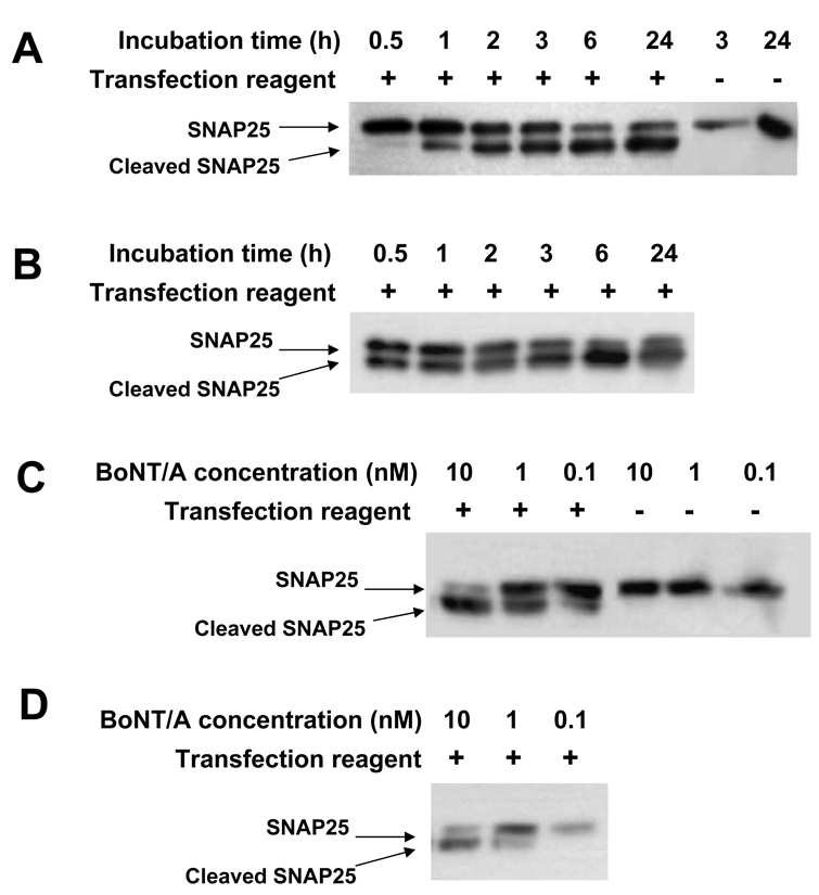
To characterize the rate at which DNA transfection reagent-enhanced intoxication occurs, N2A cells were exposed to 10 nM BoNT/A in the presence of FuGene-HD for variable times and harvested immediately. As shown in Fig. 1A, cleavage of SNAP25 could be observed in as little as 30 minutes and maximum cleavage of about 90% occurred within the first 6 hrs. In this experiment, the N2A cells were not detectably intoxicated in the absence of FuGene-HD. When the medium was changed after 1–3 hrs of BoNT/A exposure and the cells were cultured for up to 48 hrs, a small amount of additional SNAP25 cleavage occurred but never reached the 90% cleavage observed after only 6 hrs exposure to toxin/FuGene-HD (Fig. 1B). After 6 hrs toxin exposure, additional incubation of cells did not result in further SNAP25 cleavage.
Fig. 1.
DNA transfection reagent facilitates rapid BoNT/A internalization by N2A cells and the extent of intoxication is toxin concentration dependent. A. N2A cells were exposed to 10 nM BoNT/A toxin for 0.5, 1, 2, 3, 6 or 24 hrs with (+) or without (−) pre-incubation of toxin with FuGene-HD transfection reagent and immediately harvested. B. BoNT/A toxin was pre-incubated with FuGene-HD then added to N2A cells at 10 nM for 0.5, 1, 2, 3, 6 or 24 hrs. Medium was changed and all the cells were cultured until 48 hrs after initial toxin exposure. C. N2A cells were exposed to 10 nM, 1 nM and 0.1 nM of BoNT/A toxin for 24 hrs with (+) or without (−) pre-incubation with FuGene-HD transfection reagent and then harvested. D. N2A cells were exposed to 10 nM, 1 nM and 0.1 nM of BoNT/A toxin for 3 hrs in the presence of FuGene-HD transfection reagent and then harvested. Cell extracts were prepared and resolved by SDS-PAGE. The extent of BoNT/A intoxication was measured by Western blotting to assess the extent of SNAP25 cleavage.
Typically neuroblastoma cells require BoNT concentrations of 10 nM or more to cause significant intoxication while primary neurons are often sensitive to BoNT concentrations as low as 100 pM. Experiments were performed to examine the sensitivity of FuGene-HD enhanced intoxication of neuroblastoma cells to toxin concentrations. N2A cells were cultured for 24 hrs with BoNT/A concentrations ranging from 0.1 to 10 nM (Fig. 1C) and harvested 48 hrs after toxin exposure. In the absence of FuGene-HD, the N2A cells were not detectably intoxicated even at 10 nM. In the presence of FuGene-HD, some cleavage of SNAP25 was observed in N2A cells exposed to as little as 100 pM BoNT/A. In a similar experiment with only 3 hrs BoNT/A exposure, 10% and 80% of SNAP25 cleavage was detected with toxin concentrations of 1 nM and 10 nM respectively (Fig, 1D). Interestingly, the efficiency of intoxication of primary neurons was not enhanced with transfection reagents (see below), suggesting that these cells are maximally intoxicated using normal conditions. These data show that it is possible to achieve intoxication efficiencies in at least some neuroblastoma cells that approach those of primary neurons by including FuGene-HD with the toxin.
3.2. Commercial DNA transfection reagents permit BoNT/A intoxication of non-neuronal cells
In normal intoxication, BoNT is internalized through receptor-mediated endocytosis (Binz and Rummel, 2009; Humeau et al., 2000; Simpson, 1980, 1981, 2000; Verderio et al., 2007). BoNT/A uptake into cells normally requires the presence of both ganglioside and the SV2 protein receptors which leads to its specificity for neuronal cells (Dong et al., 2006). To determine whether the use of DNA transfection reagents bypass the need for surface receptors, we tested for transfection reagent enhancement of intoxication in two non-neuronal cell lines that express SNAP25 yet are not normally susceptible to BoNT/A intoxication. One cell line tested was HEK293, an embryonic kidney line, and the other was HIT-T15, a human insulinoma cell line. As expected, SNAP25 cleavage was not detected in HEK293 or HIT-T15 cells following 24 hr incubation with 10 nM of BoNT/A (Fig. 2). In contrast, inclusion of FuGene-HD during intoxication clearly facilitated the uptake of BoNT/A into these cells leading to significant SNAP25 cleavage.
Fig. 2.
DNA transfection reagent facilitates BoNT/A internalization of non-neuronal cell lines. Neuronal cell lines N2A, M17 and non-neuronal cell lines HEK293 (293), HIT-T15 were exposed to 10 nM BoNT/A for 24 hrs with (+) or without (−) pre-incubation with DNA transfection reagents, FuGene-HD or Lipofectamine 2000. Cell extracts were prepared and the extent of BoNT/A intoxication was measured by Western blotting to monitor SNAP25 cleavage.
A second lipid-based DNA transfection reagent, Lipofectamine 2000 (Invitrogen), was compared to FuGene-HD for the ability to enhance BoNT/A intoxication of neuronal and non-neuronal cell lines (Fig. 2). In the case of the M17 and N2A cells, both DNA transfection reagents significantly enhanced BoNT/A uptake into cells with FuGene-HD showing a slightly more pronounced effect. In contrast, Lipofectamine 2000 reagent proved to be the more effective transfection reagent for enhancing BoNT uptake into HIT-T15 cells. The results demonstrate that either lipid-based commercial DNA transfection reagents facilitated the uptake of BoNT/A into both neuronal and non-neuronal cells and suggest that different transfection reagents are variably efficient when used to enhance intoxication of different cell lines.
3.3. Commercial DNA transfection reagents facilitate transduction of the BoNT/A light chain protease in the absence of the BoNT/A heavy chain
The BoNT heavy chain (Hc) domain has been shown to play at least two critical roles during neuronal cell intoxication; binding to the neuronal cell receptors (Hoch et al., 1985; Kitamura et al., 1980; Lacy et al., 1998; Montecucco, 1986; Montecucco and Schiavo, 1993; Nishiki et al., 1996) and chaperoning the translocation of the BoNT light chain (Lc) protease from the endosome to the cytosol (Blocker et al., 2003; Cai et al., 2006b; Fisher and Montal, 2006; Hoch et al., 1985; Humeau et al., 2000; Schmid et al., 1993; Shone et al., 1987; Simpson, 1980, 1981). We next tested whether the BoNT Hc domain was necessary for BoNT uptake facilitated by commercial transfection reagents.
For these experiments, two different forms of the BoNT/A Lc were employed. The Lc438 form (amino acid 1 to 438 of GenBank ZP_02612822) contains the full-size protease released following proteolytic cleavage from the Hc domain during natural processing by the Clostridium botulinum microbe (Krieglstein et al., 1994). Lc424 is identical to Lc438 except that 16 amino acids are removed from the carboxyl end, a modification that does not significantly affect proteolytic activity but improves expression and solubility properties (Baldwin et al., 2004). The results shown in Fig. 3 show that the FuGene-HD and Lipofectamine 2000 DNA transfection reagents are efficiently able to promote the transduction of each recombinant BoNT/A Lc into both neuronal and non-neuronal cells. Surprisingly, a very low concentration of BoNT/A Lc was sufficient to promote internalization and cleavage of cytosolic SNAP25, a concentration similar to that needed for intoxication by BoNT holotoxin. Both FuGene-HD and Lipofectamine 2000 were effective in all four cell types tested, although as with BoNT holotoxin (Fig. 2), FuGene-HD was more effective for N2A, M17 and HEK293 while Lipofectamine 2000 was more effective with HIT-T15 cells. As expected, no SNAP25 cleavage occurred in neuronal or non-neuronal cells when the Lc was added to the medium in the absence of DNA transfection reagents.
Fig. 3.
Lipid-based DNA transfection reagents promote cellular internalization of BoNT/A Lc protease in the absence of Hc. Neuronal cell lines N2A, M17 and non-neuronal cell lines HIT-T15, HEK293 (293) were exposed for 24 hrs to 30 nM of recombinant BoNT/A LC protease, using either the full-length protease (Lc438) or the carboxyl-truncated form (Lc424). The protease was added to cell culture with (+) or without (−) pre-incubation with the DNA transfection reagents, FuGene-HD or Lipofectamine 2000. Cell extracts were prepared and the extent of BoNT/A intoxication was measured by Western blotting to monitor SNAP25 cleavage.
3.4. Polyethyleneimine polymers facilitate transduction of recombinant BoNT/A Lc protease
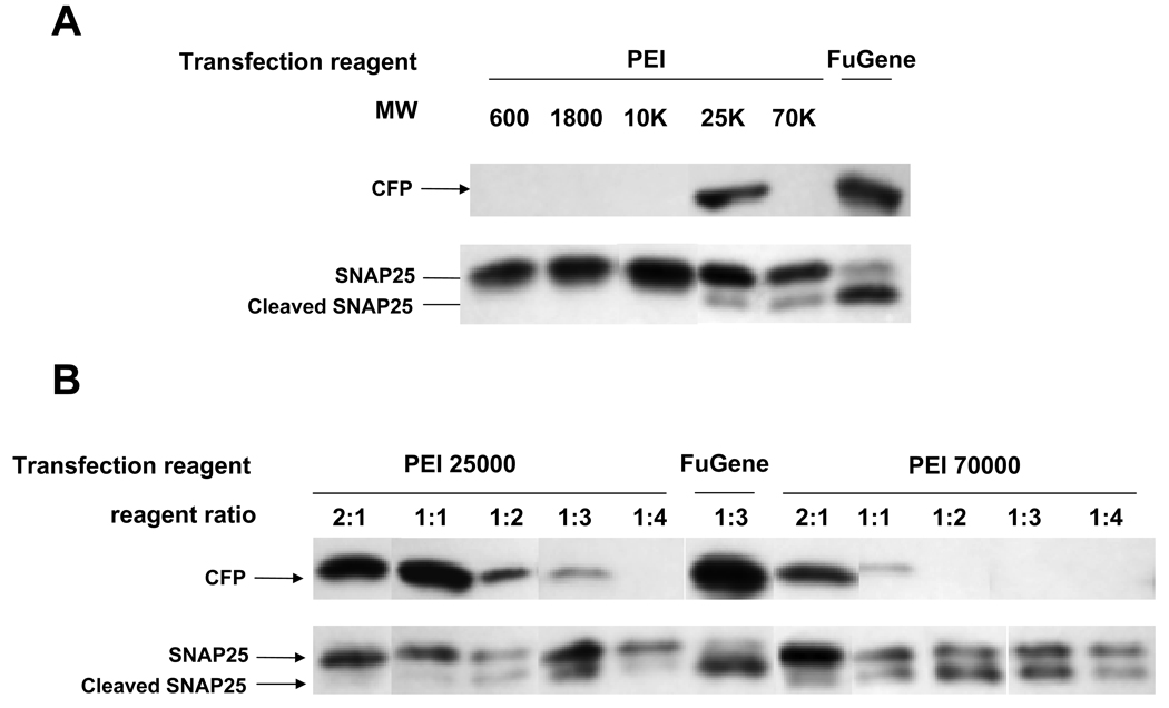
The chemical nature of both commercial DNA transfection reagents, FuGene-HD and Lipofectamine 2000, is proprietary although both are described as lipid-based and Lipofectamine 2000 as a cationic lipid reagent (Invitrogen). We next tested BoNT/A toxin transduction efficacy using cationic polyethyleneimine polymers as their chemical nature is available. The data, shown in Fig. 4A, demonstrate that high MW PEI polymers (1:1 µg toxin: µl reagent) have the ability to promote BoNT Lc transduction into N2A cells. While the lower molecular weight PEI polymers (MW 600–10000) showed no toxin transduction efficacy, the 25K and 75K MW polymers clearly facilitated intoxication by BoNT/A Lc, although not as effectively as FuGene-HD. To determine the optimal ratio of BoNT/A Lc and PEI polymer, a series of different ratios were tested using either 25K or 75K PEI polymers (Fig. 4B). The optimal ratio proved to be 1:2 using the 75K MW PEI polymer and resulted in more than 50% cleavage of SNAP25 which approached the transduction efficacy achieved with FuGene-HD.
Fig. 4.
Defined cationic polymer DNA transfection reagents promote BoNT intoxication independent of DNA transfection. A. N2A cells were incubated for 24 hrs with 20 nM recombinant BoNT/A LC (Lc438) and 0.5 µg pcDNA/CFP expression plasmid with (+) or without (−) pre-incubation with FuGene-HD (FuGene) or PEI having different average molecular weights as indicated. Pre-incubations were performed at a toxin (µg): reagent (µl) ratio of 1:1 with PEI and 1:3 with FuGEne HD. B. N2A cells were incubated for 24 hrs with 20 nM recombinant BoNT/A Lc438 (10 nM for ratios 1:3 and 1:4) and 0.25µg pcDNA/CFP following pre-incubation with FuGene-HD (FuGene) or PEI. (25000 or 75000 MW) at the indicated toxin (µg): reagent (µl) ratio. Cell extracts were prepared and the extent of BoNT/A intoxication was assessed by Western blotting to monitor SNAP25 cleavage. The efficiency of DNA transfection was assayed by Western blotting for CFP.
Interestingly, the optimum MW size and concentration of PEI polymer were distinctly different for BoNT/A Lc transduction than for DNA transfection efficacy performed simultaneously. This was tested by including a CFP expression vector during the PEI treatment step and monitoring CFP expression by Western blot (Fig. 4A and B). DNA transfection of N2A cells using PEI polymers was only effective with the 25K MW form, particularly when the ratio of plasmid to PEI polymer was high (1:1). A low level of DNA transfection of N2A cells was observed using 75K MW PEI polymer when the plasmid to reagent ratio was high (2:1). This was in distinct contrast to the BoNT/A Lc transduction efficacy which improved with lower plasmid to reagent ratios (Fig. 4B) and suggests that the two phenomena work through somewhat different mechanisms.
3.5. Lipid-based transduction of BoNT/A Lc protease is sensitive to inhibitors of ER acidification
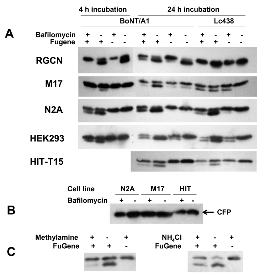
Bafilomycin is an inhibitor of vacuolar adenosine triphosphatase and prevents endosome acidification (Bowman et al., 1988). Previous studies have shown that nerve cell intoxication by BoNT is inhibited by bafilomycin (Simpson et al., 1994). To explore the role of endosome acidification in the DNA transfection reagent transduction of BoNT/A Lc, treatments were performed in the presence or absence of bafilomycin. In these experiments, cells were treated +/− 1 µM of bafilomycin A1 or control for 2 hrs prior to transfection or intoxication. Cytosolic internalization of the Lc was assessed by monitoring cleavage of SNAP25 within cells. Lc internalization was tested, in most cases, following either 4 or 24 hrs incubation with BoNT/A holotoxin or purified Lc. Pretreatment with bafilomycin was shown to completely inhibit 4 hr BoNT/A intoxication of primary rat granule cerebellar neurons (RGCN) whether DNA transfection reagents were included or not (Fig. 5A). With 24 hr BoNT/A intoxication, a small amount of SNAP25 cleavage was observed in primary cells treated with bafilomycin. Some cytoxicity due to bafilomycin treatment was visually apparent and some SNAP25 cleavage may occur as BoNT/A in the media gains access to SNAP25 released following cell lysis. Alternatively, the effect of bafilomycin may be lost during the longer intoxication period to permit entry of some Lc protease into the cell cytosol. When bafilomycin was omitted, BoNT/A treatment led to nearly complete cleavage of SNAP25. DNA transfection reagents did not promote improved BoNT/A or BoNT/A Lc intoxication of primary neurons (Fig. 5A). These neurons were also not susceptible to plasmid DNA transfection using these same reagents (data not shown).
Fig. 5.
Bafilomycin, methylamine hydrochloride and ammonium chloride inhibit DNA transfection reagent-mediated enhancement of BoNT/A holotoxin and Lc internalization into cells. Primary RCGNs, neuronal cell lines N2A, M17 and non-neuronal cell lines 293HEK, HIT-T15 were treated with bafilomycin for 2 hrs and washed with DPBS. Cells were then exposed to 10 nM of BoNT/A toxin for 4 or 24 hrs or 30 nM of Lc438 for 24 hrs, + or − pre-incubation with the FuGene-HD transfection reagent. Control cells were incubated with 10 nM of BoNT/A toxin or 30 nM of Lc438, + or − pre-incubation with FuGene-HD, without prior exposure of the cells to bafilomycin. Cell extracts were prepared and the extent of BoNT/A intoxication was assessed by Western blotting to monitor SNAP25 cleavage. B. N2A, M17 and HIT-T15 cells were treated +/− bafilomycin and then transfected 24 hrs with pcDNA/CFP in FuGene-HD as above. Cell extracts were resolved by SDS-PAGE and CFP was detected by Western blotting. C. M17 cells were treated with 10 mM methylamine hydrochloride for 1 h or 8 mM ammonium chloride (NH4Cl) for 2 hrs and then exposed to 10 nM Lc438 with (+) or without (−) FuGene-HD transfection reagent (1:3) for 4 hrs. Cell extracts were prepared and the extent of BoNT/A intoxication was assessed by Western blotting to monitor SNAP25 cleavage.
Bafilomycin inhibited SNAP25 cleavage resulting from 4 hrs or 24 hrs incubation of either M17 or N2A neuroblastoma cells with BoNT/A holotoxin, whether intoxication was enhanced by transfection reagents or not (Fig. 5A). This suggests that the enhanced intoxication obtained with these reagents occurs through the natural intoxication pathway that includes translocation of Lc from the endosome to the cytosol. More surprising, bafilomycin also inhibited functional internalization of recombinant BoNT/A Lc into both neuroblastoma and non-neuronal cells in the absence of Hc, most obviously in N2A and HIT-T15 cells (Fig. 5A). As with RGCN above, these experiments are complicated by the bafilomycin cytotoxicity, which leads to lower SNAP25 signals and increased background of SNAP25 cleavage following addition of BoNT/A Lc. This is most apparent in 293 cells which appear particularly susceptible to bafilomycin toxicity. Despite this background though, it is clear that bafilomycin blocks the enhanced cleavage of SNAP25 elicited by incubation of cells with BoNT/A Lc in the presence of transfection reagents.
Bafilomycin had no inhibitory effect on the efficiency of plasmid DNA transfection mediated by FuGene in N2A, M17 or HIT-T15 cells as assessed by CFP expression from a transfected expression vector (Fig. 5B), again indicating that DNA transfection and protein transduction employ somewhat different uptake mechanisms.
Two additional reagents, methylamine hydrochloride and ammonium chloride, have both been used previously in studies showing that BoNT intoxication requires endosomal translocation (Simpson, 1983) and each were tested for their effects on BoNT/A toxin and Lc transduction. Methylamine proved to be toxic to N2A cells at doses known to inhibit endosomal translocation and M17 cells were thus used. Figure 5C shows that 10 mM methylamine substantially reduced the amount of SNAP25 cleavage observed when BoNT/A Lc was transduced into M17 cells. A similar result was obtained using M17 cells exposed to 8 mM ammonium chloride.
3.6. Commercial DNA transfection reagents enhance cellular uptake of other BoNT serotypes
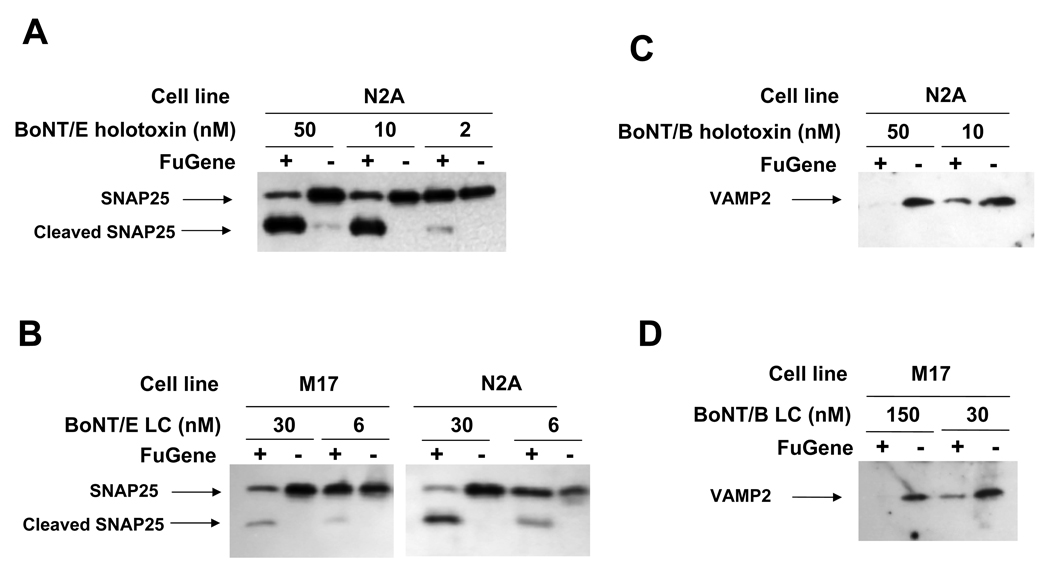
To test whether transfection reagent-mediated intoxication is unique to type A toxin, Botulinum neurotoxin serotypes B (BoNT/B) and E (BoNT/E) were also tested using this delivery system. Cleavage of SNAP25 was used as the indicator for BoNT/E holotoxin or Lc protease internalization (Fig 6A and B) while reduction of VAMP2 was used to monitor BoNT/B holotoxin or Lc internalization (Fig. 6C and D). Only trace amounts of the substrate proteins normally became cleaved following exposure of M17 or N2A neuroblastoma cells to even high concentrations of the BoNT/B or /E holotoxins or Lc proteases. In contrast, inclusion of FuGene-HD promoted substantial cleavage of the appropriate BoNT substrates for both BoNT/B and /E. Similar results were obtained when the holotoxins were replaced by purified recombinant Lc proteases of both toxin serotypes.
Fig. 6.
DNA transfection reagent enhances internalization of multiple BoNT Lc serotypes in two neuroblastoma cell lines. M17 or N2A cells were exposed for 24 hrs to 50, 10 or 2 nM of BoNT/E toxin (A) or 30 or 6 nM of GST-Lc/E (B) or 50 or 10 nM of BoNT/B toxin (C) or 150 or 30 nM of recombinant Lc/B (D), + or − pre-incubation with FuGene-HD. Cell extracts were prepared and the extent of BoNT intoxication was assessed by Western blotting to monitor SNAP25 or VAMP2 cleavage.
4. Discussions
Although BoNT intoxication is exquisitely efficient within animals and cultured primary neurons, it has not proven so efficient in established cell lines and this has inhibited research activities and cell-based drug screening efforts. Intoxication occurs following receptor-mediated internalization of BoNT and then transposition of the toxin light chain (Lc) protease from the endosome to the cytosol. The uptake and then chaperoning of the protease to the cytosol is mediated by the BoNT heavy chain (Hc). Because BoNT Hc binds to receptors found specifically on neuronal cells, non-neuronal cells have proven to be insensitive to the toxin. A variety of neuroblastoma cell lines are available and, while detectable BoNT intoxication will often take place (usually measured by SNARE protein cleavage), it is generally far less efficient than in primary neurons. We show here that BoNT holotoxin intoxication of the two neuroblastoma cells tested, M17 and N2A, can be substantially improved by the presence of several different DNA transfection reagents tested. Furthermore, we demonstrate that these reagents permit intoxication of two non-neuronal cell types, HEK293 and HIT-T15, both not normally susceptible to BoNT intoxication. Finally, we show that the transfection reagents facilitate intoxication of these neuronal and non-neuronal cells when exposed only to isolated BoNT Lc proteases.
While it has been possible to experimentally produce BoNT Lc protein within cells by DNA transfection of mammalian expression plasmids (Fernandez-Salas et al., 2004; He et al., 2008; Huang et al., 2001; Huang et al., 1998; Ji et al., 2002; Land et al., 1997; Shu et al., 2008) or deliver the Lc into cells by permeabilization (Gonelle-Gispert et al., 1999; Leung et al., 1998; Pickett et al., 2007; Sadoul et al., 1997; Wang and Nadler, 2007), these methods result in cells that are often damaged and having excessive and uneven levels of Lc protein which arrives in the cytosol by processes completely different than occur during intoxication. The reagents used in this study for delivery of BoNT to cells are widely used for DNA transfection and are either lipid-based (FuGene-HD, Lipofectamine 2000) or polycationic polymers (PEI) reagents that cause little damage to cells. Other lipid-based reagents were specifically developed for protein transduction (Zelphati et al., 2001) but have not been studied for delivery of BoNT proteases and were not tested here. An early report of intoxication after liposomal delivery of BoNT Lc to motor neurons in animals was not apparently followed up using cultured cells (de Paiva and Dolly, 1990).
The ability of the DNA transfection reagents to delivery BoNT to non-neuronal cells expected to lack the protein receptors required by BoNT during natural intoxication implies a receptor-independent process. The two cell lines studied, HEK293 and HIT-T15, are both commonly used cell lines for studies of protein secretion and contain SNARE protein substrates for BoNT. Easily detected SNARE protein cleavage was observed following incubation with 10 nM BoNT/A, but only when exposure was performed in the presence of FuGene-HD or Lipofectamine 2000.
The lack of a requirement for cell surface receptors using the transfection reagent enhanced intoxication is further supported by the ability of these reagents to promote uptake of isolated BoNT/A, /B and /E Lcs as these proteins all lack the receptor binding domains. Lipid-based transfection reagents, such as used in our studies, are known to facilitate DNA endocytosis in a wide array of cell types in a process not shown to involve protein receptors (Zabner et al., 1995; Zelphati and Szoka, 1996; Zhou and Huang, 1994) and it is likely these reagents perform a similar role in BoNT transduction. It is possible that the improved intoxication efficacy obtained with lipid-based DNA transfection reagents is aided by the enhanced SNARE protein cleavage activity that has been reported for some BoNT serotypes in the presence of charged lipid mixtures (Caccin et al., 2003). This seems unlikely as it would require that the lipid components remain associated with the BoNT Lc following uptake and translocation and promote improved substrate cleavage in the cytosol.
The ability of the transfection reagents to bypass the need for cell surface receptors during BoNT intoxication can explain why recombinant BoNT Lc was internalized into cells in the absence of the BoNT Hc receptor-binding domain. It is more difficult to explain how the Lc is transferred to the cytosol following endocytosis in the absence of the Hc translocation domain. This appears inconsistent with prior reports that BoNT Hc is required for Lc translocation from the endosome to the cytosol (Blaustein et al., 1987; Hoch et al., 1985; Koriazova and Montal, 2003). Yet our results show that isolated BoNT Lc for serotypes A, B and E are each capable of efficient internalization to the cytosol and consequent SNARE protein cleavage when delivered to neuronal and non-neuronal cells in the presence of DNA transfection reagents. The molar amount of BoNT Lc required to produce SNARE protein cleavage are similar to that required using native holotoxin implying that efficient endosomal translocation of BoNT Lc is taking place in the absence of Hc.
Surprisingly, functional transduction of BoNT Lc was sensitive to bafilomycin, indicating that endosome acidification remains a critical component of the BoNT Lc transduction process even in the absence of Hc. FuGene-HD enhanced BoNT holotoxin intoxication was also sensitive to bafilomycin. Intoxication sensitivity to bafilomycin was in contrast to DNA transfection mediated by lipid-based reagents which we (Fig. 5B) and others (Zelphati and Szoka, 1996) find to be independent of endosome acidification. The role of endosomal translocation in the BoNT Lc transduction was further supported by the observation that transduction was inhibited by two other agents, methylamine and ammonium chloride, known to inhibit endosomal acidification. Thus, the intoxication process after endosomal uptake, specifically the translocation from the endosome to the cytosol, appears to take place by a different process than for DNA transfection. We speculate that the transfection reagents may remain associated with the BoNT Lc in a manner which allows the Lc to undergo an endosomal translocation event independent of the translocation and chaperone function normally performed by the Hc. This method which permits recombinant BoNT Lc internalization through the endosome in the absence of Hc should make possible experiments to further elucidate the pathways and mechanisms of BoNT Lc translocation and intracellular transport and facilitate mutational studies relating structural features to functions within cells.
Primary neurons are the most sensitive cells to BoNT holotoxin intoxication, requiring as little as picomolar quantities of some serotypes to produce effects on the cells. Use of transfection reagents did not improve the efficiency of BoNT/A intoxication of primary rat cerebellar granule neurons. We were also unable to achieve detectable DNA transfection in these cells with these reagents. Thus we do not know whether the inability to improve intoxication efficiency was due to poor responsiveness to DNA transfection reagents in primary cerebellar neurons, or because receptor-mediated internalization is not limiting in these cells.
Typically neuroblastoma cells are sensitive to a more limited number of BoNT serotypes than primary neurons and require higher BoNT concentrations to achieve measurable intoxication (Purkiss et al., 2001). The neuroblastoma cells used in this study, N2A and M17, required nanomolar amounts of BoNT serotypes A, B and E for detectable cleavage of their SNARE protein substrates. N2A cells, which are the least sensitive of the two lines, were found to become several orders of magnitude more sensitive to these BoNT serotypes in the presence of DNA transfection reagents. The BoNT sensitivity of M17 cells was also improved substantially by these reagents. BoNT/A intoxication of a third neuroblastoma cell line, PC12, was only mildly improved by transfection reagents (not shown). DNA transfection efficiency in PC12 with these reagents was also low and may explain the poor enhancement of intoxication. The results indicate that, for neuroblastoma cells susceptible to DNA transfection, it is possible to achieve BoNT intoxication with sensitivities close to those obtained in primary neurons.
To further characterize transfection reagent enhanced intoxication, we compared the time required to achieve intoxication in the presence or absence of these reagents in N2A cells. First we showed that delivery of functional BoNT Lc to the cell cytosol was both time and dose dependent. Some SNARE protein cleavage could be detected as soon as 30 min following addition of 10 nM BoNT/A in the presence of FuGene-HD, while almost no cleavage could be detected after 24 hrs in the absence of FuGene. In a separate study, N2A cells were exposed to toxin for variable amounts of time with FuGene-HD, then washed and cultured an additional 24 hrs before being tested for SNAP25 cleavage. These results showed that exposure to BoNT/A for times longer than 2 hrs did not improve the level of SNAP25 cleavage detected a day later (data not shown). Extending the culture time beyond a day also did not improve the level of SNAP25 cleavage. These results suggest that virtually all of the N2A cells that are susceptible to BoNT/A in FuGene-HD have endocytosed toxin by two hours and that the small amount of SNAP25 that remains intact in the population probably derives from a subset of cells that have not internalized BoNT/A.
The ability to achieve BoNT intoxication of normally insensitive neuronal and non-neuronal secretory cell lines without the need to transfect or permeabilize the cells should have useful research applications. A method to intoxicate cultured cells without the need for holotoxin also reduces risks to workers and simplifies facility requirements which could be useful in the performance of high throughput screening for BoNT inhibitors using cell-based assays.
Acknowledgments
We thank Ms. Jacque Tremblay for excellent technical assistance and Dr. Xiaochuan Feng and Jong-Beak Park for providing the primary cerebellar neurons. We appreciate Dr. Jean Mukherjee for her support in our use of the Select Agent Facility. We thank Dr. Randall Kincaid at Veritas Labs for kindly providing anti-CFP rabbit antisera and recombinant BoNT/E protease, fused to GST. We also thank Dr. Saul Tzipori, PI of the Tufts Microbiology and Botulism Research Unit of the Food and Waterborne Diseases Integrated Research Network, for his support of this project. This work has been supported with Federal funds from the National Institute of Allergy and Infectious Diseases, National Institutes of Health, Department of Health and Human Services, under contract number N01-AI-30050. The costs of publication of this article were defrayed in part by the payment of page charges. This article must therefore be hereby marked “advertisement” in accordance with 18 U.S.C. Section 1734 solely to indicate this fact.
Footnotes
Publisher's Disclaimer: This is a PDF file of an unedited manuscript that has been accepted for publication. As a service to our customers we are providing this early version of the manuscript. The manuscript will undergo copyediting, typesetting, and review of the resulting proof before it is published in its final citable form. Please note that during the production process errors may be discovered which could affect the content, and all legal disclaimers that apply to the journal pertain.
References
- Baldwin MR, Bradshaw M, Johnson EA, Barbieri JT. The C-terminus of botulinum neurotoxin type A light chain contributes to solubility, catalysis, and stability. Protein Expr Purif. 2004;37:187–195. doi: 10.1016/j.pep.2004.05.009. [DOI] [PubMed] [Google Scholar]
- Binz T, Blasi J, Yamasaki S, Baumeister A, Link E, Sudhof TC, Jahn R, Niemann H. Proteolysis of SNAP-25 by types E and A botulinal neurotoxins. J Biol Chem. 1994;269:1617–1620. [PubMed] [Google Scholar]
- Binz T, Rummel A. Cell entry strategy of clostridial neurotoxins. Journal of neurochemistry. 2009;109:1584–1595. doi: 10.1111/j.1471-4159.2009.06093.x. [DOI] [PubMed] [Google Scholar]
- Blasi J, Chapman ER, Link E, Binz T, Yamasaki S, De Camilli P, Sudhof TC, Niemann H, Jahn R. Botulinum neurotoxin A selectively cleaves the synaptic protein SNAP-25. Nature. 1993;365:160–163. doi: 10.1038/365160a0. [DOI] [PubMed] [Google Scholar]
- Blaustein RO, Germann WJ, Finkelstein A, DasGupta BR. The N-terminal half of the heavy chain of botulinum type A neurotoxin forms channels in planar phospholipid bilayers. FEBS Lett. 1987;226:115–120. doi: 10.1016/0014-5793(87)80562-8. [DOI] [PubMed] [Google Scholar]
- Blocker D, Pohlmann K, Haug G, Bachmeyer C, Benz R, Aktories K, Barth H. Clostridium botulinum C2 toxin: low pH-induced pore formation is required for translocation of the enzyme component C2I into the cytosol of host cells. J Biol Chem. 2003;278:37360–37367. doi: 10.1074/jbc.M305849200. [DOI] [PubMed] [Google Scholar]
- Bowman EJ, Siebers A, Altendorf K. Bafilomycins: a class of inhibitors of membrane ATPases from microorganisms, animal cells, and plant cells. Proc Natl Acad Sci U S A. 1988;85:7972–7976. doi: 10.1073/pnas.85.21.7972. [DOI] [PMC free article] [PubMed] [Google Scholar]
- Boyd RS, Duggan MJ, Shone CC, Foster KA. The effect of botulinum neurotoxins on the release of insulin from the insulinoma cell lines HIT-15 and RINm5F. J Biol Chem. 1995;270:18216–18218. doi: 10.1074/jbc.270.31.18216. [DOI] [PubMed] [Google Scholar]
- Caccin P, Rossetto O, Rigoni M, Johnson E, Schiavo G, Montecucco C. VAMP/synaptobrevin cleavage by tetanus and botulinum neurotoxins is strongly enhanced by acidic liposomes. FEBS Lett. 2003;542:132–136. doi: 10.1016/s0014-5793(03)00365-x. [DOI] [PubMed] [Google Scholar]
- Cai F, Adrion CB, Keller JE. Comparison of extracellular and intracellular potency of botulinum neurotoxins. Infect Immun. 2006a;74:5617–5624. doi: 10.1128/IAI.00552-06. [DOI] [PMC free article] [PubMed] [Google Scholar]
- Cai S, Kukreja R, Shoesmith S, Chang TW, Singh BR. Botulinum neurotoxin light chain refolds at endosomal pH for its translocation. Protein J. 2006b;25:455–462. doi: 10.1007/s10930-006-9028-1. [DOI] [PubMed] [Google Scholar]
- DasGupta BR, Sugiyama H. A common subunit structure in Clostridium botulinum type A, B and E toxins. Biochem Biophys Res Commun. 1972;48:108–112. doi: 10.1016/0006-291x(72)90350-6. [DOI] [PubMed] [Google Scholar]
- de Paiva A, Dolly JO. Light chain of botulinum neurotoxin is active in mammalian motor nerve terminals when delivered via liposomes. FEBS Lett. 1990;277:171–174. doi: 10.1016/0014-5793(90)80836-8. [DOI] [PubMed] [Google Scholar]
- Deshpande SS, Sheridan RE, Adler M. Efficacy of certain quinolines as pharmacological antagonists in botulinum neurotoxin poisoning. Toxicon. 1997;35:433–445. doi: 10.1016/s0041-0101(96)00147-x. [DOI] [PubMed] [Google Scholar]
- Dong M, Liu H, Tepp WH, Johnson EA, Janz R, Chapman ER. Glycosylated SV2A and SV2B Mediate the Entry of Botulinum Neurotoxin E into Neurons. Mol Biol Cell. 2008;19:5226–5237. doi: 10.1091/mbc.E08-07-0765. [DOI] [PMC free article] [PubMed] [Google Scholar]
- Dong M, Tepp WH, Liu H, Johnson EA, Chapman ER. Mechanism of botulinum neurotoxin B and G entry into hippocampal neurons. J Cell Biol. 2007;179:1511–1522. doi: 10.1083/jcb.200707184. [DOI] [PMC free article] [PubMed] [Google Scholar]
- Dong M, Yeh F, Tepp WH, Dean C, Johnson EA, Janz R, Chapman ER. SV2 is the protein receptor for botulinum neurotoxin A. Science. 2006;312:592–596. doi: 10.1126/science.1123654. [DOI] [PubMed] [Google Scholar]
- Duff JT, Wright GG, Yarinsky A. Activation of Clostridium botulinum type E toxin by trypsin. J Bacteriol. 1956;72:455–460. doi: 10.1128/jb.72.4.455-460.1956. [DOI] [PMC free article] [PubMed] [Google Scholar]
- Erdal E, Bartels F, Binscheck T, Erdmann G, Frevert J, Kistner A, Weller U, Wever J, Bigalke H. Processing of tetanus and botulinum A neurotoxins in isolated chromaffin cells. Naunyn Schmiedebergs Arch Pharmacol. 1995;351:67–78. doi: 10.1007/BF00169066. [DOI] [PubMed] [Google Scholar]
- Farkas MH, Weisgraber KH, Shepherd VL, Linton MF, Fazio S, Swift LL. The recycling of apolipoprotein E and its amino-terminal 22 kDa fragment: evidence for multiple redundant pathways. J Lipid Res. 2004;45:1546–1554. doi: 10.1194/jlr.M400104-JLR200. [DOI] [PubMed] [Google Scholar]
- Fernandez-Salas E, Ho H, Garay P, Steward LE, Aoki KR. Is the light chain subcellular localization an important factor in botulinum toxin duration of action? Mov Disord. 2004;19 Suppl 8:S23–S34. doi: 10.1002/mds.20006. [DOI] [PubMed] [Google Scholar]
- Fisher A, Montal M. Characterization of Clostridial botulinum neurotoxin channels in neuroblastoma cells. Neurotox Res. 2006;9:93–100. doi: 10.1007/BF03033926. [DOI] [PubMed] [Google Scholar]
- Foran PG, Mohammed N, Lisk GO, Nagwaney S, Lawrence GW, Johnson E, Smith L, Aoki KR, Dolly JO. Evaluation of the therapeutic usefulness of botulinum neurotoxin B, C1, E, and F compared with the long lasting type A. Basis for distinct durations of inhibition of exocytosis in central neurons. J Biol Chem. 2003;278:1363–1371. doi: 10.1074/jbc.M209821200. [DOI] [PubMed] [Google Scholar]
- Gonelle-Gispert C, Halban PA, Niemann H, Palmer M, Catsicas S, Sadoul K. SNAP-25a and -25b isoforms are both expressed in insulin-secreting cells and can function in insulin secretion. Biochem J. 1999;339(Pt 1):159–165. [PMC free article] [PubMed] [Google Scholar]
- He Y, Elias CL, Huang YC, Gao X, Leung YM, Kang Y, Xie H, Chaddock JA, Tsushima RG, Gaisano HY. Botulinum neurotoxin A and neurotoxin E cleavage products of synaptosome-associated protein of 25 kd exhibit distinct actions on pancreatic islet beta-cell Kv2.1 channel gating. Pancreas. 2008;36:10–17. doi: 10.1097/mpa.0b013e31812eee28. [DOI] [PubMed] [Google Scholar]
- Hoch DH, Romero-Mira M, Ehrlich BE, Finkelstein A, DasGupta BR, Simpson LL. Channels formed by botulinum, tetanus, and diphtheria toxins in planar lipid bilayers: relevance to translocation of proteins across membranes. Proc Natl Acad Sci U S A. 1985;82:1692–1696. doi: 10.1073/pnas.82.6.1692. [DOI] [PMC free article] [PubMed] [Google Scholar]
- Huang X, Kang YH, Pasyk EA, Sheu L, Wheeler MB, Trimble WS, Salapatek A, Gaisano HY. Ca(2+) influx and cAMP elevation overcame botulinum toxin A but not tetanus toxin inhibition of insulin exocytosis. Am J Physiol Cell Physiol. 2001;281:C740–C750. doi: 10.1152/ajpcell.2001.281.3.C740. [DOI] [PubMed] [Google Scholar]
- Huang X, Wheeler MB, Kang YH, Sheu L, Lukacs GL, Trimble WS, Gaisano HY. Truncated SNAP-25 (1–197), like botulinum neurotoxin A, can inhibit insulin secretion from HIT-T15 insulinoma cells. Mol Endocrinol. 1998;12:1060–1070. doi: 10.1210/mend.12.7.0130. [DOI] [PubMed] [Google Scholar]
- Humeau Y, Doussau F, Grant NJ, Poulain B. How botulinum and tetanus neurotoxins block neurotransmitter release. Biochimie. 2000;82:427–446. doi: 10.1016/s0300-9084(00)00216-9. [DOI] [PubMed] [Google Scholar]
- Ibanez C, Blanes-Mira C, Fernandez-Ballester G, Planells-Cases R, Ferrer-Montiel A. Modulation of botulinum neurotoxin A catalytic domain stability by tyrosine phosphorylation. FEBS Lett. 2004;578:121–127. doi: 10.1016/j.febslet.2004.10.084. [DOI] [PubMed] [Google Scholar]
- Ji J, Yang SN, Huang X, Li X, Sheu L, Diamant N, Berggren PO, Gaisano HY. Modulation of L-type Ca(2+) channels by distinct domains within SNAP-25. Diabetes. 2002;51:1425–1436. doi: 10.2337/diabetes.51.5.1425. [DOI] [PubMed] [Google Scholar]
- Keller JE, Cai F, Neale EA. Uptake of botulinum neurotoxin into cultured neurons. Biochemistry. 2004;43:526–532. doi: 10.1021/bi0356698. [DOI] [PubMed] [Google Scholar]
- Kitamura M, Iwamori M, Nagai Y. Interaction between Clostridium botulinum neurotoxin and gangliosides. Biochim Biophys Acta. 1980;628:328–335. doi: 10.1016/0304-4165(80)90382-7. [DOI] [PubMed] [Google Scholar]
- Koriazova LK, Montal M. Translocation of botulinum neurotoxin light chain protease through the heavy chain channel. Nat Struct Biol. 2003;10:13–18. doi: 10.1038/nsb879. [DOI] [PubMed] [Google Scholar]
- Kornyei Z, Toth B, Tretter L, Madarasz E. Effects of retinoic acid on rat forebrain cells derived from embryonic and perinatal rats. Neurochem Int. 1998;33:541–549. doi: 10.1016/s0197-0186(98)00063-1. [DOI] [PubMed] [Google Scholar]
- Kozaki S, Miki A, Kamata Y, Ogasawara J, Sakaguchi G. Immunological characterization of papain-induced fragments of Clostridium botulinum type A neurotoxin and interaction of the fragments with brain synaptosomes. Infect Immun. 1989;57:2634–2639. doi: 10.1128/iai.57.9.2634-2639.1989. [DOI] [PMC free article] [PubMed] [Google Scholar]
- Krieglstein KG, DasGupta BR, Henschen AH. Covalent structure of botulinum neurotoxin type A: location of sulfhydryl groups, and disulfide bridges and identification of C-termini of light and heavy chains. J Protein Chem. 1994;13:49–57. doi: 10.1007/BF01891992. [DOI] [PubMed] [Google Scholar]
- Lacy DB, Tepp W, Cohen AC, DasGupta BR, Stevens RC. Crystal structure of botulinum neurotoxin type A and implications for toxicity. Nat Struct Biol. 1998;5:898–902. doi: 10.1038/2338. [DOI] [PubMed] [Google Scholar]
- Lalli G, Herreros J, Osborne SL, Montecucco C, Rossetto O, Schiavo G. Functional characterisation of tetanus and botulinum neurotoxins binding domains. J Cell Sci. 1999;112(Pt 16):2715–2724. doi: 10.1242/jcs.112.16.2715. [DOI] [PubMed] [Google Scholar]
- Land J, Zhang H, Vaidyanathan VV, Sadoul K, Niemann H, Wollheim CB. Transient expression of botulinum neurotoxin C1 light chain differentially inhibits calcium and glucose induced insulin secretion in clonal beta-cells. FEBS Lett. 1997;419:13–17. doi: 10.1016/s0014-5793(97)01411-7. [DOI] [PubMed] [Google Scholar]
- Leung SM, Chen D, DasGupta BR, Whiteheart SW, Apodaca G. SNAP-23 requirement for transferrin recycling in Streptolysin-O-permeabilized Madin-Darby canine kidney cells. J Biol Chem. 1998;273:17732–17741. doi: 10.1074/jbc.273.28.17732. [DOI] [PubMed] [Google Scholar]
- Mahrhold S, Rummel A, Bigalke H, Davletov B, Binz T. The synaptic vesicle protein 2C mediates the uptake of botulinum neurotoxin A into phrenic nerves. FEBS Lett. 2006;580:2011–2014. doi: 10.1016/j.febslet.2006.02.074. [DOI] [PubMed] [Google Scholar]
- Montecucco C. How do tetanus and botulinum toxins bind to neuronal membranes? Trends in Biochemical Sciences. 1986;11:314–317. [Google Scholar]
- Montecucco C, Papini E, Schiavo G. Bacterial protein toxins penetrate cells via a four-step mechanism. FEBS Lett. 1994;346:92–98. doi: 10.1016/0014-5793(94)00449-8. [DOI] [PubMed] [Google Scholar]
- Montecucco C, Schiavo G. Tetanus and botulism neurotoxins: a new group of zinc proteases. Trends Biochem Sci. 1993;18:324–327. doi: 10.1016/0968-0004(93)90065-u. [DOI] [PubMed] [Google Scholar]
- Nishiki T, Kamata Y, Nemoto Y, Omori A, Ito T, Takahashi M, Kozaki S. Identification of protein receptor for Clostridium botulinum type B neurotoxin in rat brain synaptosomes. J Biol Chem. 1994;269:10498–10503. [PubMed] [Google Scholar]
- Nishiki T, Tokuyama Y, Kamata Y, Nemoto Y, Yoshida A, Sato K, Sekiguchi M, Takahashi M, Kozaki S. The high-affinity binding of Clostridium botulinum type B neurotoxin to synaptotagmin II associated with gangliosides GT1b/GD1a. FEBS Lett. 1996;378:253–257. doi: 10.1016/0014-5793(95)01471-3. [DOI] [PubMed] [Google Scholar]
- Ohishi I, Sakaguchi G. Activation of botulinum toxins in the absence of nicking. Infect Immun. 1977;17:402–407. doi: 10.1128/iai.17.2.402-407.1977. [DOI] [PMC free article] [PubMed] [Google Scholar]
- Pickett JA, Campos-Toimil M, Thomas P, Edwardson JM. Identification of SNAREs that mediate zymogen granule exocytosis. Biochem Biophys Res Commun. 2007;359:599–603. doi: 10.1016/j.bbrc.2007.05.128. [DOI] [PubMed] [Google Scholar]
- Purkiss JR, Friis LM, Doward S, Quinn CP. Clostridium botulinum neurotoxins act with a wide range of potencies on SH-SY5Y human neuroblastoma cells. Neurotoxicology. 2001;22:447–453. doi: 10.1016/s0161-813x(01)00042-0. [DOI] [PubMed] [Google Scholar]
- Rummel A, Karnath T, Henke T, Bigalke H, Binz T. Synaptotagmins I and II act as nerve cell receptors for botulinum neurotoxin G. J Biol Chem. 2004;279:30865–30870. doi: 10.1074/jbc.M403945200. [DOI] [PubMed] [Google Scholar]
- Sabbieti MG, Gabrielli MG, Menghi G, Materazzi G, Marchetti L. Lectin cytochemistry on developing rat submandibular gland primary cultures. Histol Histopathol. 2004;19:853–861. doi: 10.14670/HH-19.853. [DOI] [PubMed] [Google Scholar]
- Sadoul K, Berger A, Niemann H, Weller U, Roche PA, Klip A, Trimble WS, Regazzi R, Catsicas S, Halban PA. SNAP-23 is not cleaved by botulinum neurotoxin E and can replace SNAP-25 in the process of insulin secretion. J Biol Chem. 1997;272:33023–33027. doi: 10.1074/jbc.272.52.33023. [DOI] [PubMed] [Google Scholar]
- Schiavo G, Benfenati F, Poulain B, Rossetto O, Polverino de Laureto P, DasGupta BR, Montecucco C. Tetanus and botulinum-B neurotoxins block neurotransmitter release by proteolytic cleavage of synaptobrevin. Nature. 1992;359:832–835. doi: 10.1038/359832a0. [DOI] [PubMed] [Google Scholar]
- Schiavo G, Rossetto O, Catsicas S, Polverino de Laureto P, DasGupta BR, Benfenati F, Montecucco C. Identification of the nerve terminal targets of botulinum neurotoxin serotypes A, D, and E. J Biol Chem. 1993a;268:23784–23787. [PubMed] [Google Scholar]
- Schiavo G, Santucci A, Dasgupta BR, Mehta PP, Jontes J, Benfenati F, Wilson MC, Montecucco C. Botulinum neurotoxins serotypes A and E cleave SNAP-25 at distinct COOH-terminal peptide bonds. FEBS Lett. 1993b;335:99–103. doi: 10.1016/0014-5793(93)80448-4. [DOI] [PubMed] [Google Scholar]
- Schmid MF, Robinson JP, DasGupta BR. Direct visualization of botulinum neurotoxin-induced channels in phospholipid vesicles. Nature. 1993;364:827–830. doi: 10.1038/364827a0. [DOI] [PubMed] [Google Scholar]
- Shone CC, Hambleton P, Melling J. A 50-kDa fragment from the NH2-terminus of the heavy subunit of Clostridium botulinum type A neurotoxin forms channels in lipid vesicles. Eur J Biochem. 1987;167:175–180. doi: 10.1111/j.1432-1033.1987.tb13320.x. [DOI] [PubMed] [Google Scholar]
- Shu Y, Liu X, Yang Y, Takahashi M, Gillis KD. Phosphorylation of SNAP-25 at Ser187 mediates enhancement of exocytosis by a phorbol ester in INS-1 cells. J Neurosci. 2008;28:21–30. doi: 10.1523/JNEUROSCI.2352-07.2008. [DOI] [PMC free article] [PubMed] [Google Scholar]
- Simpson LL. Kinetic studies on the interaction between botulinum toxin type A and the cholinergic neuromuscular junction. J Pharmacol Exp Ther. 1980;212:16–21. [PubMed] [Google Scholar]
- Simpson LL. The origin, structure, and pharmacological activity of botulinum toxin. Pharmacol Rev. 1981;33:155–188. [PubMed] [Google Scholar]
- Simpson LL. Ammonium chloride and methylamine hydrochloride antagonize clostridial neurotoxins. J Pharmacol Exp Ther. 1983;225:546–552. [PubMed] [Google Scholar]
- Simpson LL. Identification of the characteristics that underlie botulinum toxin potency: implications for designing novel drugs. Biochimie. 2000;82:943–953. doi: 10.1016/s0300-9084(00)01169-x. [DOI] [PubMed] [Google Scholar]
- Simpson LL, Coffield JA, Bakry N. Inhibition of vacuolar adenosine triphosphatase antagonizes the effects of clostridial neurotoxins but not phospholipase A2 neurotoxins. J Pharmacol Exp Ther. 1994;269:256–262. [PubMed] [Google Scholar]
- Verderio C, Grumelli C, Raiteri L, Coco S, Paluzzi S, Caccin P, Rossetto O, Bonanno G, Montecucco C, Matteoli M. Traffic of botulinum toxins A and E in excitatory and inhibitory neurons. Traffic (Copenhagen, Denmark) 2007;8:142–153. doi: 10.1111/j.1600-0854.2006.00520.x. [DOI] [PubMed] [Google Scholar]
- Wang L, Nadler JV. Reduced aspartate release from rat hippocampal synaptosomes loaded with Clostridial toxin light chain by electroporation: evidence for an exocytotic mechanism. Neurosci Lett. 2007;412:239–242. doi: 10.1016/j.neulet.2006.11.006. [DOI] [PMC free article] [PubMed] [Google Scholar]
- Welch MJ, Purkiss JR, Foster KA. Sensitivity of embryonic rat dorsal root ganglia neurons to Clostridium botulinum neurotoxins. Toxicon. 2000;38:245–258. doi: 10.1016/s0041-0101(99)00153-1. [DOI] [PubMed] [Google Scholar]
- Zabner J, Fasbender AJ, Moninger T, Poellinger KA, Welsh MJ. Cellular and molecular barriers to gene transfer by a cationic lipid. J Biol Chem. 1995;270:18997–19007. doi: 10.1074/jbc.270.32.18997. [DOI] [PubMed] [Google Scholar]
- Zelphati O, Szoka FC., Jr Mechanism of oligonucleotide release from cationic liposomes. Proc Natl Acad Sci U S A. 1996;93:11493–11498. doi: 10.1073/pnas.93.21.11493. [DOI] [PMC free article] [PubMed] [Google Scholar]
- Zelphati O, Wang Y, Kitada S, Reed JC, Felgner PL, Corbeil J. Intracellular delivery of proteins with a new lipid-mediated delivery system. J Biol Chem. 2001;276:35103–35110. doi: 10.1074/jbc.M104920200. [DOI] [PubMed] [Google Scholar]
- Zhou X, Huang L. DNA transfection mediated by cationic liposomes containing lipopolylysine: characterization and mechanism of action. Biochim Biophys Acta. 1994;1189:195–203. doi: 10.1016/0005-2736(94)90066-3. [DOI] [PubMed] [Google Scholar]


