Abstract
Nicotinamide, the amide derivative of vitamin B3, cooperates with retinoic acid (RA), a form of vitamin A, and 1,25-dihydroxyvitamin D3 (D3), to regulate cell differentiation and proliferation of human myeloblastic leukemia cells. In human myeloblastic leukemia cells, RA or D3 are known to cause MAPK signaling leading to myeloid or monocytic differentiation and G0 cell cycle arrest. In this process, RA or D3 induces the early expression of CD38, a receptor that causes ERK signaling and propels further differentiation. Our study demonstrates that nicotinamide in combination with RA or D3 affected induced expression levels of CD38, CD11b and CD14, suggesting a cooperative function of nicotinamide and RA or D3. Nicotinamide transiently retarded the initial RA- or D3-induced expression of CD38, which subsequently reached the same nearly 100% expression. Nicotinamide induced ERK activation and further enhanced the RA-induced ERK activation, but the D3-induced ERK activation was diminished by nicotinamide, although levels still exceeded those induced by RA, suggesting lineage-specific nicotinamide responses. Nicotinamide enhanced both RA- and D3-induced CD11b expression, inducible oxidative metabolism, and G0 cell cycle arrest, accelerating their induced occurrence in all instances. Consistent with this, the RA- or D3-induced downregulation of PARP was enhanced by nicotinamide. Nicotinamide thus regulated RA- or D3-induced differentiation and G0 arrest, causing a transient delay in certain early aspects of the progression to terminal differentiation but ultimately accelerating the occurrence of terminally, functionally differentiated G0 cells.
Key Words: Nicotinamide, Retinoic acid, Vitamin D3, Cell differentiation, Arrest, Leukemia
Introduction
Nicotinamide, the amide derivative of vitamin B3, is a precursor used by cells for the synthesis of nicotinamide adenine dinucleotide, which is known to play a major role as a co-enzyme in numerous oxidation-reduction reactions [1]. Studies by Ogata et al. [2] suggest that nicotinamide acts as an inducer of apoptosis in HL-60 cells. More interestingly, some studies show that nicotinamide represents a pharmacological agent, and has been reported to exert inhibitory effects on poly(ADP-ribose) polymerase (PARP) [3]. PARPs have been involved in DNA repair and replication [4, 5], cell viability, apoptosis and regulation of numerous cellular functions [6, 7]. Szabo [8] indicated that PARP inhibition not only prevents the development of diabetic endothelial dysfunction, but also restores normal vascular function in established diabetes. Thus, as a PARP inhibitor, nicotinamide may be useful in the therapy of diseases, where PARP is thought to play a role.
Nicotinamide has been used in a broad spectrum of diseases, such as preventing streptozotocin-induced diabetes in rats [9] or exerting protective effects on acute lung injury caused by ischemia-reperfusion [10]. Therefore, new functions of nicotinamide, such as use as anticancer tools, are of interest. It was shown that several niacin-related compounds including nicotinamide had an effect on the differentiation of leukemia cells [11, 12]. Studies by Munshi et al. [12] showed that nicotinamide inhibited retinoic acid (RA)-induced CD38 expression and differentiation. Niacin-related compounds such as isonicotinamide or vitamin B3 had histone deacetylase(HDAC) inhibitory activity, like phenyl butyrate, and affected leukemic cell differentiation [11]. Merzvinskyte et al. [13] showed that treating HL-60 cells with both sodium phenyl butyrate plus vitamin B3, prior to RA treatment caused enhanced CD11b expression, inducible oxidative metabolism, and cell cycle arrest, suggesting that niacin-related compounds may regulate cell differentiation. Nicotinamide may thus affect a variety of processes that regulate proliferation and differentiation [11, 14], including cellular response to RA or 1,25-dihydroxyvitamin D3 (D3), two well-known inducers of differentiation.
RA or D3 are known to induce HL-60 myeloblastic leukemia cells to undergo G0 cell cycle arrest and myeloid or monocytic differentiation [15]. Our previous studies have indicated that in HL-60 cells RA and D3 cause ERK phosphorylation and activation of MAPK signaling leading to myeloid or monocytic differentiation and G0 cell cycle arrest [16]. In this process RA or D3 induces the early expression of CD38, which is associated with lipid rafts upon receptor stimulation, and signals through MAPK to promote cell differentiation [17, 18]. These considerations motivate the anticipation that nicotinamide may cooperate with RA or D3 to regulate cell differentiation and proliferation.
In this study, we investigated whether nicotinamide affects processes involved in control of proliferation and differentiation that regulate RA- or D3-induced differentiation and cell cycle arrest. This would contribute to understanding the mechanism of differentiation and cell arrest for inducers used in the treatment of leukemia [19]. This study also showed interesting evidence that nicotinamide worked together with RA or D3 to regulate functional cell differentiation and cell cycle arrest. Nicotinamide has already been used in the clinic to treat various diseases [20, 21]; the finding thus has interesting clinical relevance. Nicotinamide also induced ERK activation and further enhanced the ERK activation induced by RA, but diminished the D3-induced enhanced ERK activation, suggesting that nicotinamide differentially affects HL-60 cell myeloid or monocytic differentiation. Our study provides a comprehensive scientific evaluation of the differential roles of nicotinamide in RA- or D3-induced differentiation and cell cycle arrest. The data suggest the potential advantage of combined RA/nicotinamide therapy.
Materials and Methods
Cell Culture
Human myeloblastic leukemia cells (HL-60) were grown in a humidified atmosphere of 5% CO2 at 37°C and maintained in RPMI 1640 supplemented with 5% fetal bovine serum (Invitrogen, Carlsbad, Calif., USA). The cells were cultured in constant exponential growth as previously described [22]. The experimental cultures were initiated at a cell density of 0.2 × 10⁶ cells/ml. Viability was monitored by 0.2% trypan blue (Invitrogen) exclusion and routinely exceeded 95% prior to drug administration.
Chemicals
RA (Sigma, St. Louis, Mo., USA) and D3 (Cayman, Mich., USA) were dissolved in 100% ethanol with a stock concentration of 5 and 1 mM, and used at a final concentration of 1 and 0.5 μM, respectively, as previously described [22]. Nicotinamide (Sigma, St. Louis, Mo., USA) dissolved in water with a stock concentration of 1 M was used at a final concentration of 10 mM and added at the same time as RA and D3 treatment.
CD11b, CD38, and CD14 Expression Studies by Flow Cytometry
HL-60 cells (0.5 × 10⁶) were harvested by centrifugation at 700 g for 5 min. Cells were resuspended in 100 μl PBS containing 5 μl of APC-conjugated CD11b, PE-conjugated CD38 (BD Biosciences, San Jose, Calif., USA), and FITC-conjugated CD14 (Biolegend, San Diego, Calif., USA). Following incubation for 1 h at 37°C, cells were analyzed by flow cytometry (LSRII flow cytometer, BD Biosciences). For CD11b expression, cells were analyzed by 633-nm red laser excitation and collecting emitted fluorescence through a 735 long-pass dichroic and a 660/20 band-pass filter. For CD38 and CD14, cells were analyzed by flow cytometry using 488-nm blue laser excitation, and emitted florescence was collected through a dichroic 550 long-pass and 576/26 band-pass, and a dichroic 505 long-pass and 530/30 band-pass filter, respectively. The threshold to determine percent increase of expression was set to exclude 95% of control cells.
ERK Phosphorylation
Cells (0.5 × 10⁶) were fixed by resuspension in 100 μl PBS with 2% paraformaldehyde (Alfa Aesar, Ward Hill, Mass., USA) for 10 min incubation at room temperature and then permeabilized by addition of 900 μl −20°C methanol for 20 min at −20°C. Following incubation and two washes, cells were stained with Alexa 647-conjugated phospho-p44/42 MAPK (Cell Signaling, Beverly, Mass., USA) for 1 h and analyzed by flow cytometry (BD LSRII) using 633-nm red laser excitation with emitted florescence reflected from a dichroic 735 long-pass through a 660/20 band-pass. The gate to determine percent increase in expression was set to exclude 95% of HL-60 control cells, which represents basal levels of pERK. Cell populations exceeding basal ERK upon RA treatments were detected by their positive shift above the basal levels from control cells. The percentage of positive cells is reported and thus is a measure of the shift in the pERK per cell flow-cytometric histogram.
PARP Expression by Flow Cytometry
Cells (0.5 × 10⁶) were fixed and permeabilized as described above. After washing twice with 1 ml PBS, cells were stained with anti-PARP monoclonal antibody (Trevigen, Gaithersburg, Md., USA) for 1 h at room temperature. Following incubation and washing, cells were stained with an Alexa 350-conjugated goat anti-mouse secondary antibody (Invitrogen) for 1 h and analyzed by flow cytometry using 325 nm excitation with emitted fluorescence reflected from a 505 long-pass dichroic through 440/40 band-pass filter. Results given as mean fluorescence intensity were determined by quantifying the fluorescence intensity of the gated entire cell population.
Measurement of Inducible Oxidative Metabolism
0.5 × 10⁶ cells were harvested by centrifugation and resuspended in 200 μl 37°C PBS containing 10 μM 5 (and 6)-chloromethyl-2′,7′-dichlorodihydro-fluorescein diacetate acetyl ester (H2-DCF, Molecular Probes, Eugene, Oreg., USA) and 0.4 μg/ml 12-O-tetradecanoylphorbol-13-acetate (TPA, Sigma, St. Louis, Mo., USA) with incubation for 20 min in a humidified atmosphere of 5% CO2 at 37°C. Flow-cytometric analysis was done (BD LSRII flow cytometer) using 488 nm excitation laser and emission collected through a 505-nm long-pass dichroic and 530/30 nm band-pass filter. The shift in fluorescence intensity in response to TPA was used to determine the percent cells with the capability to generate inducible superoxide. Gates to determine percent positive cells were set to exclude 95% of control cells. Control cells with or without TPA and RA-treated cells without TPA typically showed indistinguishable DCF fluorescence histograms.
Cell Cycle Analysis
0.5 × 10⁶ cells were collected by centrifugation and resuspended in cold (4°C) 200 μl hypotonic staining solution containing 50 μg/ml propidium iodine, 1 μl/ml Triton X and 1 mg/ml sodium citrate. Cells were incubated at room temperature for 1 h and analyzed by flow cytometry (BD LSRII) using 488-nm excitation and collection through a 550 long-pass dichroic and a 576/26 band-pass filter.
Statistics
Three independent repeats were conducted in all experiments. Error bars represent three repeats. StatView statistical package (SAS Institute, Version 5.0.1) was used to analyze the data via ANOVA, Fisher's PLSD.
Results
Nicotinamide Enhanced RA- or D3-Induced CD11b Expression
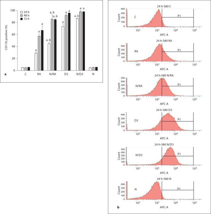
In this study, the ability of nicotinamide to regulate HL-60 cell differentiation in response to different treatments (RA, D3, or nicotinamide or their combinations) was measured using a cell surface marker and a functional differentiation marker. First, a cell surface differentiation marker, CD11b, was used to measure cell differentiation by immunofluorescence using APC-conjugated CD11b antibody. CD11b, a cell surface antigen, functions as a receptor for complement (C3bi), fibrinogen, or clotting factor X. RA or D3 induces CD11b expression in HL-60 cells [23, 24]. To determine the effects of nicotinamide (Nam) on CD11b expression, we compared CD11b expression in HL-60 untreated cells (control), and cells treated with RA, Nam plus RA, D3, Nam plus D3 and Nam alone for 24, 48 and 72 h using flow cytometry. Compared to cells with just RA or D3 treatment, cells treated with Nam plus RA or D3 showed enhanced expression of CD11b (fig. 1a, b). For example, at 24 h, the cells treated with Nam plus RA or Nam plus D3 showed a 1.6- or 2.0-fold increase in CD11b expression compared to the cells treated with RA or D3 alone. Figure 1a shows that nicotinamide increased the rate of induced CD11b expression from 24 to 72 h, indicating an acceleration of the induced differentiation. Figure 1b shows CD11b expression histograms after 24 h RA treatments. In cells treated with Nam alone, the expression of CD11b was the same as in untreated control cells, indicating that nicotinamide alone does not affect the expression of the CD11b cell surface differentiation marker. But when combined with RA or D3, nicotinamide enhanced the rate of induced CD11b expression.
Fig. 1.
Nicotinamide (Nam) enhanced RA- or D3-induced CD11b expression. a CD11b expression was enhanced by RA, D3 and Nam as indicated by flow cytometry. Untreated control (C) cells and cells treated with RA, Nam plus RA (N/RA), D3, Nam plus D3 (N/D3), and Nam alone (N) for the indicated times were stained with APC-conjugated CD11b, and the percent of cells expressing CD11b was analyzed by flow cytometry. b Representative CD11b expression histograms of untreated and treated (RA, N/RA, D3, N/D3, and N) HL-60 cells after 24-hour treatments. a CD11b expression in cells treated with RA, N/RA, D3 and N/D3 was significantly different from that in untreated cells at the corresponding time; b p ≤ 0.05, N/RA or N/D3 treatment was significantly different from treatments with RA or D3 alone.
Nicotinamide Affects RA- or D3-Induced Functional Differentiation
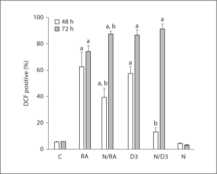
HL-60 cells undergo G0 cell cycle arrest and myeloid differentiation in response to RA or monocytic differentiation in response to D3. To confirm the regulation of cell differentiation by nicotinamide and RA or D3, a functional differentiation marker, inducible oxidative metabolism was used. Inducible oxidative metabolism is a hallmark of terminally differentiated myeloid cells. The chemically reduced and acetylated form of 2′,7′-dichlorohydrofluorescein diacetate (H2DCFDA), known as chlorofluorescein diacetate, is a cell-permeant fluorescent indicator for reactive oxygen species. H2DCFDA is nonfluorescent until the acetate groups are removed by intracellular esterases and oxidation occurs within the cell. The oxidation of the nonfluorescent H2DCFDA to the highly fluorescent DCF was used to detect the generation of reactive oxygen species. Inducible oxidative metabolism can be used as a functional marker of mature myeloid and monocytic cells. HL-60 cells treated with Nam plus RA or D3 showed slower differentiation at early time points, but underwent faster differentiation at later time points compared to treatments with RA or D3 alone (fig. 2). At 48 h, HL-60 cells treated with Nam plus RA or D3 showed reduced RA- or D3-induced differentiation compared to cells treated with RA or D3 alone (62 or 57% in RA or D3, 39 or 13% in Nam plus RA or Nam plus D3). Cells treated with Nam plus D3 resulted in a significant decrease in the percentage of cells capable of inducible oxidative metabolism compared to cells treated with Nam plus RA, suggesting that Nam may function differently in myeloid or monocytic differentiation. In contrast by 72 h of treatment, Nam plus RA or D3 increased the percentage of cells capable of inducible oxidative metabolism compared to cells treated with RA or D3 alone. It was striking that Nam first retarded and then accelerated functional differentiation. Nam thus apparently had differential effects on cells that were earlier or later in the RA or D3-induced progression to terminal myeloid or monocytic differentiation. The cells treated with Nam alone showed no differentiation, which was consistent with results from CD11b expression, suggesting that nicotinamide alone did not affect cell differentiation in HL-60 cells.
Fig. 2.
Nicotinamide (Nam) affects RA- or D3-induced functional differentiation. Cells treated with Nam plus RA (N/RA) or D3 (N/D3) underwent slower functional differentiation at the early time point (48 h), but showed faster differentiation at the later time point (72 h) compared to treatments with RA or D3 alone by using a functional differentiation marker, DCF. The percentage of positive cells for 48 and 72 h after treatment for control (C), RA, N/RA, D3, N/D3, and Nam (N) is shown. Cells were incubated in PBS containing DCF and TPA and analyzed by flow cytometry. Gates to determine percent positive cells were set to exclude 95% of control cells. a DCF expression levels in cells treated with RA, N/RA, D3 and N/D3 were significantly different from that in untreated cells; b p ≤ 0.05, N/RA or N/D3 treatment was significantly different from treatments with RA or D3 alone.
Nicotinamide Accelerated G0 Arrest and Enhanced ERK Activation in HL-60 Cells
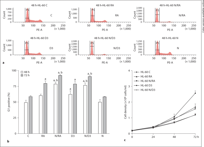
RA- or D3-treated terminally differentiated cells are growth arrested [19, 25]. To determine the effects of nicotinamide on RA- or D3-induced G1/G0 cell cycle arrest, the percentage of untreated control, RA, Nam plus RA, D3, Nam plus D3, and Nam cells, in G1/G0 was measured using flow cytometry (fig. 3a, b). G1/G0 arrest would be revealed by an enrichment of cells with G1 DNA. Previous studies [16, 26] indicated that RA or D3 induced G0 arrest, and our study here shows that Nam enhanced RA- or D3-induced G0 arrest. However, cells treated with Nam alone did not undergo cell cycle arrest. The effects of Nam on growth arrest were consistent with the effects on the differentiation after 72 h treatments, indicating the role of nicotinamide in promoting RA- or D3-induced differentiation and cell cycle arrest. To confirm that the changes in differentiation and promotion of cell cycle arrest caused by nicotinamide reflected decreased cell growth, the growth of RA-, Nam plus RA-, D3-, and Nam plus D3-treated cells was measured. Figure 3c indicated that there was curtailed cell growth associated with G0 enrichment in Nam-treated cells.
Fig. 3.
Nicotinamide (Nam) accelerated G0 arrest. a Representative histograms of percentage of cells with G1/G0 DNA after 48-hour treatments with RA, Nam plus RA (N/RA), D3, Nam plus D3 (N/D3), and Nam (N), and untreated (control, C). b Nicotinamide enhanced G0 arrest after 48- and 72-hour treatment. The percentage of G1 DNA cells at 48 and 72 h after treatment for control (C), RA, N/RA, D3, N/D3, and N is shown. Cells stained with hypotonic staining solution were incubated for 1 h and analyzed by flow cytometry. a Percentage of G1 in cells treated with RA, N/RA, D3 and N/D3 was significantly different from that in untreated cells; b p ≤ 0.05, N/RA or N/D3 treatment was significantly different from treatments with RA or D3 alone. c HL-60 cells upon nicotinamide treatments grew slowly compared to cells treated with RA or D3 alone or untreated cells.
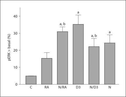
ERK activation is known to propel cell differentiation and cell cycle arrest [19]. In particular ERK drives p21waf1/cip1 expression to propel growth arrest as well as differentiation [27]. To investigate if regulation of RA- or D3-induced myeloid or monocytic differentiation and cell cycle arrest by nicotinamide was related to ERK activation, we compared ERK activation in HL-60 cells treated with RA, Nam plus RA, D3, Nam plus D3, and Nam. Dual phosphorylated ERK [T(203) EY(205)] was measured by flow cytometry. All treated cells show increases in activated ERK per cell above basal levels of control cells in all treated cells, suggesting that RA, D3, and Nam all caused ERK activation (fig. 4). Cells treated with Nam plus RA showed increased activated ERK per cell compared to cells treated with RA or Nam alone, suggesting that Nam and RA function additively for activation of ERK expression. Treating cells with Nam alone increased ERK activation, but did not produce cell differentiation. This suggested that enhanced ERK activation facilitates RA-induced differentiation, but is not the only driver needed to promote cell differentiation. In contrast, ERK activation was not as elevated in cells treated with Nam plus D3 compared to cells treated with D3 alone. These differential effects on ERK activation in cells treated with Nam plus RA and Nam plus D3 coincide with the differential effects of nicotinamide on HL-60 cell myeloid or monocytic differentiation.
Fig. 4.
Nicotinamide (Nam) affected ERK2 activation. Percentage of cells with pERK exceeding basal levels of untreated controls in RA, Nam plus RA (N/RA), D3, Nam plus D3 (N/D3), and Nam (N) treatments after 24 h is shown. HL-60 cells treated with N/RA caused enhanced ERK activation; however, cells treated with N/D3 showed decreased ERK expression after 24 treatments. Cells fixed and permeabilized were stained with Alexa 647-conjugated phospho-p44/42 MARK, and mean fluorescence intensity was determined by quantifying the fluorescence intensity of the entire cell population. a Percentage of pERK in cells treated with RA, N/RA, D3 and N/D3 was significantly different from that in untreated cells; b p ≤ 0.05, N/RA or N/D3 treatment was significantly different from treatments with RA or D3 alone.
Nicotinamide Inhibited CD38, CD14 and PARP Expression
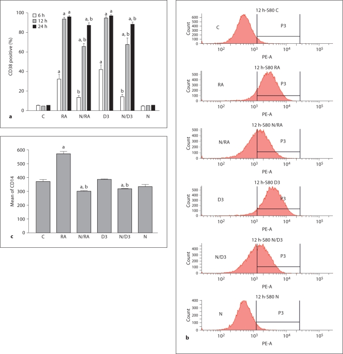
The above differentiation data showed that cells treated with Nam plus RA or D3 underwent slower differentiation at early time points (48 h), but showed faster differentiation at later time points (72 h) compared to treatments of RA or D3 alone. These different effects of nicotinamide in combination with RA or D3 may reflect differential effects of various nicotinamide targets as cells progress toward RA- or D3-induced terminal differentiation. Since nicotinamide enhanced the expression of the late marker CD11b, its effect on earlier putative regulatory progresses became of interest. CD38 is amongst the earliest membrane receptors upregulated by RA and D3, and it signals through MAPK to help propel RA- or D3-induced differentiation and cell cycle arrest in G1/G0, making it of functional significance to induced differentiation. Comparing cells treated with RA or D3 alone and cells treated with Nam plus RA or Nam plus D3, Nam retarded the RA- or D3-induced expression of CD38 (fig. 5a, b). The differences were evident at 6 and 12 h of treatment. After 6 h treatments, CD38 expression levels in the cells treated with Nam plus RA or Nam plus D3 showed a 2.4- or 3.0-fold decrease compared to the cells treated with RA or D3 alone, and at 12 h CD38 expression levels were decreased 1.4- to 1.5-fold in the cells treated with Nam plus RA or D3. But most cells (except control and Nam-treated cells) had attained similar maximal expression levels by 24 h (approximately a 1.1-fold decrease in Nam treatment groups). This suggested that cells treated with Nam plus RA or Nam plus D3 expressed CD38 more slowly. Since CD38 signals through MAPK to promote cell differentiation [17], reduction of CD38 expression by Nam slows early processes providing a rationale for slower functional cell differentiation at the 48-hour time point. In cells treated with Nam alone, there was no CD38 expression, suggesting that Nam alone does not affect CD38 expression.
Fig. 5.
Nicotinamide (Nam) inhibited CD38 and CD14 expressions. a Percent of cells expressing CD38 after RA, Nam plus RA (N/RA), D3, Nam plus D3 (N/D3), and Nam (N) treatments for the indicated times compared to untreated control cells. RA and D3 enhanced CD38 expression; however, Nam inhibited CD38 expression. b Representative CD38 expression histograms of untreated (C) and treated (RA, N/RA, D3, N/D3, N) HL-60 cells. Cells were incubated in PBS containing PE-conjugated CD38, and CD38 expression levels were analyzed by flow cytometry. c Nam inhibited RA-induced CD14 expression (72 h) by flow cytometry. a Expression levels of CD38 and CD14 in cells treated with RA, N/RA, D3, N/D3, and N were significantly different from that in untreated cells; b p ≤ 0.05, N/RA or N/D3 treatment was significantly different from treatments with RA or D3 alone.
To corroborate these CD38 results, the effects of Nam on the CD14 cell differentiation marker were determined. CD14 is a membrane-associated glycosylphosphatidylinositol-linked protein expressed at the surface of cells, and takes its name from its inclusion in the cluster group of cell surface marker proteins. CD14 can bind to lipopolysaccharide. When lipopolysaccharide binds to CD14, the cells become activated and release cytokines such as tumor necrosis factor and upregulate cell surface molecules, including adhesion molecules [28, 29]. In cells treated with Nam plus RA or D3 compared to RA or D3 alone, Nam diminished induced CD14 expression (fig. 5c). The CD14 expression levels in the cells treated with Nam alone were not significantly different compared to control cells, suggesting that Nam alone does not affect CD14 expression. The effects of Nam on CD14 expression were consistent with effects on CD38 expression.
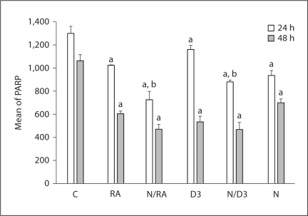
PARP is thought to function in many important cellular processes, such as cell proliferation, gene transcription, cell differentiation, apoptosis, and DNA repair [30, 31]. In particular PARP-1 interacts with RAR and regulates RA signaling [32], suggesting its potential involvement here. To test the effects of nicotinamide on PARP expression in HL-60 cells, we measured the expression of PARP by flow cytometry in cells with RA, D3 and Nam treatments. Figure 6 shows that RA or D3 decreased PARP expression and Nam enhanced the decrease, consistent with the effects it had enhancing certain aspects of induced differentiation and arrest.
Fig. 6.
Nicotinamide (Nam) inhibited PARP expression. HL-60 cells treated with Nam plus RA (N/RA) or D3 (N/D3) caused a decreased PARP expression as indicated by flow cytometry. Control (C), RA, N/RA, D3, N/D3, and Nam (N) treatments after 24 and 48 h are shown. Cells fixed and permeabilized were stained with anti-PARP primary and Alexa 350-conjugated goat anti-mouse secondary antibodies, and mean fluorescence intensity was determined by quantifying the fluorescence intensity of the entire cell population. a Mean of PARP in cells treated with RA, N/RA, D3 and N/D3 was significantly different from that in untreated cells; b p ≤ 0.05, N/RA or N/D3 treatment was significantly different from treatments with RA or D3 alone.
Discussion
The functions of nicotinamide in numerous enzymatic reactions and its relatively low toxicity in vivo have suggested that it might act as a pharmacological agent with potential effects on different pathologies, such as cancer, diabetes and infectious disease [33, 34]. Nicotinamide has been thought to be a potential therapeutic agent in the fields of medicine and nutrition; however, whether nicotinamide works together with other vitamins, such as RA or D3, and its function in cellular processes such as ERK activation involved in cell differentiation remain unclear. We found that nicotinamide modulated the effects of RA or D3 on the expression of cell surface antigens, as well as functional differentiation markers and cell cycle arrest. The differential responses of these to nicotinamide betrayed different sensitivities to nicotinamide for RA-induced myeloid and D3-induced monocytic differentiation dependent on the extent of progression along those lineages. While nicotinamide appears to retard certain early aspects of the induced differentiation, it accelerates later aspects and the occurrence of terminal differentiation and G0 arrest ultimately.
ERK1/2 has a potentially complex role in propelling cellular differentiation. We have previously [16, 17] indicated that RA or D3 cause ERK phosphorylation and activation of MAPK signaling to promote cell differentiation. In this study, we found that nicotinamide modulated RA- or D3-induced ERK1/2 activation and cell differentiation. Nam plus RA increased the amount of activated ERK compared to cells treated with RA or Nam alone, suggesting that Nam and RA function together for the activation of ERK. However, ERK activation was decreased in cells treated with Nam plus D3 compared to cells treated with D3 alone. This suggests that Nam functions differently in terms of ERK activation when combined with RA or D3. The data also suggest that Nam affects other targets as well as ERK to regulate cell differentiation.
Inhibition of PARP has been used to facilitate the death of tumor cells. PARP inhibitors may have potential utility as anticancer agents, radiosensitizers and antiviral agents [35]. Our study indicated nicotinamide enhanced RA- or D3-induced downregulation of PARP expression, indicating again that nicotinamide may work together with RA or D3 to cooperatively downregulate PARP, which is known to regulate RA signaling [32]. Nicotinamide has been used in daily doses of 1–12 g to treat various diseases [20, 21, 34]. Our study provides interesting evidence that nicotinamide could potentially cooperate with other vitamins, such as RA or D3, to regulate cell proliferation and differentiation.
CD38 is a signaling molecule that is amongst the earliest membrane receptors upregulated by RA and D3 [20, 36], and it signals through MAPK to promote RA- or D3-induced differentiation and cell cycle arrest. In addition to receptorial activity, CD38 was also confirmed to be an ectoenzyme involved in transmembrane signaling and cell adhesion [37, 38]. Our study indicated that nicotinamide transiently retarded the initial RA- or D3-induced expression of CD38, suggesting that Nam affected CD38 receptorial function. But we do not know if Nam also affects the enzymatic activity of CD38. Several studies have shown that CD38 expression levels were correlated with other molecules, such as CD31 [39] and CD157 [40, 41]. CD157 shares the same enzymatic properties as CD38, and the two molecules have been reported to be a family of molecules acting as surface receptor of immune cells [42], have 36% amino acid identity and a close chromosomal localization [41, 43]. It thus indicates a possibility of molecular intervention in the events of CD38 expression. In this study, we also compared CD157 expression on HL-60-untreated cells with the cells treated with RA, Nam plus RA, D3, Nam plus D3, and Nam (data not shown). Our data showed that CD157 expression levels were not significantly changed upon RA, D3, and Nam treatments, indicating that there is no Nam-dependent coregulation between CD38 and CD157 for the effects of Nam on the expression of CD38 observed by treating cells with RA, D3, or Nam.
Acknowledgements
We thank Dr. James Smith for assistance in flow cytometry. This work was supported by grants from the NIH (USPHS) and NY STEM award.
References
- 1.Knip M, Douek IF, Moore WPT, Gillmor HA, McLean AE, Bingley PJ, Gale EA, European Nicotinamide Diabetes Intervention Trial Group Safety of high-dose nicotinamide: a review. Diabetologia. 2000;43:1337–1345. doi: 10.1007/s001250051536. [DOI] [PubMed] [Google Scholar]
- 2.Ogata S, Takeuchi M, Okumura K, Taguchi H. Apoptosis induced by niacin-related compounds in HL-60 cells. Biosci Biotechnol Biochem. 1998;62:2351–2356. doi: 10.1271/bbb.62.2351. [DOI] [PubMed] [Google Scholar]
- 3.Daniel J, Marechal Y, van Gool F, Andris F, Leo O. Nicotinamide inhibits B lymphocyte activation by disrupting MAPK signal transduction. Biochem Pharmacol. 2007;73:831–842. doi: 10.1016/j.bcp.2006.11.024. [DOI] [PubMed] [Google Scholar]
- 4.Szabo C. Cell Death: the Role of PARP. vol 17. Boca Raton: CRC Press; 2000. pp. 439–440. [Google Scholar]
- 5.DeMurcia G, Shall S. From DNA Damage and Stress Signaling to Cell Death: Poly ADP-Ribosylation Reactions. Oxford: Oxford University Press; 2001. p. 260. [Google Scholar]
- 6.Shiokawa D, Maruta H, Tanuma S. Inhibitors of poly(ADP-ribose) polymerase suppress nuclear fragmentation and apoptotic-body formation during apoptosis in HL-60 cell. FEBS Lett. 1997;413:99–103. doi: 10.1016/s0014-5793(97)00887-9. [DOI] [PubMed] [Google Scholar]
- 7.Schreiber V, Dantzer F, Ame JC, de Murcia G. Poly(ADP-ribose): novel functions for an old molecular. Nat Rev Mol Cell Biol. 2006;7:517–528. doi: 10.1038/nrm1963. [DOI] [PubMed] [Google Scholar]
- 8.Szabo C. PARP as a drug target for the therapy of diabetic cardiovascular dysfunction. Drug News Perspect. 2002;15:197–205. doi: 10.1358/dnp.2002.15.4.840052. [DOI] [PubMed] [Google Scholar]
- 9.Dulin WE, Wyse M. Reversal of streptozotocin diabetes with nicotinamide. Proc Soc Exp Biol Med. 1969;130:992–994. doi: 10.3181/00379727-130-33707. [DOI] [PubMed] [Google Scholar]
- 10.Su CF, Liu DD, Kao SJ, Chen HI. Nicotinamide abrogates acute lung injury caused by ischemia-reperfusion. Eur Respir J. 2007;30:199–204. doi: 10.1183/09031936.00025107. [DOI] [PubMed] [Google Scholar]
- 11.Iwata K, Ogata S, Okumura K, Taguchi H. Induction of differentiation in human promyelocytic leukemia HL-60 cell line by niacin-related compounds. Biosci Biotechnol Biochem. 2003;67:1132–1135. doi: 10.1271/bbb.67.1132. [DOI] [PubMed] [Google Scholar]
- 12.Munshi CB, Graeff R, Lee HC. Evidence for a causal role of CD38 expression in granulocytic differentiation of human HL-60 cells. J Biol Chem. 2002;277:49453–49458. doi: 10.1074/jbc.M209313200. [DOI] [PubMed] [Google Scholar]
- 13.Merzvinskyte R, Treigyte G, Savickiene J, Maqnusson KE, Navakauskiene R. Effects of histone deacetylase inhibitors, sodium phenyl butyrate and vitamin B3, in combination with retinoic acid on granulocytic leukemia HL-60 cells. Ann NY Acad Sci. 2006;1091:356–367. doi: 10.1196/annals.1378.080. [DOI] [PubMed] [Google Scholar]
- 14.Vaca P, Berna G, Martin F, Soria B. Nicotinamide induces both proliferation and differentiation of embryonic stem cells into insulin-producing cells. Transplant Proc. 2003;35:2021–2023. doi: 10.1016/s0041-1345(03)00735-8. [DOI] [PubMed] [Google Scholar]
- 15.Yen A, Brown D, Fishbaugh J. Control of HL-60 monocytic differentiation. Different pathways and uncoupled expression of differentiation markers. Exp Cell Res. 1987;168:247–254. doi: 10.1016/0014-4827(87)90432-0. [DOI] [PubMed] [Google Scholar]
- 16.Yen A, Roberson MS, Varvayanis S, Lee AT. Retinoic acid induces mitogen-activated protein (MAP)/extracellular signal-regulated kinase (ERK) kinase-dependent MAP kinase activation needed to elicit HL-60 cell differentiation and growth arrest. Cancer Res. 1998;58:3163–3172. [PubMed] [Google Scholar]
- 17.Lamkin TJ, Chin V, Varvayanis S, Smith JL, Sramkoski RM, Jacobberger JW, Yen A. Retinoic acid-induced CD38 expression in HL-60 myeloblastic leukemia cells regulates cell differentiation or viability depending on expression levels. J Cell Biochem. 2006;97:1328–1338. doi: 10.1002/jcb.20745. [DOI] [PubMed] [Google Scholar]
- 18.Zubiaur M, Fernandez O, Ferrero E, Salmeron J, Malissen B, Malavas F, Sancho J. CD38 is associated with lipid rafts and upon receptor stimulation leads to Akt/protein kinase B and Erk activation in the absence of the CD3-zeta immune receptor tyrosine-based activation motifs. J Biol Chem. 2002;277:13–22. doi: 10.1074/jbc.M107474200. [DOI] [PubMed] [Google Scholar]
- 19.Koeffler HP. Induction of differentiation of human acute myelogenous leukemia cells: therapeutic implication. Blood. 1983;62:709–721. [PubMed] [Google Scholar]
- 20.Vague P, Picq R, Bernal M, Lassmann-Vague V, Vialettes B. Effect of nicotinamide treatment on the residual insulin secretion in type 1 (insulin-dependent) diabetic patients. Diabetologia. 1989;32:316–321. doi: 10.1007/BF00265549. [DOI] [PubMed] [Google Scholar]
- 21.Ungerstedt JS, Blomback M, Soderstrom T. Nicotinamide is a potent inhibitor of proinflammatory cytokines. Clin Exp Immunol. 2003;131:48–52. doi: 10.1046/j.1365-2249.2003.02031.x. [DOI] [PMC free article] [PubMed] [Google Scholar]
- 22.Brooks SC, 3rd, Kazmer S, Levin AA, Yen A. Myeloid differentiation and RB phosphorylation changes in HL-60 cells induced by RAR- and RXR-selective retinoic acid analogs. Blood. 1996;87:227–237. [PubMed] [Google Scholar]
- 23.Battle T, Yen A. Ectopic expression of CXCR5/BLR1 accelerates retinoic acid- and vitamin D(3)-induced monocytic differentiation of U937 cells. Exp Biol Med. 2002;227:753–762. doi: 10.1177/153537020222700906. [DOI] [PubMed] [Google Scholar]
- 24.Yen A, Placanica L, Bloom S, Varvayanisi S. Polyomavirus small t antigen prevents retinoic acid-induced retinoblastoma protein hypophosphorylation and redirects retinoic acid-induced G0 arrest and differentiation to apoptosis. J Virol. 2001;75:5302–5314. doi: 10.1128/JVI.75.11.5302-5314.2001. [DOI] [PMC free article] [PubMed] [Google Scholar]
- 25.Daniel CP, Parreira A, Goldman JM, McCarthy DM. The effect of 1,25-dihydroxyvitamin D3 on the relationship between growth and differentiation in HL-60 cells. Leuk Res. 1987;11:191–196. doi: 10.1016/0145-2126(87)90025-7. [DOI] [PubMed] [Google Scholar]
- 26.Wang Q, Salman H, Danilenko M, Studzinski GP. Cooperation between antioxidants and 1,25-dihydroxyvitamin D3 in induction of leukemia HL-60 cell differentiation through the JNK/AP-1/Egr-1 pathway. J Cell Physiol. 2005;204:964–974. doi: 10.1002/jcp.20355. [DOI] [PubMed] [Google Scholar]
- 27.Wang J, Yen A. A MAPK positive feedback mechanism for Blr1 signaling propels retinoic acid triggered differentiation and cell cycle arrest. J Biol Chem. 2008;283:4375–4386. doi: 10.1074/jbc.M708471200. [DOI] [PubMed] [Google Scholar]
- 28.Schumann RR, Leong SR, Flaggs GW, Gray PW, Wright SD, Mathison JC, Tobias PS, Ulevitch RJ. Structure and function of lipopolysaccharide binding protein. Science. 1990;249:1429–1431. doi: 10.1126/science.2402637. [DOI] [PubMed] [Google Scholar]
- 29.Pugin J, Ulevitch RJ, Tobias PS. Activation of endothelial cells by endotoxin: direct versus indirect pathways and the role of CD14. Prog Clin Biol Res. 1994;392:369–373. [PubMed] [Google Scholar]
- 30.Jacobson MK, Jacobson EL. ADP-Ribose Transfer Reactions: Mechanisms and Biological Significance. New York: Springer; 1989. [Google Scholar]
- 31.Poirier G, Moreau P. ADP-Ribosylation Reactions. New York: Springer; 1992. [Google Scholar]
- 32.Pavri R, Lewis B, Kim TK, Dilworth FJ, Erdjument-Bromage H, Tempst P, de Murcia G, Evans R, Chambon P, Reinberq D. PARP-1 determines specificity in a retinoid signaling pathway via direct modulation of mediator. Mol Cell. 2005;18:83–96. doi: 10.1016/j.molcel.2005.02.034. [DOI] [PubMed] [Google Scholar]
- 33.Agote RM, Finochietto P, Gamba CA, Dagrosa MA, Viaqqi ME, Franco MC, Poderoso JJ, Juvenal GJ, Pisarev MA. Nicotinamide increases thyroid radiosensitivity by stimulating nitric oxide synthase expression and the generation of organic peroxides. Horm Metab Res. 2006;38:12–15. doi: 10.1055/s-2006-924966. [DOI] [PubMed] [Google Scholar]
- 34.Cabrera-Rode E, Molina G, Arranz C, Vera M, Gonzalez P, Suarez R, Prieto M, Padron S, Leon R, Tillan J, Garcia I, Tiberti C, Rodriquez OM, Gutierrez A, Fernandez T, Govea A, Hernandez J, Chiong D, Dominquez E, Di Mario U, Diaz-Diaz O, Diaz-Horta O. Effect of standard nicotinamide in the prevention of type 1 diabetes in first degree relatives of persons with type 1 diabetes. Autoimmunity. 2006;39:333–340. doi: 10.1080/08916930600738383. [DOI] [PubMed] [Google Scholar]
- 35.Southan GJ, Szabo C. Poly(ADP-ribose) polymerase inhibitors. Curr Med Chem. 2003;10:321–340. doi: 10.2174/0929867033368376. [DOI] [PubMed] [Google Scholar]
- 36.Deaglio S, Capobianco A, Bergui L, Dürig J, Morabito F, Dührsen U, Malavasi F. CD38 is a signaling molecule in B-cell chronic lymphocytic leukemia cells. Blood. 2003;102:2146–2155. doi: 10.1182/blood-2003-03-0989. [DOI] [PubMed] [Google Scholar]
- 37.Howard M, Grimaldi JC, Bazan JF, Lund FE, Santos-Argumedo L, Parkhouse RM, Walseth TF, Lee HC. Formation and hydrolysis of cyclic ADP-ribose catalyzed by lymphocyte antigen CD38. Science. 1993;262:1056–1059. doi: 10.1126/science.8235624. [DOI] [PubMed] [Google Scholar]
- 38.Zocchi E, Franco L, Cuida L, Benatti U, Bargellesi A, Malavasi F, Lee HC, De Flora A. A single protein immunologically identified as CD38 displays NAD+ glycohydrolase, ADP-ribosyl cyclase and cyclic ADP-ribose hydrolase activities at the outer surface of human erythrocytes. Biochem Biophys Res Commun. 1993;196:1459–1465. doi: 10.1006/bbrc.1993.2416. [DOI] [PubMed] [Google Scholar]
- 39.Gallay N, Anani L, Lopez A, Colombat P, Binet C, Domenech J, Weksler BB, Malavasi F, Herault O. The role of platelet/endothelial cell adhesion molecule 1 (CD31) and CD38 antigens in marrow microenvironmental retention of acute myelogenous leukemia cells. Cancer Res. 2007;67:8624–8632. doi: 10.1158/0008-5472.CAN-07-0402. [DOI] [PubMed] [Google Scholar]
- 40.Ferrero E, Saccucci F, Malavasi F. The human CD38 gene: polymorphism, CpG island, and linkage to the CD157 (BST-1) gene. Immunogenetics. 1999;49:597–604. doi: 10.1007/s002510050654. [DOI] [PubMed] [Google Scholar]
- 41.Deaglio S, Malavasi F. The CD38/CD157 mammalian gene family: an evolutionary paradigm for other leukocyte surface enzymes. Purinergic Signal. 2006;2:431–441. doi: 10.1007/s11302-006-9002-6. [DOI] [PMC free article] [PubMed] [Google Scholar]
- 42.Malavasi F, Deaglio S, Ferrero E, Funaro A, Sancho J, Ausiello CM, Ortolan E, Vaisitti T, Zubiaur M, Fedele G, Aydin S, Tibaldi EV, Durelli I, Lusso R, Cozno F, Horenstein AL. CD38 and CD157 as receptors of the immune system: a bridge between innate and adaptive immunity. Mol Med. 2006;12:334–341. doi: 10.2119/2006-00094.Malavasi. [DOI] [PMC free article] [PubMed] [Google Scholar]
- 43.Dong C, Willerford D, lt FW, Cooper MD. Genomic organization and chromosomal localization of the mouse Bp3 gene, a member of the CD38/ADP-ribosyl cyclase family. Immunogenetics. 1996;45:35–43. doi: 10.1007/s002510050164. [DOI] [PubMed] [Google Scholar]


