Abstract
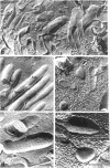
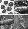
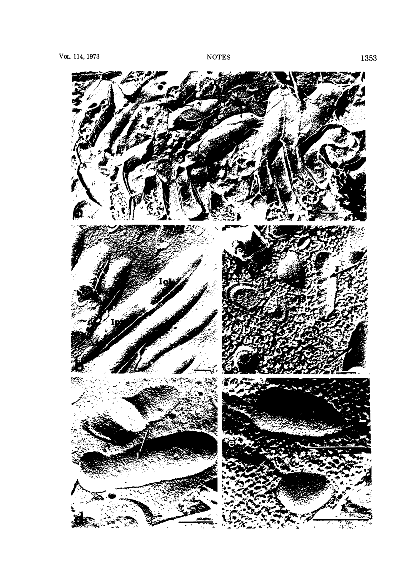
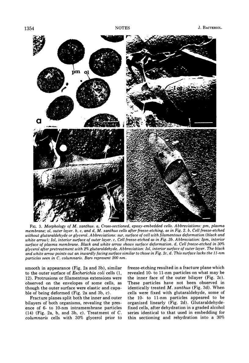
Ultrastructural studies of gliding bacteria demonstrate 10- to 11-nm beads on the inner surface of the outer bilayer of Cytophaga columnaris. These were not found in Myxococcus xanthus. On treatment with glutaraldehyde and ethanol, the beads appear in linear arrays.
Full text
PDF
Images in this article
Selected References
These references are in PubMed. This may not be the complete list of references from this article.
- Bayer M. E., Remsen C. C. Structure of Escherichia coli after freeze-etching. J Bacteriol. 1970 Jan;101(1):304–313. doi: 10.1128/jb.101.1.304-313.1970. [DOI] [PMC free article] [PubMed] [Google Scholar]
- Brown D. T., Waite M. R., Pfefferkorn E. R. Morphology and morphogenesis of Sindbis virus as seen with freeze-etching techniques. J Virol. 1972 Sep;10(3):524–536. doi: 10.1128/jvi.10.3.524-536.1972. [DOI] [PMC free article] [PubMed] [Google Scholar]
- Burchard R. P., Dworkin M. Light-induced lysis and carotenogenesis in Myxococcus xanthus. J Bacteriol. 1966 Feb;91(2):535–545. doi: 10.1128/jb.91.2.535-545.1966. [DOI] [PMC free article] [PubMed] [Google Scholar]
- Burchard R. P. Gliding motility mutants of Myxococcus xanthus. J Bacteriol. 1970 Nov;104(2):940–947. doi: 10.1128/jb.104.2.940-947.1970. [DOI] [PMC free article] [PubMed] [Google Scholar]
- De Petris S. Ultrastructure of the cell wall of Escherichia coli and chemical nature of its constituent layers. J Ultrastruct Res. 1967 Jul;19(1):45–83. doi: 10.1016/s0022-5320(67)80059-5. [DOI] [PubMed] [Google Scholar]
- Doetsch R. N., Hageage G. J. Motility in procaryotic organisms: problems, points of view, and perspectives. Biol Rev Camb Philos Soc. 1968 Aug;43(3):317–362. doi: 10.1111/j.1469-185x.1968.tb00963.x. [DOI] [PubMed] [Google Scholar]
- Holt S. C., Leadbetter E. R. Fine structure of Sporocytophaga myxococcoides. Arch Mikrobiol. 1967 Jun 21;57(3):199–213. doi: 10.1007/BF00405947. [DOI] [PubMed] [Google Scholar]
- Jahn T. L., Bovee E. C. Protoplasmic movements within cells. Physiol Rev. 1969 Oct;49(4):793–862. doi: 10.1152/physrev.1969.49.4.793. [DOI] [PubMed] [Google Scholar]
- Kleinig H. Membranes from Myxococcus fulvus (Myxobacterales) containing carotenoid glucosides. I. Isolation and composition. Biochim Biophys Acta. 1972 Aug 9;274(2):489–498. doi: 10.1016/0005-2736(72)90194-0. [DOI] [PubMed] [Google Scholar]
- LUFT J. H. Improvements in epoxy resin embedding methods. J Biophys Biochem Cytol. 1961 Feb;9:409–414. doi: 10.1083/jcb.9.2.409. [DOI] [PMC free article] [PubMed] [Google Scholar]
- MURRAY R. G., STEED P., ELSON H. E. THE LOCATION OF THE MUCOPEPTIDE IN SECTIONS OF THE CELL WALL OF ESCHERICHIA COLI AND OTHER GRAM-NEGATIVE BACTERIA. Can J Microbiol. 1965 Jun;11:547–560. doi: 10.1139/m65-072. [DOI] [PubMed] [Google Scholar]
- Nanninga N. Ultrastructure of the cell envelope of Escherichia coli B after freeze-etching. J Bacteriol. 1970 Jan;101(1):297–303. doi: 10.1128/jb.101.1.297-303.1970. [DOI] [PMC free article] [PubMed] [Google Scholar]
- Pate J. L., Ordal E. J. The fine structure of Chondrococcus columnaris. 3. The surface layers of Chondrococcus columnaris. J Cell Biol. 1967 Oct;35(1):37–51. doi: 10.1083/jcb.35.1.37. [DOI] [PMC free article] [PubMed] [Google Scholar]
- Pinto da Silva P., Branton D. Membrane splitting in freeze-ethching. Covalently bound ferritin as a membrane marker. J Cell Biol. 1970 Jun;45(3):598–605. doi: 10.1083/jcb.45.3.598. [DOI] [PMC free article] [PubMed] [Google Scholar]
- Reichenbach H., Voelz H., Dworkin M. Structural changes in Stigmatella aurantiaca during myxospore induction. J Bacteriol. 1969 Feb;97(2):905–911. doi: 10.1128/jb.97.2.905-911.1969. [DOI] [PMC free article] [PubMed] [Google Scholar]
- Remsen C. C. The fine structure of frozen-etched Bacillus cereus spores. Arch Mikrobiol. 1966 Sep 8;54(3):266–275. doi: 10.1007/BF00408999. [DOI] [PubMed] [Google Scholar]
- Schmidt-Lorenz W., Kühlwein H. Intracelluläre Bewegungsorganellen der Myxobakterien. Arch Mikrobiol. 1968;60(1):95–98. [PubMed] [Google Scholar]
- Verma J. P., Martin H. H. Uber die Oberflächenstruktur von Myxobakterien. I. Chemie und Morphologie der Zellwände von Cytophaga hutchinsonii und Sporocytophaga myxococcoides. Arch Mikrobiol. 1967;59(4):355–380. [PubMed] [Google Scholar]
- Voelz H. The physical organization of the cytoplasm in Myxococcus zanthus and the fine structure of its components. Arch Mikrobiol. 1967 Jun 6;57(2):181–195. doi: 10.1007/BF00408700. [DOI] [PubMed] [Google Scholar]


