

Abstract
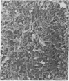
We examined structural changes in bovine kidney tubular basement membrane (TBM) following in vitro nonenzymatic glycosylation (NEG). Isolated TBM was incubated for 2 wk at 37 degrees C in the absence of sugar or in the presence of either glucose or ribitol under conditions that minimized degradation and oxidative damage. NEG and crosslink formation in glycated TBM were confirmed by decreased solubility, increased amounts of low mobility material by SDS-PAGE, and increased specific fluorescence compared to controls. Morphological analysis using high resolution, low voltage scanning electron microscopy (LV-SEM) revealed a complex three-dimensional meshwork of interconnecting strands with intervening openings. Glycated TBM underwent distinct morphological changes, including a 58% increase in the amount of image surface area occupied by openings. This was due to an apparent increase in the number of large openings (diameters > 12.5 nm), whereas the number of small openings (diameters < 12.5 nm) remained unchanged. These findings corroborate earlier physiological studies, which established that the loss of glomerular permselectivity seen in patients with diabetic nephropathy is due to the formation of large pores in the kidney filtration barrier of which the BM is a major component. We conclude that NEG and crosslink formation among BM components lead to modifications of BM ultrastructure, which could play a role in loss of barrier function in diabetic microangiopathy and nephropathy.
Full text
PDF


Images in this article


Selected References
These references are in PubMed. This may not be the complete list of references from this article.
- Baynes J. W. Role of oxidative stress in development of complications in diabetes. Diabetes. 1991 Apr;40(4):405–412. doi: 10.2337/diab.40.4.405. [DOI] [PubMed] [Google Scholar]
- Butkowski R. J., Hagen S. A method for preparation of renal tubular basement membrane. Connect Tissue Res. 1990;25(2):121–130. doi: 10.3109/03008209009006986. [DOI] [PubMed] [Google Scholar]
- Carlson E. C., Brendel K., Hjelle J. T., Meezan E. Ultrastructural and biochemical analyses of isolated basement membranes from kidney glomeruli and tubules and brain and retinal microvessels. J Ultrastruct Res. 1978 Jan;62(1):26–53. doi: 10.1016/s0022-5320(78)80028-8. [DOI] [PubMed] [Google Scholar]
- Carlson E. C., Meezan E., Brendel K., Kenney M. C. Ultrastructural analyses of control and enzyme-treated isolated renal basement membranes. Anat Rec. 1981 Aug;200(4):421–436. doi: 10.1002/ar.1092000405. [DOI] [PubMed] [Google Scholar]
- Charonis A. S., Reger L. A., Dege J. E., Kouzi-Koliakos K., Furcht L. T., Wohlhueter R. M., Tsilibary E. C. Laminin alterations after in vitro nonenzymatic glycosylation. Diabetes. 1990 Jul;39(7):807–814. doi: 10.2337/diab.39.7.807. [DOI] [PubMed] [Google Scholar]
- Charonis A. S., Tsilbary E. C. Structural and functional changes of laminin and type IV collagen after nonenzymatic glycation. Diabetes. 1992 Oct;41 (Suppl 2):49–51. doi: 10.2337/diab.41.2.s49. [DOI] [PubMed] [Google Scholar]
- Cohen M. P., Urdanivia E., Surma M., Wu V. Y. Increased glycosylation of glomerular basement membrane collagen in diabetes. Biochem Biophys Res Commun. 1980 Jul 31;95(2):765–769. doi: 10.1016/0006-291x(80)90852-9. [DOI] [PubMed] [Google Scholar]
- Dyer D. G., Blackledge J. A., Thorpe S. R., Baynes J. W. Formation of pentosidine during nonenzymatic browning of proteins by glucose. Identification of glucose and other carbohydrates as possible precursors of pentosidine in vivo. J Biol Chem. 1991 Jun 25;266(18):11654–11660. [PubMed] [Google Scholar]
- Haitoglou C. S., Tsilibary E. C., Brownlee M., Charonis A. S. Altered cellular interactions between endothelial cells and nonenzymatically glucosylated laminin/type IV collagen. J Biol Chem. 1992 Jun 25;267(18):12404–12407. [PubMed] [Google Scholar]
- Hayase F., Nagaraj R. H., Miyata S., Njoroge F. G., Monnier V. M. Aging of proteins: immunological detection of a glucose-derived pyrrole formed during maillard reaction in vivo. J Biol Chem. 1989 Mar 5;264(7):3758–3764. [PubMed] [Google Scholar]
- Hironaka K., Makino H., Yamasaki Y., Ota Z. Renal basement membranes by ultrahigh resolution scanning electron microscopy. Kidney Int. 1993 Feb;43(2):334–345. doi: 10.1038/ki.1993.51. [DOI] [PubMed] [Google Scholar]
- Inoué S., Leblond C. P., Laurie G. W. Ultrastructure of Reichert's membrane, a multilayered basement membrane in the parietal wall of the rat yolk sac. J Cell Biol. 1983 Nov;97(5 Pt 1):1524–1537. doi: 10.1083/jcb.97.5.1524. [DOI] [PMC free article] [PubMed] [Google Scholar]
- Kent M. J., Light N. D., Bailey A. J. Evidence for glucose-mediated covalent cross-linking of collagen after glycosylation in vitro. Biochem J. 1985 Feb 1;225(3):745–752. doi: 10.1042/bj2250745. [DOI] [PMC free article] [PubMed] [Google Scholar]
- Kubosawa H., Kondo Y. Ultrastructural organization of the glomerular basement membrane as revealed by a deep-etch replica method. Cell Tissue Res. 1985;242(1):33–39. doi: 10.1007/BF00225560. [DOI] [PubMed] [Google Scholar]
- Laemmli U. K. Cleavage of structural proteins during the assembly of the head of bacteriophage T4. Nature. 1970 Aug 15;227(5259):680–685. doi: 10.1038/227680a0. [DOI] [PubMed] [Google Scholar]
- Laurie G. W., Leblond C. P., Inoue S., Martin G. R., Chung A. Fine structure of the glomerular basement membrane and immunolocalization of five basement membrane components to the lamina densa (basal lamina) and its extensions in both glomeruli and tubules of the rat kidney. Am J Anat. 1984 Apr;169(4):463–481. doi: 10.1002/aja.1001690408. [DOI] [PubMed] [Google Scholar]
- Lubec G., Pollak A. Reduced susceptibility of nonenzymatically glucosylated glomerular basement membrane to proteases: is thickening of diabetic glomerular basement membranes due to reduced proteolytic degradation? Ren Physiol. 1980;3(1-6):4–8. doi: 10.1159/000172733. [DOI] [PubMed] [Google Scholar]
- Makino H. Three-dimensional ultrastructure of rat acellular glomerulus by scanning electron microscopy. J Electron Microsc (Tokyo) 1988;37(6):294–304. [PubMed] [Google Scholar]
- Monnier V. M. Toward a Maillard reaction theory of aging. Prog Clin Biol Res. 1989;304:1–22. [PubMed] [Google Scholar]
- Monnier V. M., Vishwanath V., Frank K. E., Elmets C. A., Dauchot P., Kohn R. R. Relation between complications of type I diabetes mellitus and collagen-linked fluorescence. N Engl J Med. 1986 Feb 13;314(7):403–408. doi: 10.1056/NEJM198602133140702. [DOI] [PubMed] [Google Scholar]
- Moran S. M., Myers B. D. Pathophysiology of protracted acute renal failure in man. J Clin Invest. 1985 Oct;76(4):1440–1448. doi: 10.1172/JCI112122. [DOI] [PMC free article] [PubMed] [Google Scholar]
- Myers B. D. Pathophysiology of proteinuria in diabetic glomerular disease. J Hypertens Suppl. 1990 Mar;8(1):S41–S46. doi: 10.1097/00004872-199003001-00009. [DOI] [PubMed] [Google Scholar]
- Myers B. D., Winetz J. A., Chui F., Michaels A. S. Mechanisms of proteinuria in diabetic nephropathy: a study of glomerular barrier function. Kidney Int. 1982 Apr;21(4):633–641. doi: 10.1038/ki.1982.71. [DOI] [PubMed] [Google Scholar]
- Njoroge F. G., Monnier V. M. The chemistry of the Maillard reaction under physiological conditions: a review. Prog Clin Biol Res. 1989;304:85–107. [PubMed] [Google Scholar]
- Osterby R., Parving H. H., Hommel E., Jørgensen H. E., Løkkegaard H. Glomerular structure and function in diabetic nephropathy. Early to advanced stages. Diabetes. 1990 Sep;39(9):1057–1063. doi: 10.2337/diab.39.9.1057. [DOI] [PubMed] [Google Scholar]
- Ota Z., Makino H., Takaya Y., Ofuji T. Molecular sieve in renal glomerular and tubular basement membranes as revealed by electron microscopy. Ren Physiol. 1980;3(1-6):317–323. doi: 10.1159/000172777. [DOI] [PubMed] [Google Scholar]
- Pawley J. B., Erlandsen S. L. The case for low voltage high resolution scanning electron microscopy of biological samples. Scanning Microsc Suppl. 1989;3:163–178. [PubMed] [Google Scholar]
- Sawada H. The fine structure of the bovine Descemet's membrane with special reference to biochemical nature. Cell Tissue Res. 1982;226(2):241–255. doi: 10.1007/BF00218356. [DOI] [PubMed] [Google Scholar]
- Sell D. R., Monnier V. M. Isolation, purification and partial characterization of novel fluorophores from aging human insoluble collagen-rich tissue. Connect Tissue Res. 1989;19(1):77–92. doi: 10.3109/03008208909016816. [DOI] [PubMed] [Google Scholar]
- Shikata K., Makino H., Ichiyasu A., Ota Z. Three-dimensional meshwork structure of glomerular basement membrane revealed by chemical treatment. J Electron Microsc (Tokyo) 1990;39(3):182–185. [PubMed] [Google Scholar]
- Shirato I., Tomino Y., Koide H., Sakai T. Fine structure of the glomerular basement membrane of the rat kidney visualized by high-resolution scanning electron microscopy. Cell Tissue Res. 1991 Oct;266(1):1–10. doi: 10.1007/BF00678705. [DOI] [PubMed] [Google Scholar]
- Steffes M. W., Sutherland D. E., Goetz F. C., Rich S. S., Mauer S. M. Studies of kidney and muscle biopsy specimens from identical twins discordant for type I diabetes mellitus. N Engl J Med. 1985 May 16;312(20):1282–1287. doi: 10.1056/NEJM198505163122003. [DOI] [PubMed] [Google Scholar]
- Tanaka S., Avigad G., Eikenberry E. F., Brodsky B. Isolation and partial characterization of collagen chains dimerized by sugar-derived cross-links. J Biol Chem. 1988 Nov 25;263(33):17650–17657. [PubMed] [Google Scholar]
- Timpl R., Dziadek M. Structure, development, and molecular pathology of basement membranes. Int Rev Exp Pathol. 1986;29:1–112. [PubMed] [Google Scholar]
- Trüeb B., Flückiger R., Winterhalter K. H. Nonenzymatic glycosylation of basement membrane collagen in diabetes mellitus. Coll Relat Res. 1984 Aug;4(4):239–251. doi: 10.1016/s0174-173x(84)80032-1. [DOI] [PubMed] [Google Scholar]
- Tsilibary E. C., Charonis A. S., Reger L. A., Wohlhueter R. M., Furcht L. T. The effect of nonenzymatic glucosylation on the binding of the main noncollagenous NC1 domain to type IV collagen. J Biol Chem. 1988 Mar 25;263(9):4302–4308. [PubMed] [Google Scholar]
- WADDELL W. J. A simple ultraviolet spectrophotometric method for the determination of protein. J Lab Clin Med. 1956 Aug;48(2):311–314. [PubMed] [Google Scholar]
- Yamasaki Y., Makino H., Hironaka K., Hayashi Y., Shikata K., Ota Z. Three-dimensional architecture of rat glomerular basement membrane by ultra-high resolution scanning electron microscopy. Acta Med Okayama. 1990 Dec;44(6):333–335. doi: 10.18926/AMO/30431. [DOI] [PubMed] [Google Scholar]
- Yurchenco P. D., Ruben G. C. Basement membrane structure in situ: evidence for lateral associations in the type IV collagen network. J Cell Biol. 1987 Dec;105(6 Pt 1):2559–2568. doi: 10.1083/jcb.105.6.2559. [DOI] [PMC free article] [PubMed] [Google Scholar]