Abstract
The purpose of this study was to conduct a comprehensive evaluation of the vascular supply to the femoral head, including the vessels that give rise to the terminal perfusing branches. Using a casting agent, we highlighted the anatomy of the external iliac and ischiatic arteries with their associated branches after anatomic dissection of 24 hips from 12 Leghorn chickens. We confirmed published findings regarding perfusion of the femoral head and identified 3 previously undescribed arterial branches to this structure. The first branch (the acetabular branch of the femoralis artery) was supplied by the femoralis artery and directly perfused the acetabulum and femoral head. The second branch (the lateral retinacular artery) was a tributary of the femoralis artery that directly supplied the femoral head. Finally, we found that the middle femoral nutrient artery supplies a previously undescribed ascending intraosseous branch (the ascending branch of the middle femoral nutrient artery) that perfuses the femoral head. Precise understanding of the major vascular branches to the femoral head would allow for complete or selective ligation of its blood supply and enable the creation of a reproducible bipedal model of femoral head osteonecrosis.
Like humans, chickens are bipedal animals that rely on the hip joint to absorb the majority of the body's weight. This anatomy, in concert with their high activity level, makes chickens an attractive model for the study of osteonecrosis of the femoral head in humans. The vast majority of animal research on osteonecrosis of the femoral head has been performed on quadrupedal animals,3,4,10,19,25,26,28,29,31,36,37,41,51,52 thus limiting its application to bipedal species because most quadruped models fail to progress to end-stage mechanical collapse similar to that in humans.6
Avascular necrosis is the death of bone that occurs from ischemia due to disruption of the vascular supply to bone through direct or indirect mechanisms.38 Avascular necrosis should be differentiated from the broader term of osteonecrosis, which refers to bone death in general.32 Causes of femoral head osteonecrosis include direct and indirect disruption of vascular supply (traumatic injury, intravascular coagulation, extrinsic compression) as well as changes in cellular differentiation and cellular apoptosis.4,7,12,15,17,18,24,30-32,38,49,50 Accordingly, causes of osteonecrosis are both traumatic and nontraumatic.16,31,32
The arterial anatomy in the chicken hindlimb has been outlined by several authors.20,22,27,35,42,44,45 Briefly, the external iliac and ischiatic artery arise from the abdominal aorta to provide blood supply to the chicken hind limb. The external iliac artery has 2 main branches—the femoralis and femoral circumflex arteries—that distribute blood to the chicken hindlimb. The ischiatic artery provides 3 main branches: the trochanteric artery, superior femoral nutrient artery, and middle femoral nutrient artery. Although the terminal vascular supply to the femoral head of Leghorn and Broiler chickens has been described,46,47 the origin of these terminal arteries with reference to the ischiatic and femoralis arteries and their respective branches has not been addressed. The current study will describe the blood vessels that feed these terminal branches to the chicken femoral head.
Materials and Methods
The Animal Care and Use Committee at the University of Virginia approved the experimental protocol. Twelve White Leghorns (Gallus gallus domesticus) were used at 18 wk of age with a weight of approximately 2 kg. Heparin (400 U/kg) was injected into the wing vein, and then the chicken was euthanized (1 mL Euthasol IV; Delmarva Laboratories, Midlothian, VA). The thoracic and abdominal aorta was exposed immediately. A polyethylene cannula was placed in the aorta, extending from the thorax to a point just superior to the external iliac artery, and the aorta ligated. Casting compound (Microfil, Flow Tech, Carver, MA) mixed according to the manufacturer's instructions was used to fill the vasculature at a pressure of 150 mm Hg. To this end, red MV130 compound (Flow Tech) was mixed with an equal quantity by weight of MV Diluent (Flow Tech) and catalyzed with 5% by weight of MV Curing Agent (Flow Tech) to produce a viscosity range from 20 to 30 centipoise. The solution had a working time of 20 min that began with the addition of the curing agent. Immediately after injection, the chickens were refrigerated at 4 °C and allowed to cure overnight to form a 3-dimensional cast of the vasculature. The pelvis and hindlimbs were harvested and fixed in formalin for at least 72 h.
The specimens were immersed in decalcifying solution (Cal-EX, Fisher Scientific, Pittsburgh, PA) for 7 d to decalcify the bone. Blunt dissection was performed to expose the branches of the external iliac artery and ischiatic artery of the hip. The femoral shaft was incised along the lateral cortex to expose the nutrient arteries in the marrow cavity. Half of the specimens were cleared by using glycerin, to determine the terminal branches of the ischiatic and external iliac arteries and to further clarify observations noted during blunt dissection. The specimens were placed in a 50% mixture of water and glycerin; the glycerin concentration was raised at 24-h intervals to 75%, 85%, and finally pure glycerin. Branches supplying blood to the femoral head from the external iliac and ischiatic arteries were carefully examined, identified, and photographed.
Results
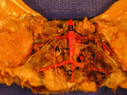
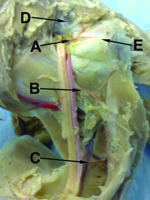
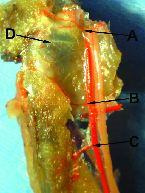
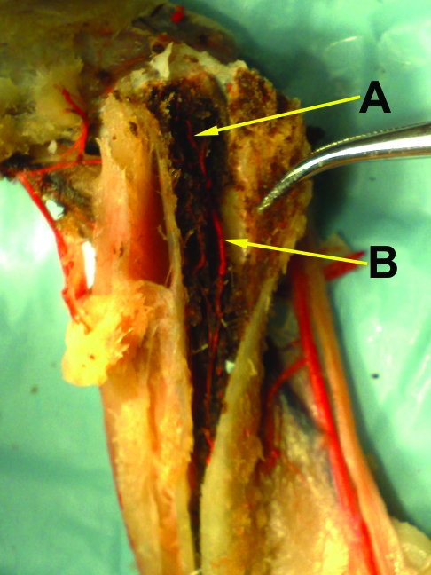
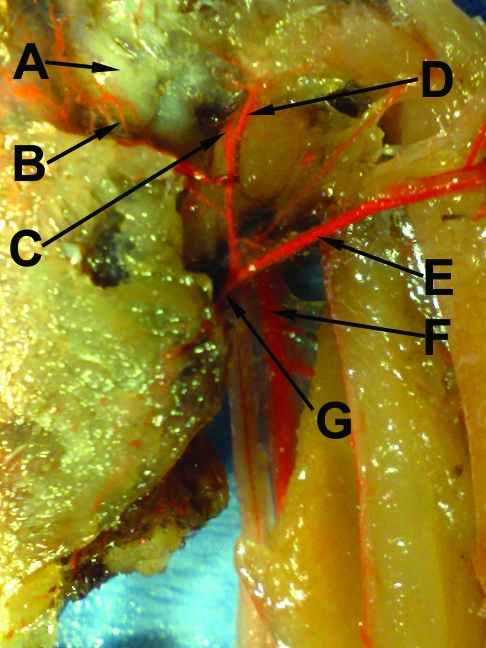
In all 24 hindlimbs, the abdominal aorta gave rise to the external iliac and ischiatic arteries (Figure 1). The ischiatic artery runs distally with the ischiatic nerve posteromedial to the femur and gives off the trochanteric artery, superior femoral nutrient artery, and middle femoral nutrient artery (Figures 2 and 3). The middle femoral nutrient artery penetrates the femur and gives rise to 2 arteries within the bone marrow cavity: the ascending and descending branches (Figure 4). The external iliac provides 2 main branches to the hind limb: the femoralis artery and femoral circumflex artery. The femoral circumflex artery perfuses the musculature of the thigh. We noted 2 previously undescribed branches of the femoralis artery providing blood to the acetabulum (acetabular branch, Figure 5) and femoral head (lateral retinacular artery, Figure 5).
Figure 1.
Abdominal aorta giving rise to the external iliac and ischiatic arteries. A, Abdominal aorta; B, external iliac artery; C, ischiatic artery.
Figure 2.
Branches of the ischiatic artery. A, Trochanteric artery; B, superior femoral nutrient artery; C, middle femoral nutrient artery; D, femoral head; E, greater trochanter.
Figure 3.
Branches of the ischiatic artery after glycerin clearing. A, Trochanteric artery; B, superior femoral nutrient artery; C, middle femoral nutrient artery; D, greater trochanter.
Figure 4.
Branches of middle femoral nutrient artery. (A) Ascending Branch of Middle Femoral Nutrient Artery, (B) Descending Branch of Middle Femoral Nutrient Artery.
Figure 5.
Branches of the external iliac artery after glycerin clearing. A vascular circle around the femoral neck and head is visible. A, Femoral head; B, acetabular branch of the femoralis artery; C, continuation of femoralis artery; D, lateral retinacular artery; E, femoral circumflex artery; F, ischiatic artery; G, external iliac artery.
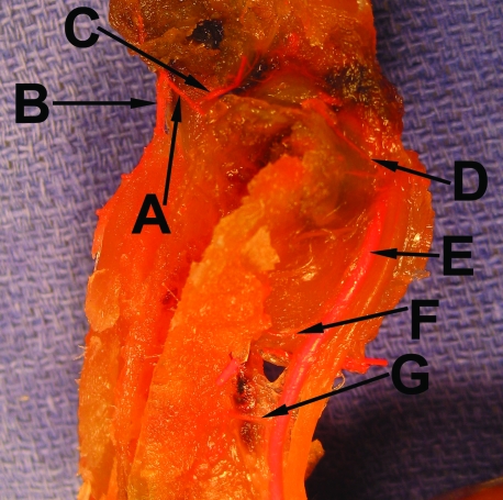
Half of the specimens were cleared by using glycerin to determine the terminal branches of the ischiatic and external iliac arteries. The branches of the ischiatic artery include the trochanteric, superior femoral nutrient, and middle femoral nutrient arteries (Figures 3, 6). The femoralis artery gives rise to the acetabular branch, which perfuses the acetabulum and femoral head (Figure 5). The femoralis artery provides an additional previously undescribed branch to the femoral head, which we named the lateral retinacular artery (Figure 5). The femoralis artery then continues caudally and ventrally, crossing the proximal femur, and terminates in the proximal thigh (Figure 5). We noted that the acetabular branch of the femoralis artery, lateral retinacular artery, and trochanteric artery from the ischiatic artery form a vascular ring around the femoral neck and perfuse the femoral head and neck (Figures 5, 6).
Figure 6.
View of proximal to mid-femur. A vascular ring around the femoral neck and head is visible. A, Femoralis artery; B, femoral circumflex artery; C, acetabular branch of the femoralis artery; D, trochanteric artery; E, ischiatic artery; F, superior femoral nutrient artery; G, middle femoral nutrient artery.
Discussion
As described previously for the terminal vasculature42,44,45,46 and as we confirmed in the present study, the chicken femoral head is perfused by 2 different major arteries, the femoralis and ischiatic arteries. In the present study, we noted that the femoralis artery supplies 2 previously undescribed branches: the acetabular branch of femoralis artery and the lateral retinacular artery. The ischiatic artery gives off 3 branches: the trochanteric artery, superior femoral nutrient artery, and middle femoral nutrient artery. The acetabular branch of femoralis artery, lateral retinacular artery, and trochanteric artery form a vascular ring that perfuses the chicken femoral head and neck. In addition, the middle femoral nutrient artery gives rise to a common femoral medullary cavity branch that subdivides into 2 branches, including the ascending branch of the middle femoral nutrient artery, which provides blood supply directly to the femoral head. Therefore, blood supply to the chicken femoral head is provided by the lateral retinacular artery, acetabular branch of the femoralis artery, trochanteric artery (from the ischiatic artery), and the ascending branch of the middle femoral nutrient artery. The femoral circumflex arteries, which arise from the external iliac artery, do not appear to be an important source of blood to the femoral head in the chicken.
In comparison with the chicken, the human femoral head predominantly is supplied by the superior retinacular vessels of the deep branch of the medial femoral circumflex artery, with an important anastamosis14 occurring between the deep medial femoral circumflex and inferior gluteal arteries.2,11,13,14,34,40 By contrast, the blood supply to the chicken femoral head is from an anastamotic ring, with contributions from 3 arteries (trochanteric, lateral retinacular, and acetabular branch) and additional blood supply provided by the intraosseous ascending branch of the middle femoral nutrient artery. These arteries likely supply the terminal intraosseous branches.44,45 We were unable to confirm the presence of an arterial supply to the chicken femoral head through the ligamentum teres. However, the contribution from blood vessels passing through the ligamentum teres is not reliably present in domestic fowls 15 wk of age and older.45 Similarly, in humans, the blood supply from the foveal artery is rarely significant.2,40
Osteonecrosis of the femoral head often affects young patients with devastating consequences, including complete collapse of the femoral head in more than 80% of patients if left untreated.32,33 Causes of femoral head osteonecrosis include direct and indirect disruption of vascular supply, as well as changes in cellular differentiation and cellular apoptosis.4,7,12,15,17,18,30-32,38,50 Vascular supply is disrupted directly through fractures of the femoral neck and dislocations.8,15,21,39 In addition, blood supply can be inhibited by intravascular coagulation, as with sickle cell disease or hypercoagulable disorders.5,12,18,38 Extrinsic pressure on the delicate intraosseous vascular canals can be caused by adipocyte hypertrophy secondary to glucocorticoids7,31,38 or by lipid storage disease such as Gaucher disease.1 Other etiologies of osteonecrosis include alcohol intake,50 lupus,1 HIV,30 Caisson disease,32 and Legg–Calve–Perthe disease.48 Ultimately, the final pathway is terminal ischemia, with death of both osteocytes and surrounding bone marrow.52 Treatment options are primarily surgical and include core decompression, total hip arthroplasty, osteotomies, and vascularized bone grafts.17,23 Similar in etiology to the human form of the disease, femoral head necrosis is the most common cause of lameness among broiler chickens.9
Numerous animals, including sheep,29 rats,3 goats,43,52 rabbits,25,31,51 dogs,10,26 and pigs,19,37,41 have been proposed to create a viable research model to study human osteonecrosis of the femoral head. Methods to produce osteonecrosis have included but are not limited to corticosteroid administration,4,7,31,36,51 vessel ligation or destruction,3,19,37,41 ethanol injection,29 cryogenic insults,26 hip dislocation,28 and combined methods.6,52 For example, 79% of rabbits treated with corticosteroids developed osteonecrosis of the femoral head with associated intraosseous hypertension and decreased femoral head blood flow.31 Corticosteroid use induces osteonecrosis in chickens49 but not geese.4 Unfortunately, most animal models have shown histologic changes consistent with osteonecrosis without progressing to end-stage collapse.6 An emu model of femoral head osteonecrosis was developed by combining ablation of the femoral head blood supply with direct cryogenic insults.6 More than 80% of the emus progressed to osseous structural failure, with 6 emus developing a ‘crescent sign’ on histology.6 Although promising as a model for human femoral head osteonecrosis, the emu is a large animal with less availability than the chicken, and mechanical collapse in the emu model required a combined insult (freezing and vessel ligation). The effects of cryogenic insults to the surrounding tissue may not mimic the true pathology of human femoral head osteonecrosis.
An ideal animal model for human femoral head osteonecrosis would be an animal that is readily available, bipedal, and active. Because most previous studies have involved quadrupeds or have relied on nonphysiologic means to produce osteonecrosis, the clinical utility of the models for testing treatments for human disease is questionable. Osteonecrosis in an experimental model ideally would be produced by simply disrupting the vascular supply to the femoral head rather than by relying on nonphysiologic methods. The chicken femoral head may be less susceptible to traumatic osteonecrosis because it has multiple sources of blood supply, compared with the human femoral head, which has only one main source of blood supply. However, with our now-complete knowledge of the vascular supply to the chicken femoral head, we propose that surgical disruption of the trochanteric, acetabular branch of femoralis, lateral retinacular, and middle femoral nutrient arteries would be sufficient to reliably produce osteonecrosis with progression to end-stage mechanical collapse by virtue of the chicken's high activity level and bipedalism.
Acknowledgments
The study was supported by institutional training grant T32 AR050960 from NIH-NIAMS and a grant from Smith and Nephew.
References
- 1.Abu-Shakra M, Buskila D, Shoenfeld Y. 2003. Osteonecrosis in patients with SLE. Clin Rev Allergy Immunol 25:13–24 [DOI] [PubMed] [Google Scholar]
- 2.Beaty JH, Kasser JR. Rockwood and Wilkins fractures in children, 6th ed Philadelphia (PA): Lippincott, Williams, and Wilkins; 2006 [Google Scholar]
- 3.Boss JH, Misselevich I, Peskin B, Zinman C, Levin D, Norman D, Reis DN. 2003. Postosteonecrotic osteoarthritis-like disorder of the femoral head of rats. J Comp Pathol 129:235–239 [DOI] [PubMed] [Google Scholar]
- 4.Bouteiller G, Arlet J, Blasco A, Vigonia F, Elefterion A. 1983. Is osteonecrosis of the femoral head avascular? Bone blood flow measurements after long-term treatment with corticosteroids. Metab Bone Dis Relat Res 4:313–318 [DOI] [PubMed] [Google Scholar]
- 5.Buck J, Davies SC. 2005. Surgery in sickle cell disease. Hematol Oncol Clin North Am 19:897–902 [DOI] [PubMed] [Google Scholar]
- 6.Conzemius MG, Brown TD, Zhang Y, Robinson RA. 2002. A new animal model of femoral head osteonecrosis: one that progresses to human-like mechanical failure. J Orthop Res 20:303–309 [DOI] [PubMed] [Google Scholar]
- 7.Cui Q, Wang GJ, Su CC, Balian G. 1997. The Otto Aufranc Award. Lovastatin prevents steroid-induced adipogenesis and osteonecrosis. Clin Orthop Relat Res 344:8–19 [PubMed] [Google Scholar]
- 8.Davidovitch RI, Jordan CJ, Egol KA, Vrahas MS. 2010. Challenges in the treatment of femoral neck fractures in the nonelderly adult. J Trauma 68:236–242 [DOI] [PubMed] [Google Scholar]
- 9.Dinev I. 2009. Clinical and morphological investigations on the prevalence of lameness associated with femoral head necrosis in broilers. Br Poult Sci 50:284–290 [DOI] [PubMed] [Google Scholar]
- 10.Eisenberg MJ. 1992. Avascular necrosis in a canine model. AJR Am J Roentgenol 158:1172–1173 [DOI] [PubMed] [Google Scholar]
- 11.Gautier E, Ganz K, Krügel N, Gill T, Ganz R. 2000. Anatomy of the medial femoral circumflex artery and its surgical implications. J Bone Joint Surg Br 82:679–683 [DOI] [PubMed] [Google Scholar]
- 12.Glueck CJ, Freiberg RA, Wang P. 2003. Role of thrombosis in osteonecrosis. Curr Hematol Rep 2:417–422 [PubMed] [Google Scholar]
- 13.Gray H. 1995. Gray's anatomy, 15th ed Lyndhurst (NJ): Barnes and Noble [Google Scholar]
- 14.Grose AW, Gardner MJ, Sussmann PS, Helfet DL, Lorich DG. 2008. The surgical anatomy of the blood supply to the femoral head: description of the anastomosis between the medial femoral circumflex and inferior gluteal arteries at the hip. J Bone Joint Surg Br 90:1298–1303 [DOI] [PubMed] [Google Scholar]
- 15.Herrera-Soto JA, Price CT. 2009. Traumatic hip dislocations in children and adolescents: pitfalls and complications. J Am Acad Orthop Surg 17:15–21 [DOI] [PubMed] [Google Scholar]
- 16.Hungerford DS. 1981. Pathogenetic considerations in ischemic necrosis of bone. Can J Surg 24:583–587, 590 [PubMed] [Google Scholar]
- 17.Jones LC, Hungerford DS. 2004. Osteonecrosis: etiology, diagnosis, and treatment. Curr Opin Rheumatol 16:443–449 [DOI] [PubMed] [Google Scholar]
- 18.Jones LC, Mont MA, Le TB, Petri M, Hungerford DS, Wang P, Glueck CJ. 2003. Procoagulants and osteonecrosis. J Rheumatol 30:783–791 [PubMed] [Google Scholar]
- 19.Kim HK, Su PH. 2002. Development of flattening and apparent fragmentation following ischemic necrosis of the capital femoral epiphysis in a piglet model. J Bone Joint Surg Am 84-A:1329–1334 [DOI] [PubMed] [Google Scholar]
- 20.Koch T. 1973. Anatomy of the chicken and domestic birds, p 111–113 Ames (IA): Iowa State University Press [Google Scholar]
- 21.Lafforgue P. 2006. Pathophysiology and natural history of avascular necrosis of bone. Joint Bone Spine 73:500–507 [DOI] [PubMed] [Google Scholar]
- 22.Levinsohn EM, Packard DS, Jr, West EM, Hootnick DR. 1984. Arterial anatomy of chicken embryo and hatchling. Am J Anat 169:377–405 [DOI] [PubMed] [Google Scholar]
- 23.Lieberman JR, Berry DJ, Mont MA, Aaron RK, Callaghan JJ, Rajadhyaksha AD, Urbaniak JR. 2003. Osteonecrosis of the hip: management in the 21st century. Instr Course Lect 52:337–355 [PubMed] [Google Scholar]
- 24.Li X, Cui Q, Kao C, Wang GJ, Balian G. 2003. Lovastatin inhibits adipogenic and stimulates osteogenic differentiation by suppressing PPARY2 and increasing CBFA1/RUNX2 expression in bone marrow mesenchymal cell cultures. Bone 33:652–659 [DOI] [PubMed] [Google Scholar]
- 25.Li Y, Han R, Geng C, Wang Y, Wei L. 2009. A new osteonecrosis animal model of the femoral head induced by microwave heating and repaired with tissue-engineered bone. Int Orthop 33:573–580 [DOI] [PMC free article] [PubMed] [Google Scholar]
- 26.Liu GZ, Yang TT, Han JW, Wu J, Wen YH, Gao J, Ma BA. 2009. [An improved liquid nitrogen freezing method for establishing canine models of femoral head necrosis]. Nan Fang Yi Ke Da Xue Xue Bao. 29:1602–1604 [Article in Chinese]. [PubMed] [Google Scholar]
- 27.Lucas AM, Stettenheim PR. 1972. Subgross morphology, p 469–475 In: Avian anatomy integument, part II. Washington (DC): United States Department of Agriculture [Google Scholar]
- 28.Malizos KN, Darryl L, Quarks AV, Seaber WS, Rizk JR. 1993. An experimental canine model of osteonecrosis: characterization of the repair process. J Orthop Res 11:350–357 [DOI] [PubMed] [Google Scholar]
- 29.Manggold J, Sergi C, Becker K, Lukoschek M, Simank HG. 2002. A new animal model of femoral head necrosis induced by intraosseous injection of ethanol. Lab Anim 36:173–180 [DOI] [PubMed] [Google Scholar]
- 30.Matos MA, Alencar RW, Matos SS. 2007. Avascular necrosis of the femoral head in HIV-infected patients. Braz J Infect Dis 11:31–34 [DOI] [PubMed] [Google Scholar]
- 31.Miyanishi K, Yamamoto T, Irisa T, Yamashita A, Jingushi S, Noguchi Y, Iwamoto Y. 2002. Bone marrow fat cell enlargement and a rise in intraosseous pressure in steroid-treated rabbits with osteonecrosis. Bone 30:185–190 [DOI] [PubMed] [Google Scholar]
- 32.Mont MA, Jones LC, Hungerford DS. 2006. Nontraumatic osteonecrosis of the femoral head: 10 years later. J Bone Joint Surg Am 88:1117–1132 [DOI] [PubMed] [Google Scholar]
- 33.Mont MA, Marulanda GA, Jones LC, Saleh KJ, Gordon N, Hungerford DS, Steinberg ME. 2006. Systematic analysis of classification systems for osteonecrosis of the femoral head. J Bone Joint Surg Am 88 Suppl 3:16–26 [DOI] [PubMed] [Google Scholar]
- 34.Netter F. 2003. Atlas of human anatomy, 3rd ed Yardley (PA): Icon Learning Systems [Google Scholar]
- 35.Nishidat T. 1963. [Comparative and topographical anatomy of the fowl. X. The blood vascular system of the hindlimb in the fowl. Part I. The artery]. Nippon Juigaku Zasshi 25:93–106 [Article in Japanese]. [DOI] [PubMed] [Google Scholar]
- 36.Okazaki S, Nishitani Y, Nagoya S, Kaya M, Yamashita T, Matsumoto H. 2009. Femoral head osteonecrosis can be caused by disruption of the systemic immune response via the Toll-like receptor 4 signalling pathway. Rheumatology (Oxford) 48:227–232 [DOI] [PubMed] [Google Scholar]
- 37.Rowe SM, Lee JJ, Chung JY, Moon ES, Song EK, Seo HY. 2006. Deformity of the femoral head following vascular infarct in piglets. Acta Orthop 77:33–38 [DOI] [PubMed] [Google Scholar]
- 38.Rueda JC, Duque MA, Mantilla RD, Iglesias-Gamarra A. 2009. Osteonecrosis and antiphospholipid syndrome. J Clin Rheumatol 15:130–132 [DOI] [PubMed] [Google Scholar]
- 39.Schmidt AH, Asnis SE, Haidukewych G, Koval KJ, Thorngren KG. 2005. Femoral neck fractures. Instr Course Lect 54:417–445 [PubMed] [Google Scholar]
- 40.Sevitt S, Thompson RG. 1965. The distribution and anastomoses of arteries supplying the head and neck of the femur. J Bone Joint Surg Br 47:560–573 [PubMed] [Google Scholar]
- 41.Shapiro F, Connolly S, Zurakowski D, Menezes N, Olear E, Jimenez M, Flynn E, Jaramillo D. 2009. Femoral head deformation and repair following induction of ischemic necrosis: a histologic and magnetic resonance imaging study in the piglet. J Bone Joint Surg Am 91:2903–2914 [DOI] [PMC free article] [PubMed] [Google Scholar]
- 42.Steeves HR 3rd, Siegel PB. 1968. Comparative histology of the vascular system in the domestic and game cock. Experientia 24:937–938 [DOI] [PubMed] [Google Scholar]
- 43.Tang TT, Lu B, Yue B, Xie XH, Xie YZ, Dai KR, Lu JX, Lou JR. 2007. Treatment of osteonecrosis of the femoral head with hBMP2 gene-modified tissue-engineered bone in goats. J Bone Joint Surg Br 89:127–129 [DOI] [PubMed] [Google Scholar]
- 44.Thorp BH. 1986. Vascular pattern of the developing proximal femur in the domestic fowl. Res Vet Sci 40: 231–235 [PubMed] [Google Scholar]
- 45.Thorp BH. 1988. Pattern of vascular canals in the bone extremities of the pelvic appendicular skeleton in broiler type fowl. Res Vet Sci 44:112–124 [PubMed] [Google Scholar]
- 46.Thorp BH, Duff RI. 1988. Effect of unilateral weightbearing on pelvic limb development in broiler fowls: vascular studies. Res Vet Sci 44:164–174 [PubMed] [Google Scholar]
- 47.Thorp BH, Lynch M, Duff RI. 1986. Embedding of skeletal tissue in plastic for vascular and histological study to demonstrate delayed enchondral ossification in leghorn-type fowl. Res Vet Sci 40:236–240 [PubMed] [Google Scholar]
- 48.Wall EJ. 1999. Legg–Calvé–Perthes disease. Curr Opin Pediatr 11:76–79 [DOI] [PubMed] [Google Scholar]
- 49.Wang GJ, Cui Q, Balian G. 2000. The Nicolas Andry award. The pathogenesis and prevention of steroid-induced osteonecrosis. Clin Orthop Relat Res 370:295–310 [DOI] [PubMed] [Google Scholar]
- 50.Wang Y, Li Y, Mao K, Li J, Cui Q, Wang GJ. 2003. Alcohol-induced adipogenesis in bone and marrow: a possible mechanism for osteonecrosis. Clin Orthop Relat Res 410:213–224 [DOI] [PubMed] [Google Scholar]
- 51.Wen Q, Ma L, Chen YP, Yang L, Luo W, Wang XN. 2008. A rabbit model of hormone-induced early avascular necrosis of the femoral head. Biomed Environ Sci 21:398–403 [DOI] [PMC free article] [PubMed] [Google Scholar]
- 52.Yang S, Wu X, Mei R, Yang C, Li J, Xu W, Ye S. 2008. Biomaterial-loaded allograft-threaded cage for the treatment of femoral head osteonecrosis in a goat model. Biotechnol Bioeng 100:560–566 [DOI] [PubMed] [Google Scholar]


