

Abstract
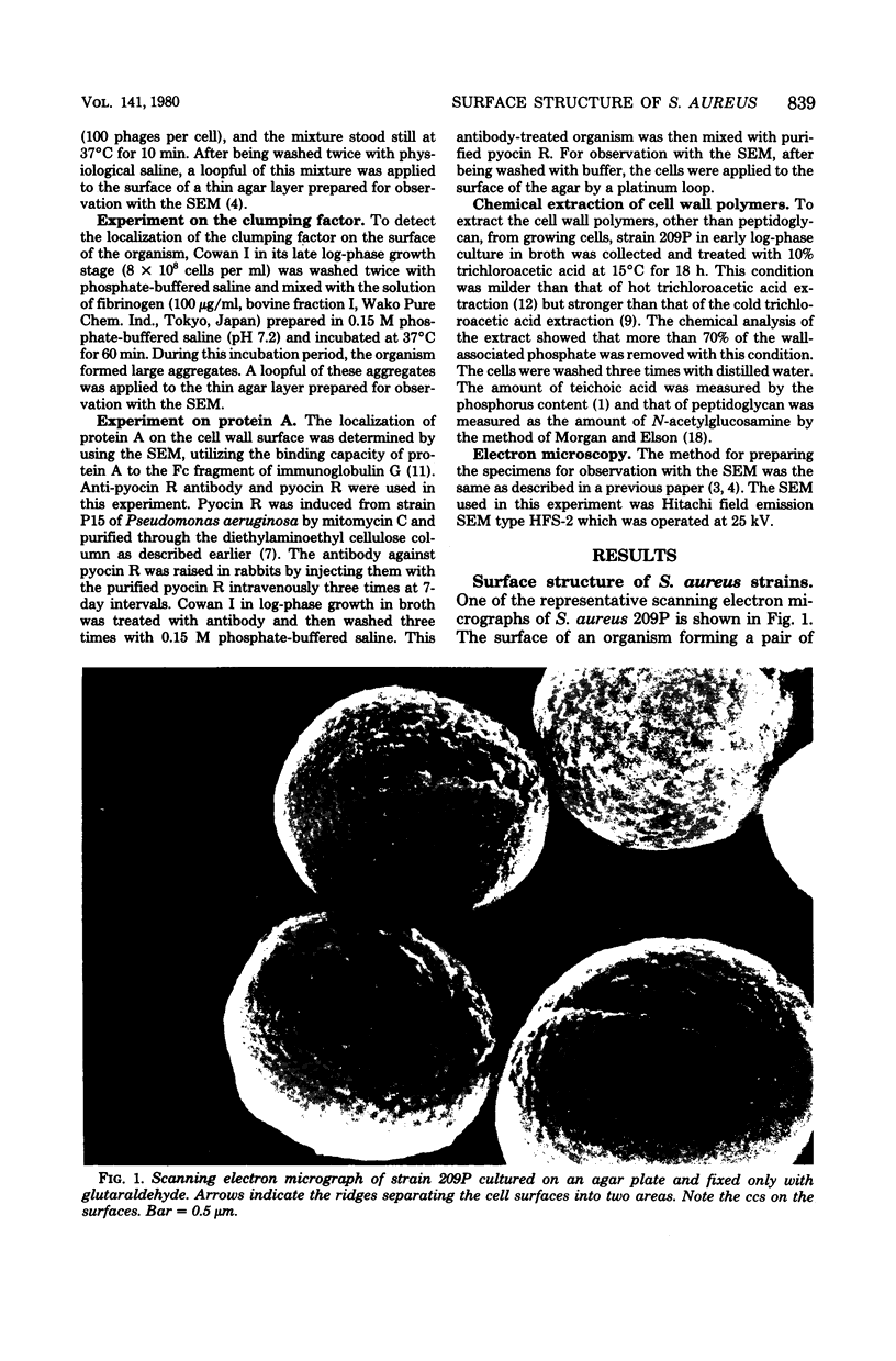
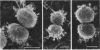
The surface of several laboratory strains of Staphylococcus aureus were observed with a scanning electron microscope, and the presence of two morphologically characteristic structures--a ridge separating cell surface into old and new surfaces and a concentric circular structure--are described. These two structures seemed to be present universally on the surfaces of cells of the genus Staphylococcus. The removal of the circular structures by a mild treatment of the cell with trichloroacetic acid suggested that this structure seemed to represent circularly arranged teichoic acid. With experiments using morphologically recognizable markers among three of the cell wall components, clumping factor, phage receptor, and protein A, the clumping factor was proven to be specifically localized on the old surface; and more phage receptors were detected on the old surface than on the new surface, but protein A was present all over the cell surface. This indicated that the clumping factor and most of the phage receptors appeared on the cell wall surface in a late stage of the cell growth cycle, but protein A was present in an early stage of the growth. The idea of aging of the cell wall is discussed.
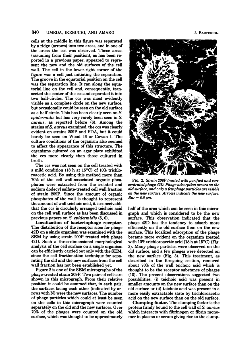
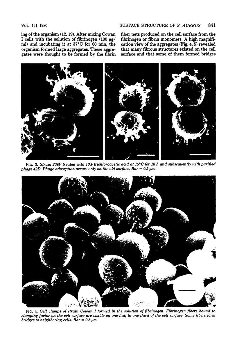
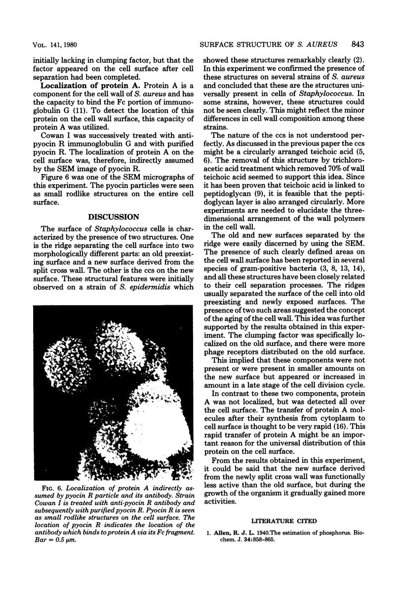
Full text
PDF


Images in this article


Selected References
These references are in PubMed. This may not be the complete list of references from this article.
- Allen R. J. The estimation of phosphorus. Biochem J. 1940 Jun;34(6):858–865. doi: 10.1042/bj0340858. [DOI] [PMC free article] [PubMed] [Google Scholar]
- Amako K., Umeda A. An improved method for observation of bacterial growth using the scanning electron microscope. J Electron Microsc (Tokyo) 1977;26(2):155–159. [PubMed] [Google Scholar]
- Amako K., Umeda A. Bacterial surfaces as revealed by the high resolution scanning electron microscope. J Gen Microbiol. 1977 Jan;98(1):297–299. doi: 10.1099/00221287-98-1-297. [DOI] [PubMed] [Google Scholar]
- Amako K., Umeda A. Concentric circular structure of the cell wall of Staphylococcus epidermidis revealed by the scanning electron microscope. J Electron Microsc (Tokyo) 1978;27(2):147–148. [PubMed] [Google Scholar]
- Amako K., Umeda A. Regular arrangement of wall polymers in staphylococci. J Gen Microbiol. 1979 Aug;113(2):421–424. doi: 10.1099/00221287-113-2-421. [DOI] [PubMed] [Google Scholar]
- Amako K., Umeda A. Scanning electron microscopy of Staphylococcus. J Ultrastruct Res. 1977 Jan;58(1):34–40. doi: 10.1016/s0022-5320(77)80005-1. [DOI] [PubMed] [Google Scholar]
- Amako K., Yasunaka K. Effects of sodium dodecyl sulphate on the structure of purified pyocin sheaths. J Gen Microbiol. 1974 Feb;80(2):443–450. doi: 10.1099/00221287-80-2-443. [DOI] [PubMed] [Google Scholar]
- Burdett I. D., Higgins M. L. Study of pole assembly in Bacillus subtilis by computer reconstruction of septal growth zones seen in central, longitudinal thin sections of cells. J Bacteriol. 1978 Feb;133(2):959–971. doi: 10.1128/jb.133.2.959-971.1978. [DOI] [PMC free article] [PubMed] [Google Scholar]
- Coley J., Tarelli E., Archibald A. R., Baddiley J. The linkage between teichoic acid and peptidoglycan in bacterial cell walls. FEBS Lett. 1978 Apr 1;88(1):1–9. doi: 10.1016/0014-5793(78)80594-8. [DOI] [PubMed] [Google Scholar]
- Coyette J., Ghuysen J. M. Structure of the cell wall of Staphylococcus aureus, strain Copenhagen. IX. Teichoic acid and phage adsorption. Biochemistry. 1968 Jun;7(6):2385–2389. doi: 10.1021/bi00846a048. [DOI] [PubMed] [Google Scholar]
- Forsgren A., Sjöquist J. "Protein A" from Staphylococcus aureus. 3. Reaction with rabbit gamma-globulin. J Immunol. 1967 Jul;99(1):19–24. [PubMed] [Google Scholar]
- Hawiger J., Hammond D. K., Timmons S. Human fibrinogen possesses binding site for staphyococci on Aalpha and Bbeta polypeptide chains. Nature. 1975 Dec 18;258(5536):643–645. doi: 10.1038/258643a0. [DOI] [PubMed] [Google Scholar]
- Higgins M. L., Shockman G. D. Study of cycle of cell wall assembly in Streptococcus faecalis by three-dimensional reconstructions of thin sections of cells. J Bacteriol. 1976 Sep;127(3):1346–1358. doi: 10.1128/jb.127.3.1346-1358.1976. [DOI] [PMC free article] [PubMed] [Google Scholar]
- Kolenbrander P. E., Hohman R. J. Electron microscopic study of cell surface rings during cell division and morphogenesis of Arthrobacter crystallopoietes. J Bacteriol. 1977 Jun;130(3):1345–1356. doi: 10.1128/jb.130.3.1345-1356.1977. [DOI] [PMC free article] [PubMed] [Google Scholar]
- Koyama T., Yamada M., Matsuhashi M. Formation of regular packets of Staphylococcus aureus cells. J Bacteriol. 1977 Mar;129(3):1518–1523. doi: 10.1128/jb.129.3.1518-1523.1977. [DOI] [PMC free article] [PubMed] [Google Scholar]
- Movitz J. A study on the biosynthesis of protein A in Staphylococcus aureus. Eur J Biochem. 1974 Oct 1;48(1):131–136. doi: 10.1111/j.1432-1033.1974.tb03750.x. [DOI] [PubMed] [Google Scholar]
- Osborn M. J., Rick P. D., Lehmann V., Rupprecht E., Singh M. Structure and biogenesis of the cell envelope of gram-negative bacteria. Ann N Y Acad Sci. 1974 May 10;235(0):52–65. doi: 10.1111/j.1749-6632.1974.tb43256.x. [DOI] [PubMed] [Google Scholar]
- REISSIG J. L., STORMINGER J. L., LELOIR L. F. A modified colorimetric method for the estimation of N-acetylamino sugars. J Biol Chem. 1955 Dec;217(2):959–966. [PubMed] [Google Scholar]