Abstract
Whereas genetic factors contribute crucially to brain function, early-life events, including stress, exert long-lasting influence on neuronal function. Here, we focus on the hippocampus as the target of these early-life events because of its crucial role in learning and memory. Using a novel immature-rodent model, we describe the deleterious consequences of chronic early-life ‘psychological’ stress on hippocampus-dependent cognitive tasks. We review the cellular mechanisms involved and discuss the roles of stress-mediating molecules, including corticotropin releasing hormone, in the process by which stress impacts the structure and function of hippocampal neurons.
Keywords: Neonatal, Rat, Human, Stress, Memory, Hippocampus, Corticotropin releasing hormone, CRF, Hypothalamic pituitary adrenal axis, Neuroplasticity
1. Early-life events interact with genetic factors to influence hippocampal function long-term
Brain function and dysfunction throughout life are determined by the interaction of genetic factors with ‘acquired’ environmental events, signals and stimuli [100]. Events that occur early in life are capable of exerting effects that persist throughout adulthood. Here, we focus on the hippocampus as the target of these early-life events because of its crucial role in learning, memory storage and retrieval, and general cognitive function [105,41,65]. Indeed, early-life events, via complex interactions with genetic factors [106,120], have been suspected to be a major determinant of smaller hippocampal volume and long-term cognitive dysfunction in pre-term infants [111,17] and may play a role in certain affective and dementing disorders in the adult and aging human [100,26,120]. This review focuses on the mechanisms by which certain early-life events influence populations of hippocampal neurons acutely, and impact the function and integrity of the hippocampus long-term.
2. Studying early-life stress may be used to probe the molecular mechanisms involved in experience-evoked hippocampal neuroplasticity
Stress may provide a salient example of early-life experience that might exert long-lasting influence on the brain, because (1) over 50% of the world’s children are exposed to stress [139] and (2) evidence from both human and animal studies suggests that early-life stress has profound effects on cognitive function and emotional health.
Stress has been shown to influence the hippocampus in a number of important ways. Whereas acute mild stress rapidly enhances synaptic efficacy and learning and memory processes [131,45,91,123], chronic or severe activation of the stress response early in life has been shown to be potentially injurious in both humans [6,120,147] and experimental animals [120,31]. For example, severe childhood psychological stress (neglect and abuse) correlates with a higher incidence of learning disabilities later in life, including those learning and memory functions requiring an intact hippocampus [121,50]. Further, MRI studies have suggested that adults subjected to abuse (a measure of chronic stress) early in life have lower average hippocampal volumes [24,134 but see 43]. Functional MRI data [40] suggest that infants and children subjected to severe chronic emotional stress (such as deprivation) have abnormal neuronal network function compared with age-matched controls. Primate and rat studies are in general agreement with these human data. For example, rearing monkeys without maternal or sibling contact led to learning and memory deficits later in life [121]. Taken together, these observations support the notion that early-life stress is a powerful modulator of hippocampal function, and may thus be a useful tool for studying the principles of experience-evoked hippocampal neuroplasticity.
3. Stress: definitions
Stress has been described as a potential threat, arising from outside or from within the organism [128,120], and has also been defined operationally as a physiological or psychological threat that activates the ‘stress-response’ machinery [108]. Stress triggers molecular cascades that allow rapid behavioral, autonomic and cognitive CNS responses to stressful circumstances, followed by prompt re-establishment of functional steady-state. This involves not only rapid secretion of effector molecules such as nor-epinephrine and glucocorticoids), but also a more protracted, coordinated change in programmed gene expression [100,7].
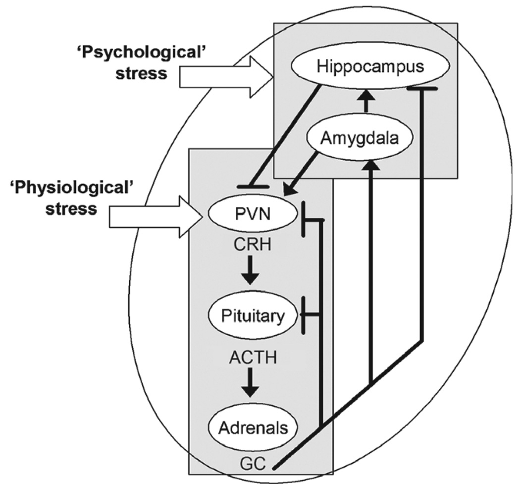
Physiological stressors (also referred to as ‘reactive,’ ‘actual,’ and ‘physical’) activate the neuroendocrine, hypothalamic–pituitary–adrenal axis [42,61] (Fig. 1). Neurochemical signals conveying a potential threat reach the hypothalamus, causing secretion of the stress-activated neuropeptide, corticotropin releasing hormone (CRH), from neurons in the paraventricular nucleus, to elicit rapid secretion of adrenocorticotropin (ACTH) from the anterior pituitary gland. ACTH induces release of glucocorticoids from the adrenal glands. Glucocorticoids bind with specific receptors (glucocorticoid receptors or GR) in hippocampus and elsewhere in the CNS to shut off the hormonal stress response and re-establish steady state [138,62]. Work by several groups has shown that stress-induced secretion of ACTH and glucocorticoids requires CRH [141,154,14]. Notably, this chain of events is already functional during the neonatal and infancy periods in the rodent [145,155,154,142,39,127] and in both pre-term and full-term human neonates [54,17].
Fig. 1.
A simplified diagram of the circuits involved in ‘physiological’ and ‘psychological’ stress responses. Physiological stress signals activate the neuroendocrine hypothalamic–pituitary–adrenal axis (outlined by the rectangular box). Physiological stress signals converge on the hypothalamic paraventricular nucleus (PVN) causing the release of corticotropin-releasing hormone (CRH) into the hypothalamo–pituitary portal system. CRH elicits secretion of adrenocorticotropic hormone (ACTH) from the pituitary and ACTH induces the release of glucocorticoids (GC) from the adrenal gland. GCs cross the blood–brain barrier and interact with type-2 corticoid receptors (GR) in the hippocampus, PVN and pituitary to ‘shut-off’ the neuroendocrine response to stress (indicated by the blunt-ended lines). In contrast, activation of GR in the central nucleus of the amygdala increases CRH expression in this region [30,113] and is generally considered to facilitate stress responses. Psychological stress engages additional brain regions and circuits, involving, at a minimum, the hippocampal formation. Arrows denote facilitatory projections.
Psychological stressors (also termed ‘anticipated’ and ‘emotional’) [42,61] activate higher-order limbic pathways that contribute to the ‘central’ stress circuit [72, reviewed in 60,12,7] (Fig. 2). This circuit includes the amygdala [67,86,112,56,117] and the hippocampus [100,55,78,61]. During acute stress, ‘limbic’ or ‘psychological’ stress signals converge on the amygdala central nucleus, inducing immediate early gene expression and influencing, for example, facilitation of memory consolidation [102,117]. From amygdala outflow nuclei [112], selected stress signals project to the hippocampus (via mono- and poly-synaptic projections), evoking IEG expression [55] and enhancing synaptic efficacy [146,20].
Fig. 2.
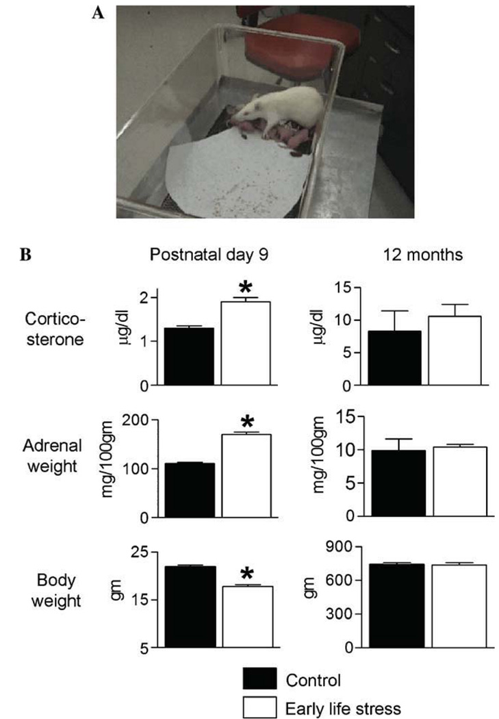
The design and consequences of a chronic, ‘psychological’ early-life stress paradigm. (A) Photograph of a cage at the moment of onset of the stress paradigm (postnatal day 2). A single paper towel is provided to the dam for creating a ‘nest’. The dam will rapidly shred it for this purpose. (B) Parameters indicative of stress immediately after the early-life stress period (postnatal day 9; left column) and in adult male rats (12 months of age; right column). Elevated basal corticosterone levels, higher adrenal gland weights, and modestly lower body weight are found in chronically stressed 9-day old rats. These changes are no longer apparent in adult rats. Black bar, control group; white bar, early-life stress group. *P< 0.05. (Modified from 31 with permission.)
4. Acute versus chronic stress differ in their effects on hippocampal function
In addition to categorizing stress as physiological or psychological, the duration of the activation of stress responses is important for the consequences of a stressor. Thus, acute stress followed by a rapid ‘shut-off’ of the stress response may influence the hippocampus quite differently than exposure to chronic stress, as discussed below [21,69,101].
5. Recognized effects of stress on hippocampal integrity and function: adult studies
The role of stress in influencing the structure and function of hippocampal neurons has been the focus of a significant body of research [100,44,28,51,7,88,78,20,70, 150,31]. Acute stress can promote hippocampus-mediated cognitive function and synaptic transmission [100,149,78, 71,21,68,81 but see 151]. The short-term effects of this facilitation may be mediated by glucocorticoids [44,81]. Additionally, local release of CRH from hippocampal neurons during acute stress [37] may prime long-term potentiation [20]. The effects of acute stress on hippocampal neuronal function differ strikingly from those discovered after chronic or repeated stress.
Chronic stress may contribute to deficits in hippocampus-dependent learning and memory [92,135] including those found during senescence [76,100,153]. In addition, chronic stress in adults negatively affects long-term potentiation (LTP) in hippocampal subfields [109,3], including the commissural-associational projections in field CA3 [109]. The structural foundation of these functional deficits may derive from the actions of chronic stress in adults on dendritic trees. For example, atrophy or remodeling of apical dendrites in CA3 has been demonstrated [94,144] and confirmed in other species, such as tree shrews [79] and non-human primates [126].
The mechanisms for stress-induced structural remodeling, and functional deficits in CA3 are not fully understood. However, the glucocorticoid stress hormones as well as glutamate-mediated excitotoxicity have been shown to contribute to these effects [71,95]. More recently, other molecules and signaling cascades have been found to contribute to the dendritic remodeling evoked by stress, including tissue plasminogen activator [110].
Whereas the effects of stress on hippocampal structure and function in adults may be profound, they are generally reversible [92,57,114]. The reasons are unclear, but may stem from the fact that the adult brain has been fully ‘programmed,’ as far as gene expression levels or ‘set-points’ are concerned. Therefore, stress may perturb gene expression transiently, perhaps via epigenetic effects [97,143], but the withdrawal of the inciting stimulus leads to re-instatement of the original, fully programmed ‘steady state.’ In addition, whereas neuronal loss has been suspected to occur upon chronic stress in the adult [125,90], most current evidence supports the notion that the structural effects of chronic stress on hippocampal neurons involve dendritic modifications, without cell loss or profound dendritotoxicity. Thus, upon withdrawal of the molecular processes initiated by stress, recovery of both function and structure occurs in mature hippocampus [109,56,114]. Taken together, these findings support the notion that, in the fully-mature hippocampal formation, both the salubrious and the deleterious effects of stress may be transient.
6. Enduring effects of early-life stress on developing hippocampus
In the rodent, maturation and full differentiation of the hippocampal formation take place during early postnatal life [for review see 7,77]. For example, during the first postnatal weeks, neuronal birth, differentiation and migration are ongoing [4,53,16]. Neurogenesis of granule cells peaks during the second week of life in rodents [15] and during the third month in humans [129]. In addition, synaptogenesis and the establishment of enduring connectivity patterns continue for years in the human, and for weeks in the rodent [7]. Therefore, it is theoretically possible that processes triggered by a potent stimulus such as stress may interrupt or corrupt the functional and structural maturation of the hippocampal network in an irreversible manner [147,82,31]. This review describes evidence for this process, and discusses established and emerging underlying mechanisms.
The importance of stress experienced early in life is a result of its significant, long-lasting effects on hippocampal structure and function. The effects of psychological stress early in life may be considered a ‘double-edged sword,’ in that the nature and magnitude of the stressor determines whether its long-term consequences will enhance or impair hippocampal function [27]. Mild stress early in life results in beneficial long-term consequences: Hippocampus-mediated memory function is facilitated in adult rodents and non-human primates reared with enhanced maternal care [89,121,49] or in enriched environments [75,149].
In contrast, chronic or severe stress early in life can have long-term adverse effects on hippocampal integrity and performance in humans (reviewed in [120,147]) and experimental animals [120,31]. These effects may manifest in impaired, or progressively deteriorating cognitive performance [98,18]. In addition, they may be evident already during early adult life [66], or emerge later in life [6,74,83,31]. These enduring and potentially progressive effects of chronic early-life stress on hippocampal function may thus contribute substantially to the burden of human cognitive dysfunction. Therefore, it is important to uncover the mechanisms involved, as a first step for potential intervention. For obvious reasons, these mechanistic studies cannot be undertaken in humans and require suitable animal models.
7. Animal models for chronic, psychological stress early in life
7.1. The timing of suitable chronic stress models
The critical period of vulnerability to early-life experience, including stress, in humans is difficult to determine, and likely includes both late gestation and early infancy [124,7]. In the rat, the critical developmental period for stress-related hippocampal plasticity is better understood. To modulate hippocampal learning and memory functions and gene expression permanently, stimuli that modulate the hypothalamic–pituitary–adrenal axis must commence early during the 1st week of life. This has been established, for example, for the handling paradigm that leads to permanent alteration of hippocampal GR expression if conducted during postnatal day (P)2–P21, or even P1–P5 [63], but not starting on P8 [103]. Avishai-Eliner et al. [8] studied the time-course of the molecular consequences of early-life handling. They found that persistently altered gene expression in hippocampus, as well as enduring alterations of the hormonal stress responses occurred if the procedure started on P2 and ended by P9. Indeed, altered gene expression was found in the hypothalamus already by P9. However, hippocampal gene expression changes emerged between P23 and P45. Thus, available data support the notions that (1) the critical period for stress-evoked hippocampal plasticity encompasses roughly the first 10 postnatal days. (2) Importantly, full manifestation of stress-induced hippocampal changes occurs later, and may be reversible during the 2nd and 3rd postnatal week [49]. Thus, recent studies demonstrate that the long-term deleterious hippocampal effects of sub-optimal neonatal experience can be prevented by later intervention, including environmental enrichment [23] or pharmacological treatment [49].
7.2. Hippocampal developmental stages in rodents and humans
Direct comparison of ‘hippocampal age’ between human and rat is difficult. Whereas a composite of neuro-anatomical data suggest that the hippocampus of a P5–P7 rat might be generally comparable to that of a fullterm human neonate [7], patterns of expression of several genes involved in the stress signal cascade suggest that perhaps for some stress-related functions, rat hippocampus might be more mature than is suggested by anatomical evidence. For example, CRF1 receptor expression in hippocampus peaks on P6 [12] and glucocorticoid receptors are robustly expressed already prenatally [118,154]. In addition, the diverse mechanisms of stress-evoked hippocampal plasticity probably involve multiple afferent, efferent and intrinsic systems that mature differentially in rat and human, further complicating direct comparisons. In summary, chronic stress during a critical developmental period (around P2– P9 in rat) may set in motion long-lasting hippocampal changes. These may not manifest until later, and seem to be preventable by interventions during the 2nd–3rd postnatal weeks [23,49].
7.3. Animal models of ‘psychological’ stress early in life that approximate the human condition
Several useful and informative models of early-life stress have been previously developed. Many of these models rely on the critical role of maternal influence on the activation of the molecular stress-mediators in the immature rat. For example, short daily periods (15 min) of separation from the mother have been used to model mild stress and may have positive effects on hippocampal function. These positive effects are likely mediated by the nurturing input from the mother upon return of the pups to the cage [89,8,48,49]. In contrast, repeated prolonged (3 h) separation from the mother [113] or a single 24 h separation [46] have been considered models of more severe acute/subacute stress.
However, absence of the mother influences critical physiological parameters such as nutrition (and thus cannot be used chronically), and eliminates maternal contact and grooming [46]. Importantly, maternal separation fails to reproduce the chronic early-life psychological stress that is found in humans and typically results from abnormal interactions with a present mother. This led to the creation of the chronic, early-life stress model described here [52,9]. In this paradigm, pups are reared in cages with limited bedding material for one week, encompassing the critical developmental period (P2–P9) when stress influences hippocampal developmental programs [8,7]. The reduced nesting material (one paper towel, Fig. 2A) limits the dam’s ability to create a satisfactory nest. As a result, maternal care becomes erratic and unpredictable, which contributes to emergence of physiological and molecular states of chronic stress in the pups [52,9]. At the end of this week of chronic stress, on P9, pups have enlarged adrenal glands, increased basal corticosterone levels, and a modest loss of body weight [9,31] (Fig. 2B). After this week-long stress, pups and dams are returned to ‘normal’ cages, weaned on P21, and allowed to mature. By the time they reach adulthood, all of the physiological parameters indicative of stress dissipate, such that adrenal weight and basal plasma corticosterone are similar to those of ‘normally’ reared controls (Fig. 2B). In summary, this model of limited nesting material creates a condition of chronic, but transient, early-life stress, which disappears, and thus permits investigating the potential consequences of neonatal stress on adult hippocampal structure and function.
8. Consequences of chronic early-life emotional stress on hippocampal function and structure
8.1. Impaired hippocampus-dependent learning and memory
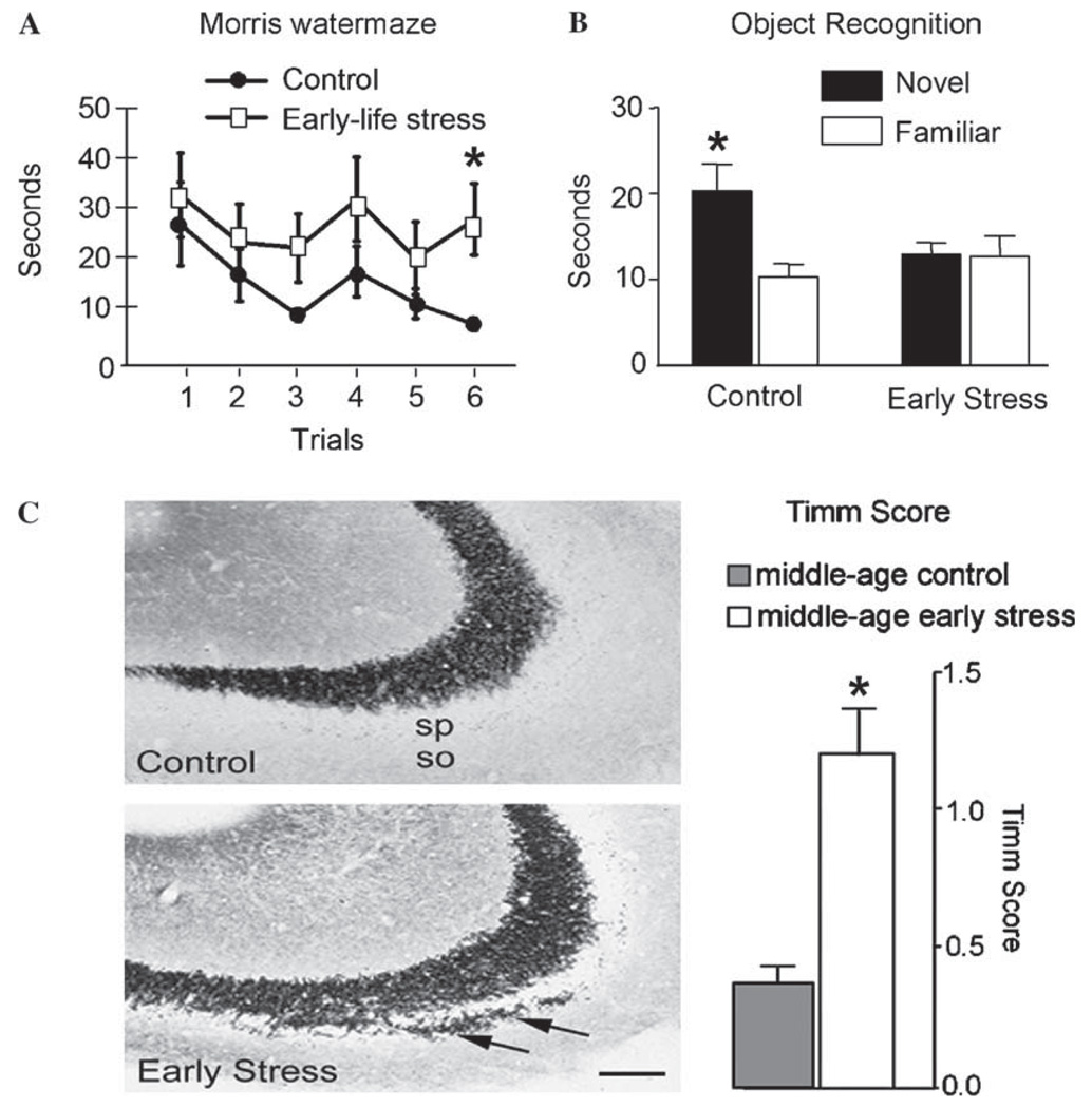
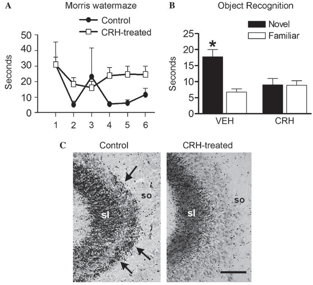
Several groups have probed the consequences of early-life stress on hippocampal function. For example, Huot et al., tested 4-month old rats that had been subjected to recurrent maternal separation, and found modest reduction in performance in the Morris watermaze [66]. Using the chronic, early-life psychological stress described above, Brunson et al. [31], found remarkable deficits of hippocampal function that emerged during middle age. The authors tested rats at 4 months and at middle age (12 months), using behavioral tasks generally considered to require an intact hippocampus [41,28,25]. When tested at 12 months, severe spatial memory impairments were apparent in the early-life stressed group in the Morris watermaze task [31] (Fig. 3A). In essence, these rats failed to reduce the time they required to locate a hidden platform based on spatial cues, suggesting significant deficits in learning and memory function. The variant of the watermaze used by the group was designed to reduce the stressful elements of this test [105]. However, the impairment demonstrated by the early-stress group may have been a result of different capabilities for ‘coping’ with the adverse elements (requiring rats to swim in room-temperature water) of this test. To exclude such potential sensitivity to stress, Brunson and colleagues [31] employed a second test that is generally devoid of stressful elements, namely the object recognition procedure [104,41,28]. The object recognition test was carried out over 2 days, after a habituation procedure. During the 1st day, two objects were placed in random locations in a cage, leading to exploration of these objects by the rat. The test trial took place 24 h later and consisted of a 5min epoch in which animals were presented with a duplicate of an object from the previous day and a novel object. On both days, the duration of exploration of each object was recorded [42,28]. In this test, middle-aged rats stressed early in life failed to distinguish a novel object from one they had seen the previous day, while age-matched controls spent about twice as long exploring the novel test object [31] (Fig. 3B). These data indicate that hippocampal cognitive function is impaired in middle-aged rats stressed early in life.
Fig. 3.
Functional and structural consequences of early-life psychological stress between P2 and P9 on the hippocampus of adult male rats. (A) Escape latencies (time to reach the hidden platform) on the testing day (day 3) are shown after 2 training days. Early-life stressed rats (white squares) require significantly longer time to locate the hidden platform in the Morris watermaze test when compared to age-matched controls (black circles; paired t test, P < 0.05). T test analyses of each trial demonstrate significantly different escape latencies at specific trials (marked by *). (B) Using the object recognition paradigm, control rats are able to discriminate between familiar and novel objects (remembering the familiar object from the day before and exploring it for significantly shorter time). In contrast, early-life stress rats spend the same amount of time exploring the familiar and novel objects, indicating impairment of recognition memory. (Modified from [31] with permission.) (C) Sections of CA3 pyramidal cell Welds from control and early-life stress rats subjected to Timm’s stain for visualizing the high zinc content of mossy fiber terminals (axons of the CA3 innervating granule cells). In early-life stressed rats, these terminals are abnormally abundant within CA3 stratum oriens (so; arrows). Quantification of the Timm’s stained sections confirmed that mossy fiber sprouting is significantly increased in early-life stress animals. sp, stratum pyramidale. Scale bar, 50 µm. *P < 0.05. (Modified from [31], with permission.)
8.2. Disruption of LTP induction accompanies the profound memory deficits after early-life stress
Electrophysiological studies were carried out by the same group, to uncover the cellular basis of the profound reduction of learning and memory functions in middle-aged rats that experienced a single week of chronic stress early in life [31]. Specifically, LTP was evaluated because this form of synaptic plasticity has been implicated in memory formation [116,22,96,132]. Indeed, LTP was impaired in both CA3 and CA1 hippocampal areas of 12-month old rats stressed early in life [31]. In area CA3, the hippocampal field considered most vulnerable to chronic stress in adults [125,100], both LTP as well as basic synaptic physiology were abnormal [31]. Interestingly, abnormal LTP, although normal synaptic physiology, was found in area CA1 [31].
8.3. Structural foundation of the disrupted synaptic function and plasticity after early-life stress
What could be the basis for disrupting synaptic physiology, most profoundly in area CA3, in rats stressed early in life? In considering the developmental aspects of hippocampal circuitry, it is notable that the connectivity of area CA3 is still evolving during the first 2 weeks of life, the time-frame of the early-life stress paradigm employed by Brunson et al., [31]. For example, axons of the granule cells, the mossy fiber system are generated throughout the period during which psychological stress was induced (during P2– P9) [5,59], whereas most other axonal systems in the hippocampus are established earlier (ibid). Because of its delayed development, this system may be particularly affected by early-life stress. The mossy fibers innervate the CA3 pyramidal cells via a powerful excitatory array of synapses [59]; they also innervate interneurons as described in [1]. Therefore, altered maturation of the mossy fibers and their giant synapses on CA3 pyramidal cells may affect the integrity and synaptic physiology of the latter.
In accord with this notion, repeated episodes of maternal separation-stress (3 h each) reduced the density of mossy fibers in 4-month old rats [66]. Brunson and colleagues [31] also described that the projections of mossy fibers in early-stressed, middle-aged rats were abnormal (Fig. 3C). However, in these animals this granule cell axon system was expanded and extended further into the CA3 stratum oriens to contact basal dendrites of CA3 pyramidal cells. The series of events that result from these (presumably early) effects of stress on mossy fiber growth and on the connectivity within area CA3 have not been defined at this point. Improved understanding of how early-life stress influences these processes should lead to deciphering the cascade of events that culminates in highly abnormal CA3 physiology that eventually influences long-term potentiation also in CA1.
9. Molecules and mechanisms that may mediate the effects of chronic early-life stress on the developing hippocampus
9.1. Glucocorticoid stress hormones
Logical mechanisms that may interfere with the developmental connectivity programs within CA3 during the early-postnatal stress period include molecules that may be active in the immature, stressed hippocampus [120,10]. Major candidates include systemic glucocorticoids (GC). GC are released from the adrenal glands by stress, cross the blood–brain barrier readily, and activate hippocampal GC receptors (GRs) [99,44]. Indeed, saturation of GRs by ‘stress-levels’ of GC can lead to hippocampal neuronal injury [125,140] but these receptors reside primarily in rodent CA1 [115,62, but see primate data in 122], whereas stress-induced structural changes involve mainly CA3 [125,99,100]. In addition, GC administration early in life does not reproduce the effects of stress on hippocampal function and integrity when given in a non-stressful manner [87], suggesting that other factors may also contribute to the mechanisms by which early-life stress influences hippocampal development and function throughout life.
9.2. Corticotropin releasing hormone
An additional potential mediator of the effects of early-life stress on hippocampal neurons, particularly in CA3, is the neuropeptide CRH. This peptide is involved in propagation and integration of ‘psychological’ stress responses in several brain regions, including amygdala and hippocampus [80,84], a role consistent with the finding that administration of the synthetic peptide via the lateral ventricles reproduces the spectrum of behavioral and neuroendocrine responses to stress [80]. The notion that hippocampal CRH in immature rat may participate in the mechanisms by which early-life stress influences learning and memory long-term is supported by several lines of evidence. These include (i) the age-specific abundance of CRH-expressing neurons in the developing hippocampus and a parallel age-specific developmental pattern of the CRH receptors; (ii) the release of hippocampal CRH by stress; (iii) the chronic upregulation of hippocampal CRH expression by early-life stress, and (iv) the excitotoxic actions of this peptide on hippocampal dendrites and neurons (see Section 10).
9.3. Interactions among glucocorticoid- and CRH-mediated mechanisms
Although the previous paragraphs discussed actions of stress-released glucocorticoid hormones and CRH as discreet entities, it is quite likely that the steroid and peptide hormones converge on the same cells, and contribute in an additive or synergistic manner to the consequences of early-life stress. In addition, stress-evoked activation of the CRH receptors may influence GC or GR levels and vice versa. For example, both early-life glucocorticoid administration [73] as well as CRH administration [31] may result in hippocampal-dependent learning and memory dysfunction later in life.
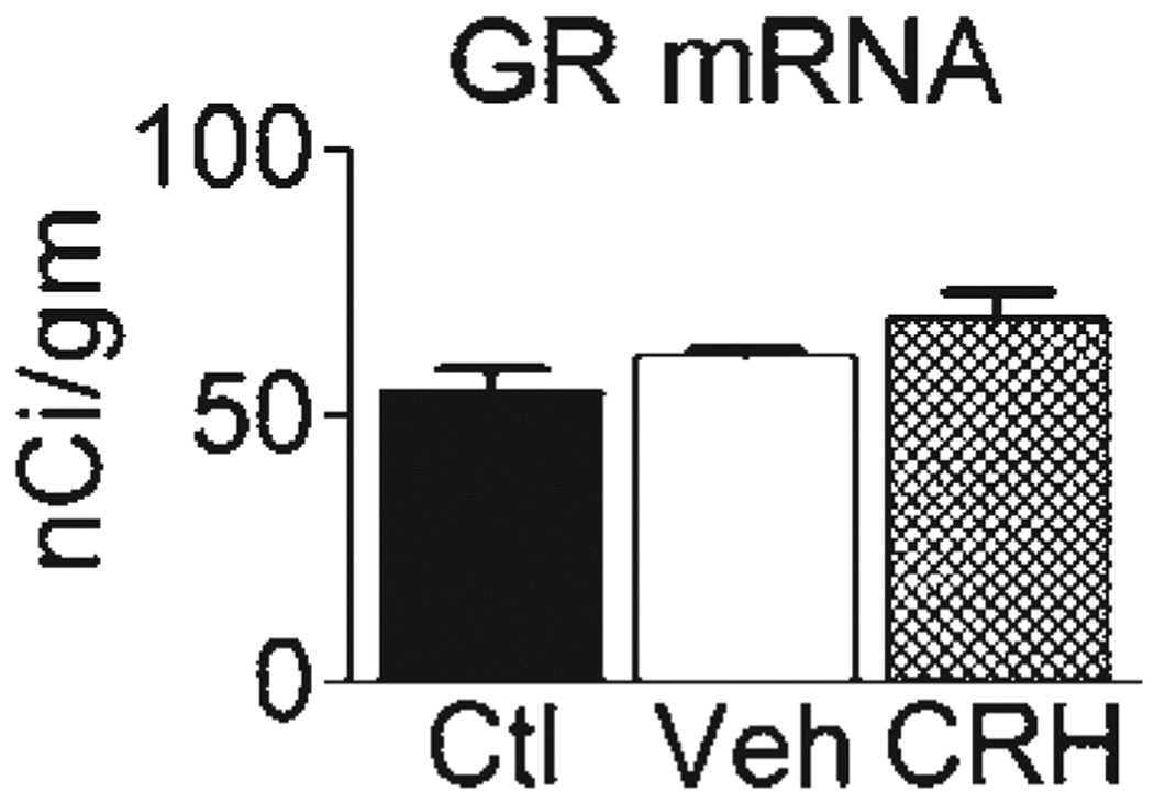
Interactions among GC- and CRH-mediated mechanisms are supported by the fact that icv administration of CRH to P10 rats reduces GR levels in hippocampal CA1 acutely (at 4 h, Brunson et al., unpublished). These CRH-induced changes to GR expression in hippocampus do not persist to adulthood (Fig. 4). Whereas there is much information about the long-term effects of early-life glucocorticoids on levels of its two receptors in the hippocampus, GR [47,107,148] and mineralocorticoid receptor (MR; 148,130), little is known about the effects of these hormones on either hippocampal CRH or the CRH receptor, CRF1. In adult rats, administration of the synthetic glucocorticoid hormone, dexamethasone, does not influence CRH levels in the hippocampus [32].
Fig. 4.
Administration of CRH early in life does not significantly influence GR-mRNA expression in hippocampal CA1 long-term. Adult male rats that were treated with CRH early in life express similar levels of hippocampal GR mRNA when compared to vehicle-treated controls. Note that a trend towards increased GR mRNA levels is found in vehicle-treated versus naive controls. Ctl, adult group, not infused at all on P10; Veh, adult rats infused with vehicle (water) on P10; CRH, adult rats treated with CRH early in life (see [28] for infusion procedures and details).
10. CRH-containing neurons and CRH receptors are abundant in developing hippocampus
Early work in adult hippocampus described relatively few CRH-containing interneurons [137,119]. However, in developing hippocampus, neurochemical, and quantitative stereological methods were used to characterize in detail CRH-expressing neuronal populations throughout postnatal development [152,36]. These experiments revealed progressively increasing numbers of CRH-expressing GABAergic interneurons within the pyramidal cell layer that peaked on P11–P18, then declined to adult levels. For example, in CA3, the number of CRH-expressing neurons was approximately 4000 on P18 and declined to approximately 2700 by P60. These CRH-expressing interneurons are primarily basket cells that synapse on somata of hippocampal pyramidal neurons that express the CRH-receptor, CRF1 [38]. This positions the CRH-expressing neurons to significantly influence pyramidal cell activity and hippocampal information flow. The majority of CA3 pyramidal cells express CRF1, the principal receptor responsible for mediating synaptic actions of CRH on these hippocampal cells [33,11,29], and this expression is highest during early postnatal life [12]. The developmental profiles of CRH and CRF1 expression in hippocampus are consistent with a key role for the peptide in mediating the effects of early-life stress on hippocampal neurons.
11. Stress induces release of CRH into the hippocampal intercellular space, and increases the set-point of CRH expression, so that subsequent stress causes a higher CRH secretion
Psychological stress induces the release of endogenous hippocampal CRH [37], as is also found in the amygdala [117]. CRH released by stress leads to the activation, measured by immediate early gene expression, of CA3 pyramidal cells. This requires binding of the peptide to CRF1, because expression and phosphorylation of transcription factors after restraint stress was abolished by selective CRF1 receptor antagonists [37]. These data implicate endogenous CRH in the mechanisms by which stress influences hippocampal pyramidal neurons.
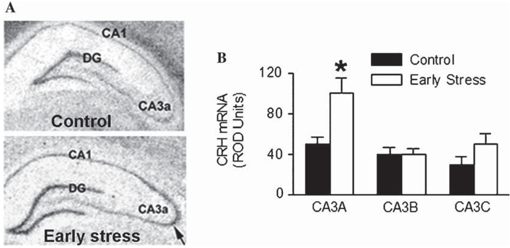
Interestingly, psychological stress increases the expression levels of hippocampal CRH. This has been found in both adult [133] and immature [55] rat hippocampal interneurons, and may be a result of ‘release-coupled synthesis’ [34]. A similar mechanism has been described in immature hypothalamus and amygdala [154,56]. Remarkably, Brunson and colleagues found that early-life chronic stress leads to enduring, life-long elevation of CRH expression within the hippocampal CA3 area (Fig. 5; Brunson et al., unpublished).
Fig. 5.
Elevated CRH mRNA expression in adult (12-month old) rats that had experienced early-life stress. (A) Photomicrographs of coronal brain sections from control and early-life stress animals after in situ hybridization for CRH mRNA. (B) Relative optical density (ROD) of the CRH signal was analyzed in hippocampal subfields. CRH mRNA expression is significantly enhanced in hippocampal subfield CA3a of early-life stressed animals when compared to that of controls (arrow). DG, dentate gyrus. *P< 0.05.
12. CRH impacts the function and structure of hippocampal neurons
Transient release of hippocampal CRH, such as that occurring after mild, acute stress enhances LTP and improves hippocampal-mediated memory consolidation [85,84,20]. In addition, electrophysiological data indicate that synthetic CRH acts as an excitatory neuropeptide that reduces after-hyperpolarization [2], and interacts with glutamatergic neurotransmission to promote excitability in vitro [64].
Whereas low (‘physiological’) levels of CRH enhance synaptic communication and efficacy, ‘pathologic’ CRH levels may have harmful effects on hippocampal function and structure [35]. Administration of high picomolar doses of CRH into the cerebral ventricles of immature rats may lead to seizures that involve the amygdala and hippocampus [12], and these may be followed by injury to CA3 pyramidal neurons [13]. CRH may contribute to neuronal excitotoxicity under several pathological conditions [136,93]. Notably, learning and memory deficits have been found in CRH-overexpressing mice [58]. These data support the notion that early-life ‘pathologic’ release of CRH may contribute to the impaired functional integrity of the hippocampus following early-life chronic psychological stress.
13. Early-life administration of CRH reproduces the long-term cognitive hippocampal deficits found after chronic early-life stress
An interesting observation, supporting a causal role for CRH in the mechanisms by which early-life stress impacts hippocampal learning and memory function, was made by Brunson et al. [28]. These authors essentially reproduced the effects of early-life stress on the cognitive function of middle-aged rats, i.e., deficits in spatial memory acquisition skills in the Morris watermaze test and memory retrieval in the relatively stress-free object-recognition test, by administering CRH into the brain of immature rats [28] (Figs. 6A and B). When administered icv (‘centrally’), CRH reaches the hippocampus [19] where it influences neurons directly [64,20]. Similar to the effects of early-life stress, these CRH-evoked deficits emerged later in adulthood, and were progressive. In addition, these progressive hippocampal functional deficits were accompanied by similar structural changes to those after early-life stress, i.e., excessive growth of mossy fibers (Fig. 6C). As mentioned above, the synapses formed by the aberrant mossy fibers on CA3 pyramidal cells are excitatory (glutamatergic), potentially promoting progressive excitotoxic injury to these cells. It should be mentioned that the long-term hippocampal deficits described above occurred also in CRH-treated animals that were never able to generate stress levels of plasma GC. These rats were adrenalectomized prior to CRH administration and provided with hormonal supplements to generate low physiological GC levels. This observation suggests that ‘stress-levels’ of these hormones were not required for CRH to exert its adverse effects on the hippocampal formation [28].
Fig. 6.
Administration of synthetic CRH into the brain recapitulates the structural and functional effects of early-life stress. (A) Adult rats that were treated with CRH early in life suffer from hippocampus-dependent memory dysfunction in the Morris watermaze. CRH-treated rats (white squares) take significantly longer to locate the hidden platform in the watermaze when compared to controls (black circles) [F(2,132) = 5.53, P< 0.01]. (B) Impaired memory is further evident by the performance of CRH-treated rats on the non-aversive, relatively stress-free object recognition test. Here, CRH-treated rats fail to distinguish the familiar object from the novel object, indicating impairment of recognition memory. (C) Sections of CA3A pyramidal cell regions from control and CRH-treated adult (12-month old), subjected to Timm’s stain for visualizing zinc-rich mossy fiber terminals. In CRH-treated rats, these terminals are abnormally abundant within the CA3 stratum oriens (so; arrows). sl, stratum lucidum. Scale bar = 50 µm. *P < 0.05. (Modified from [28] with permission.)
14. Concluding remarks
The effects of chronic stress on hippocampal function may be reversible if experienced during adulthood. In contrast, early-life chronic stress carries enormous significance for hippocampal function and integrity throughout life. The mechanisms underlying these effects are complex and, in light of recent studies discussed in this review, likely involve the stress-activated peptide, CRH. CRH is abundant in immature hippocampus, where its ‘physiological’ release enhances synaptic function. Large doses of synthetic CRH (as presumed to be released during chronic stress) administered early in life lead to progressive hippocampal dysfunction that reproduces deficits resulting from early-life stress. The longevity of the consequences of early-life stress may be explained, in part, by the enduring elevated expression and levels of hippocampal CRH in early-stressed animals.
Finally, the consequences of early-life stress carry major implications for human health: chronic emotional stress affects a large proportion of infants and children around the world, that experience poverty, parental loss, war, famine or child abuse and neglect. Therefore, discovering the mechanisms by which stress elicits adverse effects on the developing hippocampus should lead to truly exciting approaches to prevention of cognitive deficits related to these early-life events.
Acknowledgments
The authors thank Michele Hinojosa for excellent editorial assistance. Authors’ research is supported by National Institutes of Health Grants NS39307/MH 73136 and NS28912.
References
- 1.Acsady L, Kamondi A, Sik A, Freund T, Buzsaki G. GABAergic cells are the major postsynaptic targets of mossy fibers in the rat hippocampus. J. Neurosci. 1998;18:3386–3403. doi: 10.1523/JNEUROSCI.18-09-03386.1998. [DOI] [PMC free article] [PubMed] [Google Scholar]
- 2.Aldenhoff JB, Gruol DL, Rivier J, Vale W, Siggins GR. Corticotropin releasing factor decreases postburst hyperpolarizations and excites hippocampal neurons. Science. 1983;221:875–877. doi: 10.1126/science.6603658. [DOI] [PubMed] [Google Scholar]
- 3.Alfarez DN, Joels M, Krugers HJ. Chronic unpredictable stress impairs long-term potentiation in rat hippocampal CA1 area and dentate gyrus in vitro. Eur. J. Neurosci. 2003;17:1928–1934. doi: 10.1046/j.1460-9568.2003.02622.x. [DOI] [PubMed] [Google Scholar]
- 4.Altman J, Bayer SA. Prolonged sojourn of developing pyramidal cells in the intermediate zone of the hippocampus and their settling in the stratum pyramidale. J. Comp. Neurol. 1990;301:343–364. doi: 10.1002/cne.903010303. [DOI] [PubMed] [Google Scholar]
- 5.Amaral DG, Dent JA. Development of the mossy fibers of the dentate gyrus: I. A light and electron microscopic study of the mossy fibers and their expansions. J. Comp. Neurol. 1981;195:51–86. doi: 10.1002/cne.901950106. [DOI] [PubMed] [Google Scholar]
- 6.Ammerman RT, Cassisi JE, Hersen M, van Hassel VB. Consequences of physical abuse and neglect in children. Clin. Psychol. Rev. 1986;6:291–310. [Google Scholar]
- 7.Avishai-Eliner S, Brunson KL, Sandman CA, Baram TZ. Stressed out? Or in (utero) Trends Neurosci. 2002;25:518–524. doi: 10.1016/s0166-2236(02)02241-5. [DOI] [PMC free article] [PubMed] [Google Scholar]
- 8.Avishai-Eliner A, Eghbal-Ahmadi M, Tabatchnik E, Brunson KL, Baram TZ. Down-regulation of hypothalamic corticotropin-releasing hormone messenger ribonucleic acid precedes early-life experience-induced changes in hippocampal glucocorticoid receptor mRNA. Endocrinology. 2001;142:89–97. doi: 10.1210/endo.142.1.7917. [DOI] [PMC free article] [PubMed] [Google Scholar]
- 9.Avishai-Eliner S, Gilles ES, Eghbal-Ahmadi M, Bar-El Y, Baram TZ. Altered regulation of gene and protein expression of hypothalamic-pituitary-adrenal axis components in an immature rat model of chronic stress. J. Neuroendocrinol. 2001;13:799–807. doi: 10.1046/j.1365-2826.2001.00698.x. [DOI] [PMC free article] [PubMed] [Google Scholar]
- 10.Baram TZ. Long-term neuroplasticity and functional consequences of single versus recurrent early-life seizures. Ann. Neurol. 2003;54:701–705. doi: 10.1002/ana.10833. [DOI] [PMC free article] [PubMed] [Google Scholar]
- 11.Baram TZ, Chalmers DT, Chen C, Koutsoukos Y, De Souza EB. The CRF1 receptor mediates the excitatory actions of corticotropin releasing factor (CRF) in the developing rat brain: in vivo evidence using a novel selective, non-peptide CRF receptor antagonist. Brain Res. 1997;770:89–95. doi: 10.1016/s0006-8993(97)00759-2. [DOI] [PMC free article] [PubMed] [Google Scholar]
- 12.Baram TZ, Hatalski CG. Neuropeptide-mediated excitability: a key triggering mechanism for seizure generation in the developing brain. Trends Neurosci. 1998;21:471–476. doi: 10.1016/s0166-2236(98)01275-2. [DOI] [PMC free article] [PubMed] [Google Scholar]
- 13.Baram TZ, Ribak CE. Peptide-induced infant status epilepticus causes neuronal death and synaptic reorganization. Neuroreport. 1995;6:277–280. doi: 10.1097/00001756-199501000-00013. [DOI] [PMC free article] [PubMed] [Google Scholar]
- 14.Baram TZ, Yi S, Avishai-Eliner S, Schultz L. Development neurobiology of the stress response: multilevel regulation of corticotropin-releasing hormone function. Ann. N. Y. Acad. Sci. 1997;814:252–265. doi: 10.1111/j.1749-6632.1997.tb46161.x. [DOI] [PMC free article] [PubMed] [Google Scholar]
- 15.Bayer SA. Development of the hippocampal region in the rat. I. Neurogenesis examined with 3H–thymidine autoradiography. J. Comp. Neurol. 1980;190:87–114. doi: 10.1002/cne.901900107. [DOI] [PubMed] [Google Scholar]
- 16.Bender RA, Lauterborn JC, Gall CM, Cariaga W, Baram TZ. Enhanced CREB phosphorylation in immature dentate gyrus granule cells precedes neurotrophin expression and indicates a specific role of CREB in granule cell differentiation. Eur. J. Neurosci. 2001;13:679–686. doi: 10.1046/j.1460-9568.2001.01432.x. [DOI] [PMC free article] [PubMed] [Google Scholar]
- 17.Bhutta AT, Anand KJS. Abnormal cognition and behavior in pre-term neonates linked to smaller brain volumes. Trends Neurosci. 2001;24:129–132. doi: 10.1016/s0166-2236(00)01747-1. [DOI] [PubMed] [Google Scholar]
- 18.Bi X, Yong AP, Zhou J, Ribak CE, Lynch G. Rapid induction of intraneuronal neurofibrillary tangles in apolipoprotein E-deficient mice. Proc. Natl. Acad. Sci. USA. 2001;98:8832–8837. doi: 10.1073/pnas.151253098. [DOI] [PMC free article] [PubMed] [Google Scholar]
- 19.Bittencourt JC, Sawchenko PE. Do centrally administered neuropeptides access cognate receptors? An analysis in the central corticotropin-releasing factor system. J. Neurosci. 2000;20:1142–1156. doi: 10.1523/JNEUROSCI.20-03-01142.2000. [DOI] [PMC free article] [PubMed] [Google Scholar]
- 20.Blank T, Nijholt I, Eckart K, Spiess J. Priming of long-term potentiation in mouse hippocampus by corticotropin-releasing factor and acute stress: implications for hippocampus-dependent learning. J. Neurosci. 2002;22:3788–3794. doi: 10.1523/JNEUROSCI.22-09-03788.2002. [DOI] [PMC free article] [PubMed] [Google Scholar]
- 21.Blank T, Nijholt I, Spiess J. Molecular determinants mediating effects of acute stress on hippocampus-dependent synaptic plasticity and learning. Mol. Neurobiol. 2004;29:131–138. doi: 10.1385/MN:29:2:131. [DOI] [PubMed] [Google Scholar]
- 22.Bliss TV, Collingridge GL. A synaptic model of memory: long-term potentiation in the hippocampus. Nature. 1993;361:31–39. doi: 10.1038/361031a0. [DOI] [PubMed] [Google Scholar]
- 23.Bredy TW, Humpartzoomian RA, Cain DP, Meaney MJ. Partial reversal of the effect of maternal care on cognitive function through environmental enrichment. Neuroscience. 2003;118:571–576. doi: 10.1016/s0306-4522(02)00918-1. [DOI] [PubMed] [Google Scholar]
- 24.Bremner JD, Randall P, Vermetten E, Staib L, Bronen RA, Mazure C, Capelli S, McCarthy G, Innis RB, Charney DS. Magnetic resonance imaging-based measurement of hippocampal volume in posttraumatic stress disorder related to childhood physical and sexual abuse—a preliminary report. Biol. Psychiatry. 1997;41:23–32. doi: 10.1016/s0006-3223(96)00162-x. [DOI] [PMC free article] [PubMed] [Google Scholar]
- 25.Broadbent NJ, Squire LR, Clark RE. Spatial memory, recognition memory, and the hippocampus. Proc. Natl. Acad. Sci. USA. 2004;101:14515–14520. doi: 10.1073/pnas.0406344101. [DOI] [PMC free article] [PubMed] [Google Scholar]
- 26.Brunson KL, Avishai-Eliner S, Hatalski CG, Baram TZ. Neurobiology of the stress response early in life: evolution of the concept and the role of corticotropin releasing hormone. Mol. Psychiatry. 2001;6:647–656. doi: 10.1038/sj.mp.4000942. [DOI] [PMC free article] [PubMed] [Google Scholar]
- 27.Brunson KL, Chen Y, Avishai-Eliner S, Baram TZ. Stress and the developing hippocampus: a double-edged Sword? Mol. Neurobiol. 2003;27:121–136. doi: 10.1385/MN:27:2:121. [DOI] [PMC free article] [PubMed] [Google Scholar]
- 28.Brunson KL, Eghbal-Ahmadi M, Bender R, Chen Y, Baram TZ. Long-term, progressive hippocampal cell loss and dysfunction induced by early-life administration of corticotropin-releasing hormone reproduce the effects of early-life stress. Proc. Natl. Acad. Sci. USA. 2001;98:8856–8861. doi: 10.1073/pnas.151224898. [DOI] [PMC free article] [PubMed] [Google Scholar]
- 29.Brunson KL, Grigoriadis DE, Lorang MT, Baram TZ. Corticotropin-releasing hormone (CRH) down-regulates the function of its receptor (CRF1) and induces CRF1 expression in hippocampal and cortical regions of the immature rat brain. Exp. Neurol. 2002;176:75–86. doi: 10.1006/exnr.2002.7937. [DOI] [PMC free article] [PubMed] [Google Scholar]
- 30.Brunson KL, Khan N, Eghbal-Ahmadi M, Baram TZ. Corticotropin (ACTH) acts directly on amygdala neurons to down-regulate corticotropin-releasing hormone gene expression. Ann. Neurol. 2001;49:304–312. [PMC free article] [PubMed] [Google Scholar]
- 31.Brunson KL, Kramar E, Lin B, Chen Y, Colgin LL, Yanagihara TK, Lynch G, Baram TZ. Mechanisms of late-onset cognitive decline after early-life stress. J. Neurosci. 2005;25:9328–9338. doi: 10.1523/JNEUROSCI.2281-05.2005. [DOI] [PMC free article] [PubMed] [Google Scholar]
- 32.Calogero AE, Liapi C, Chrousos GP. Hypothalamic and suprahy-pothalamic effects of prolonged treatment with dexamethasone in the rat. J. Endocrinol. Invest. 1991;14:277–286. doi: 10.1007/BF03346812. [DOI] [PubMed] [Google Scholar]
- 33.Chalmers DT, Lovenberg TW, De Souza EB. Localization of novel corticotropin-releasing factor receptor (CRF2) mRNA expression to specific subcortical nuclei in rat brain: comparison with CRF1 receptor mRNA expression. J. Neurosci. 1995;15:6340–6350. doi: 10.1523/JNEUROSCI.15-10-06340.1995. [DOI] [PMC free article] [PubMed] [Google Scholar]
- 34.Chavkin C. In: Dynorphins are endogenous opioid peptides released from granule cells to act neurohumorly and inhibit excitatory neurotransmission in the hippocampus. Agnati LF, Fuxe K, Nicholson C, Sykova E, editors. Amsterdam: Elsevier; 2000. pp. 363–367. [DOI] [PubMed] [Google Scholar]
- 35.Chen Y, Bender RA, Brunson KL, Pomper JK, Grigoriadis DE, Wurst W, Baram TZ. Modulation of dendritic differentiation by corticotropin-releasing factor in the developing hippocampus. Proc. Natl. Acad. Sci. USA. 2004;101:15782–15787. doi: 10.1073/pnas.0403975101. [DOI] [PMC free article] [PubMed] [Google Scholar]
- 36.Chen Y, Bender R, Frotscher M, Baram TZ. Novel and transient populations of corticotropin-releasing hormone-expressing neurons in developing hippocampus suggest unique functional roles: a quantitative spatiotemporal analysis. J. Neurosci. 2001;21:7171–7181. doi: 10.1523/JNEUROSCI.21-18-07171.2001. [DOI] [PMC free article] [PubMed] [Google Scholar]
- 37.Chen Y, Brunson KL, Adelmann G, Bender RA, Frotscher M, Baram TZ. Hippocampal corticotropin releasing hormone: pre- and post-synaptic location and release by stress. Neuroscience. 2004;126:533–540. doi: 10.1016/j.neuroscience.2004.03.036. [DOI] [PMC free article] [PubMed] [Google Scholar]
- 38.Chen Y, Brunson KL, Muller MB, Cariaga W, Baram TZ. Immunocytochemical distribution of corticotropin-releasing hormone receptor type-1 (CRF1)-like immunoreactivity in the mouse brain: light microscopy analysis using an antibody directed against the C-terminus. J. Comp. Neurol. 2000;420:305–323. doi: 10.1002/(sici)1096-9861(20000508)420:3<305::aid-cne3>3.0.co;2-8. [DOI] [PMC free article] [PubMed] [Google Scholar]
- 39.Chen Y, Hatalski CG, Brunson KL, Baram TZ. Rapid phosphorylation of the CRE binding protein precedes stress-induced activation of the corticotropin releasing hormone gene in medial parvocellular hypothalamic neurons of the immature rat. Brain Res. Mol. Brain Res. 2001;96:39–49. doi: 10.1016/s0169-328x(01)00265-0. [DOI] [PMC free article] [PubMed] [Google Scholar]
- 40.Chugani HT, Behen ME, Muzik O, Juhasz C, Nagy F, Chugani DC. Local brain functional activity following early deprivation: a study of postinstitutionalized Romanian orphans. Neuroimage. 2001;14:1290–1301. doi: 10.1006/nimg.2001.0917. [DOI] [PubMed] [Google Scholar]
- 41.Clark RE, Zola SM, Squire LR. Impaired recognition memory in rats after damage to the hippocampus. J. Neurosci. 2000;20:8853–8860. doi: 10.1523/JNEUROSCI.20-23-08853.2000. [DOI] [PMC free article] [PubMed] [Google Scholar]
- 42.Dayas CV, Buller KM, Crane JW, Xu Y, Day TA. Stressor categorization: acute physical and psychological stressors elicit distinctive recruitment patterns in the amygdala and in medullary noradrenergic cell groups. Eur. J. Neurosci. 2001;14:1143–1152. doi: 10.1046/j.0953-816x.2001.01733.x. [DOI] [PubMed] [Google Scholar]
- 43.De Bellis MD, Keshavan MS, Shifflett H, Lyengar S, Beers SR, Hall J, Moritz G. Brain structures in pediatric maltreatment-related posttraumatic stress disorder: a sociodemographically matched study. Biol. Psychiatry. 2002;52:1066–1078. doi: 10.1016/s0006-3223(02)01459-2. [DOI] [PubMed] [Google Scholar]
- 44.De Kloet ER, Oitzl MS, Joels M. Stress and cognition: are corticosteroids good or bad guys? Trends Neurosci. 1999;22:422–426. doi: 10.1016/s0166-2236(99)01438-1. [DOI] [PubMed] [Google Scholar]
- 45.Diamond DM, Bennett MC, Fleshner M, Rose GM. Inverted-U relationship between the level of peripheral corticosterone and the magnitude of hippocampal primed burst potentiation. Hippocampus. 1992;2:421–430. doi: 10.1002/hipo.450020409. [DOI] [PubMed] [Google Scholar]
- 46.Eghbal-Ahmadi M, Avishai-Eliner S, Hatalski CG, Baram TZ. Differential regulation of the expression of corticotropin-releasing factor receptor type 2 (CRF2) in hypothalamus and amygdala of the immature rat by sensory input and food intake. J. Neurosci. 1999;19:3982–3991. doi: 10.1523/JNEUROSCI.19-10-03982.1999. [DOI] [PMC free article] [PubMed] [Google Scholar]
- 47.Felszeghy K, Gaspar E, Nyakas CZ. Long-term selective down-regulation of brain glucocorticoid receptors after neonatal dexamethasone treatment in rats. J. Neuroendocrinol. 1996;8:493–499. doi: 10.1046/j.1365-2826.1996.04822.x. [DOI] [PubMed] [Google Scholar]
- 48.Fenoglio KA, Brunson KL, Avishai-Eliner S, Chen Y, Baram TZ. Region-specific onset of handling-induced changes in corticotropin-releasing factor factor and glucocorticoid receptor expression. Endocrinology. 2004;145:2702–2706. doi: 10.1210/en.2004-0111. [DOI] [PMC free article] [PubMed] [Google Scholar]
- 49.Fenoglio KA, Brunson KL, Avishai-Eliner S, Stone BA, Kapadia BJ, Baram TZ. Enduring, handling-evoked enhancement of hippocampal memory function and glucocorticoid receptor expression involves activation of the corticotropin-releasing factor type 1 receptor. Endocrinology. 2005;146:4090–4096. doi: 10.1210/en.2004-1285. [DOI] [PMC free article] [PubMed] [Google Scholar]
- 50.Fortin NJ, Agster KL, Eichenbaum HB. Critical role of the hippocampus in memory for sequences of events. Nat. Neurosci. 2002;5:458–462. doi: 10.1038/nn834. [DOI] [PMC free article] [PubMed] [Google Scholar]
- 51.Gesing A, Bilang-Bleuel A, Droste SK, Linthorst AC, Holsboer F, Reul JM. Psychological stress increases hippocampal mineralocorticoid receptor levels: involvement of corticotropin-releasing hormone. J. Neurosci. 2001;21:4822–4829. doi: 10.1523/JNEUROSCI.21-13-04822.2001. [DOI] [PMC free article] [PubMed] [Google Scholar]
- 52.Gilles EE, Schultz L, Baram TZ. Abnormal corticosterone regulation in an immature rat model of continuous chronic stress. Pediatric Neurol. 1996;15:114–119. doi: 10.1016/0887-8994(96)00153-1. [DOI] [PMC free article] [PubMed] [Google Scholar]
- 53.Gould E, Cameron HA. Regulation of neuronal birth, migration and death in the rat dentate gyrus. Dev. Neurosci. 1996;18:22–35. doi: 10.1159/000111392. [DOI] [PubMed] [Google Scholar]
- 54.Gunnar MR, Connors J, Isensee J, Wall L. Adrenocortical activity and behavioral distress in human newborns. Dev. Psychobiol. 1988;21:297–310. doi: 10.1002/dev.420210402. [DOI] [PubMed] [Google Scholar]
- 55.Hatalski CG, Brunson KL, Tantayanubutr B, Chen Y, Baram TZ. Neuronal activity and stress differentially regulate hippocampal and hypothalamic corticotropin-releasing hormone expression in the immature rat. Neuroscience. 2000;101:571–580. doi: 10.1016/s0306-4522(00)00386-9. [DOI] [PMC free article] [PubMed] [Google Scholar]
- 56.Hatalski CG, Guirguis C, Baram TZ. Corticotropin releasing factor mRNA expression in the hypothalamic paraventricular nucleus and the central nucleus of the amygdala is modulated by repeated acute stress in the immature rat. J. Neuroendocrinol. 1998;10:663–669. doi: 10.1046/j.1365-2826.1998.00246.x. [DOI] [PMC free article] [PubMed] [Google Scholar]
- 57.Heine VM, Maslam S, Zareno J, Joels M, Lucassen PJ. Suppressed proliferation and apoptotic changes in the rat dentate gyrus after acute and chronic stress are reversible. Eur. J. Neurosci. 2004;19:131–144. doi: 10.1046/j.1460-9568.2003.03100.x. [DOI] [PubMed] [Google Scholar]
- 58.Heinrichs SC, Stenzel-Poore MP, Gold LH, Battenberg E, Bloom FE, Koob GF, Vale WW, Pich EM. Learning impairment in transgenic mice with central overexpression of corticotropin-releasing factor. Neuroscience. 1996;74:303–311. doi: 10.1016/0306-4522(96)00140-6. [DOI] [PubMed] [Google Scholar]
- 59.Henze DA, Urban NN, Barrionuevo G. The multifarious hippocampal mossy fiber pathway. Neuroscience. 2000;98:407–427. doi: 10.1016/s0306-4522(00)00146-9. [DOI] [PubMed] [Google Scholar]
- 60.Herman JP, Cullinan WE. Neurocircuitry of stress: central control of the hypothalamo-pituitary-adrenocortical axis. Trends Neurosci. 1997;20:78–84. doi: 10.1016/s0166-2236(96)10069-2. [DOI] [PubMed] [Google Scholar]
- 61.Herman JP, Figueiredo H, Mueller NK, Ulrich-Lai Y, Ostrander MM, Choi DC, Cullinan WE. Central mechanisms of stress integration: hierarchical circuitry controlling hypothalamo-pituitary-adrenocortical responsiveness. Front. Neuroendocrinol. 2003;24:151–180. doi: 10.1016/j.yfrne.2003.07.001. [DOI] [PubMed] [Google Scholar]
- 62.Herman JP, Schafer MK, Young EA, Thompson R, Douglass J, Akil H, Watson SJ. Evidence for hippocampal regulation of neuro-endocrine neurons of the hypothalamo-pituitary-adrenocortical axis. J. Neurosci. 1989;9:3072–3082. doi: 10.1523/JNEUROSCI.09-09-03072.1989. [DOI] [PMC free article] [PubMed] [Google Scholar]
- 63.Hess JL, Denenberg VH, Zarrow MX, Pfeifer D. Modification of the corticosterone response curve as a function of handling in infancy. Physiol. Behav. 1969;4:109–111. [Google Scholar]
- 64.Hollrigel GS, Chen K, Baram TZ, Soltesz I. The pro-convulsant actions of corticotropin-releasing hormone in the hippocampus of infant rats. Neuroscience. 1998;84:71–79. doi: 10.1016/s0306-4522(97)00499-5. [DOI] [PMC free article] [PubMed] [Google Scholar]
- 65.Hollup SA, Kjelstrup KG, Hoff J, Moser MB, Moser EI. Impaired recognition of the goal location during spatial navigation in rats with hippocampal lesions. J. Neurosci. 2001;21:4505–4513. doi: 10.1523/JNEUROSCI.21-12-04505.2001. [DOI] [PMC free article] [PubMed] [Google Scholar]
- 66.Huot RL, Plotsky PM, Lenox RH, McNamara RK. Neonatal maternal separation reduces hippocampal mossy fiber density in adult Long Evans rats. Brain Res. 2002;950:52–63. doi: 10.1016/s0006-8993(02)02985-2. [DOI] [PubMed] [Google Scholar]
- 67.Imaki T, Shibasaki Y, Hotta M, Demura H. Intracerebroventricular administration of corticotropin-releasing factor induces c-fos mRNA expression in brain regions related to stress responses: comparison with pattern of c-fos mRNA induction after stress. Brain Res. 1993;616:114–125. doi: 10.1016/0006-8993(93)90199-w. [DOI] [PubMed] [Google Scholar]
- 68.Irvine GI, Abraham WC. Enriched environment exposure alters the input–output dynamics of synaptic transmission in area CA1 of freely moving rats. Neurosci. Lett. 2005 Sep 7; doi: 10.1016/j.neulet.2005.08.031. Epub ahead of print. [DOI] [PubMed] [Google Scholar]
- 69.Joels M, Karst H, Alfarez D, Heine VM, Qin Y, van Riel E, Verkuyl M, Lucassen PJ, Krugers HJ. Effects of chronic stress on structure and cell function in rat hippocampus and hypothalamus. Stress. 2004;7:221–231. doi: 10.1080/10253890500070005. [DOI] [PubMed] [Google Scholar]
- 70.Joels M, Velzing E, Nair W, Verkuyl JM, Karst H. Acute stress increases calcium current amplitude in rat hippocampus: temporal changes in physiology and gene expression. Eur. J. Neurosci. 2003;18:1315–1324. doi: 10.1046/j.1460-9568.2003.02845.x. [DOI] [PubMed] [Google Scholar]
- 71.Joels M, Verkuyl JM, Van Riel E. Hippocampal and hypothalamic function after chronic stress. Ann. N. Y. Acad. Sci. 2003;1007:367–378. doi: 10.1196/annals.1286.036. [DOI] [PubMed] [Google Scholar]
- 72.Kalin NH, Takahashi LK, Chen FL. Restraint stress increases corticotropin-releasing hormone mRNA content in the amygdala and paraventricular nucleus. Brain Res. 1994;656:182–186. doi: 10.1016/0006-8993(94)91382-x. [DOI] [PubMed] [Google Scholar]
- 73.Kamphuis PJ, Gardoni F, Kamal A, Croiset G, Bakker JM, Cattabeni F, Gispen WH, van Bel F, Di Luca M, Wiegant VM. Long-lasting effects of neonatal dexamethasone treatment on spatial learning and hippocampal synaptic plasticity: involvement of the NMDA receptor complex. FASEB J. 2003;17:911–913. doi: 10.1096/fj.02-0333fje. [DOI] [PubMed] [Google Scholar]
- 74.Kaplan Z, Iancu I, Bodner E. A review of psychological debriefing after extreme stress. Psychiatr. Serv. 2001;52:824–827. doi: 10.1176/appi.ps.52.6.824. [DOI] [PubMed] [Google Scholar]
- 75.Kempermann G, Kuhn HG, Gage FH. More hippocampal neurons in adult mice living in an enriched environment. Nature. 1997;386:493–495. doi: 10.1038/386493a0. [DOI] [PubMed] [Google Scholar]
- 76.Kerr DS, Campbell LW, Applegate MD, Brodish A, Land-field PW. Chronic stress-induced acceleration of electrophysiologic and morphometric biomarkers of hippocampal aging. J. Neurosci. 1991;11:1316–1324. doi: 10.1523/JNEUROSCI.11-05-01316.1991. [DOI] [PMC free article] [PubMed] [Google Scholar]
- 77.Khazipov R, Esclapez M, Caillard O, Bernard C, Khalilov I, Tyzio R, Hirsch J, Dzhala V, Berger B, Ben-Ari Y. Early development of neuronal activity in the primate hippocampus in utero. J. Neurosci. 2001;21:9770–9781. doi: 10.1523/JNEUROSCI.21-24-09770.2001. [DOI] [PMC free article] [PubMed] [Google Scholar]
- 78.Kim JJ, Diamond DM. The stressed hippocampus, synaptic plasticity and lost memories. Nat. Rev. Neurosci. 2002;3:453–462. doi: 10.1038/nrn849. [DOI] [PubMed] [Google Scholar]
- 79.Kole MH, Czeh B, Fuchs E. Homeostatic maintenance in excitability of tree shrew hippocampal CA3 pyramidal neurons after chronic stress. Hippocampus. 2004;14:742–751. doi: 10.1002/hipo.10212. [DOI] [PubMed] [Google Scholar]
- 80.Koob GF. The role of CRF in behavioral responses to stress. Ciba Found. Symp. 1993;172:277–289. doi: 10.1002/9780470514368.ch14. [DOI] [PubMed] [Google Scholar]
- 81.Krugers HJ, Alfarez DN, Karst H, Parashkouhi K, van Gemert N, Joels M. Corticosterone shifts different forms of synaptic potentiation in opposite directions. Hippocampus. 2005;15:697–703. doi: 10.1002/hipo.20092. [DOI] [PubMed] [Google Scholar]
- 82.Ladd CO, Huot RL, Thrivikraman KV, Nemeroff CB, Plotsky PM. Long-term adaptations in glucocorticoid receptor and mineralocorticoid receptor mRNA and negative feedback on the hypothalamo-pituitary-adrenal axis following neonatal maternal separation. Biol. Psychiatry. 2004;55:367–375. doi: 10.1016/j.biopsych.2003.10.007. [DOI] [PubMed] [Google Scholar]
- 83.Landfield PW, Waymire JC, Lynch G. Hippocampal aging and adrenocorticoids: quantitative correlations. Science. 1978;202:1098–1102. doi: 10.1126/science.715460. [DOI] [PubMed] [Google Scholar]
- 84.Lee EH, Huang AM, Tsuei KS, Lee WY. Enhanced hippocampal corticotropin-releasing factor gene expression associated with memory consolidation and memory storage in rats. Chin. J. Physiol. 1996;39:197–203. [PubMed] [Google Scholar]
- 85.Lee EH, Hung HC, Lu KT, Chen WH, Chen HY. Protein synthesis in the hippocampus associated with memory facilitation by corticotropin-releasing factor in rats. Peptides. 1992;13:927–937. doi: 10.1016/0196-9781(92)90051-4. [DOI] [PubMed] [Google Scholar]
- 86.Lee Y, Davis M. Role of the hippocampus, the bed nucleus of the stria terminalis, and the amygdala in the excitatory effect of corticotropin-releasing hormone on the acoustic startle reflex. J. Neurosci. 1997;17:6434–6446. doi: 10.1523/JNEUROSCI.17-16-06434.1997. [DOI] [PMC free article] [PubMed] [Google Scholar]
- 87.Leverenz JB, Wilkinson CW, Wamble M, Corbin S, Grabber JE, Raskind MA, Peskind ER. Effect of chronic high-dose exogenous cortisol on hippocampal neuronal number in aged nonhuman primates. J. Neurosci. 1999;19:2356–2361. doi: 10.1523/JNEUROSCI.19-06-02356.1999. [DOI] [PMC free article] [PubMed] [Google Scholar]
- 88.Linthorst AC, Penalva RG, Flachskamm C, Holsboer F, Reul JM. Forced swim stress activates rat hippocampal serotonergic neurotransmission involving a corticotropin-releasing hormone receptor-dependent mechanism. Eur. J. Neurosci. 2002;16:2441–2452. doi: 10.1046/j.1460-9568.2002.02400.x. [DOI] [PubMed] [Google Scholar]
- 89.Liu D, Diorio J, Tannenbaum B, Caldji C, Francis D, Freed-man A, Sharma S, Pearson D, Plotsky PM, Meaney MJ. Maternal care, hippocampal glucocorticoid receptors, and hypothalamic-pituitary-adrenal response to stress. Science. 1997;277:1659–1662. doi: 10.1126/science.277.5332.1659. [DOI] [PubMed] [Google Scholar]
- 90.Lucassen PJ, Vollmann-Honsdorf GK, Gleisberg M, Czeh B, De Kloet ER, Fuchs E. Chronic psychosocial stress differentially affects apoptosis in hippocampal subregions and cortex of the adult tree shrew. Eur. J. Neurosci. 2001;14:161–166. doi: 10.1046/j.0953-816x.2001.01629.x. [DOI] [PubMed] [Google Scholar]
- 91.Luine V, Martinez C, Villegas M, Magarinos AM, McEwen BS. Restraint stress reversibly enhances spatial memory performance. Physiol. Behav. 1996;59:27–32. doi: 10.1016/0031-9384(95)02016-0. [DOI] [PubMed] [Google Scholar]
- 92.Luine V, Villegas M, Martinez C, McEwen BS. Repeated stress causes reversible impairments of spatial memory performance. Brain Res. 1994;639:167–170. doi: 10.1016/0006-8993(94)91778-7. [DOI] [PubMed] [Google Scholar]
- 93.Maecker H, Desai A, Dash R, Rivier J, Vale W, Sapolsky R. Astressin a novel and potent CRF antagonist, is neuroprotective in hippocampus when administered after a seizure. Brain Res. 1997;744:166–170. doi: 10.1016/s0006-8993(96)01207-3. [DOI] [PubMed] [Google Scholar]
- 94.Magarinos AM, McEwen BS. Stress-induced atrophy of apical dendrites of hippocampal CA3c neurons: comparison of stressors. Neuroscience. 1995;69:83–88. doi: 10.1016/0306-4522(95)00256-i. [DOI] [PubMed] [Google Scholar]
- 95.Magarinos AM, McEwen BS. Stress-induced atrophy of apical dendrites of hippocampal CA3c neurons: involvement of glucocorticoid secretion and excitatory amino acid receptors. Neuroscience. 1995;69:89–98. doi: 10.1016/0306-4522(95)00259-l. [DOI] [PubMed] [Google Scholar]
- 96.Malenka RC, Nicoll RA. Long-term potentiation—a decade of progress? Science. 1999;285:1870–1874. doi: 10.1126/science.285.5435.1870. [DOI] [PubMed] [Google Scholar]
- 97.Marutha Ravindran CR, Ticku MK. Changes in methylation pattern of NMDA receptor NR2B gene in cortical neurons after chronic ethanol treatment in mice. Brain Res. Mol. Brain Res. 2004;121:19–27. doi: 10.1016/j.molbrainres.2003.10.025. [DOI] [PubMed] [Google Scholar]
- 98.Mattson MP, Chan SL. Dysregulation of cellular calcium homeostasis in Alzheimer’s disease: bad genes and bad habits. J. Mol. Neurosci. 2001;17:205–224. doi: 10.1385/JMN:17:2:205. [DOI] [PubMed] [Google Scholar]
- 99.McEwen BS. Protective and damaging effects of stress mediators. N. Engl. J. Med. 1998;338:171–179. doi: 10.1056/NEJM199801153380307. [DOI] [PubMed] [Google Scholar]
- 100.McEwen BS. Stress and hippocampal plasticity. Annu. Rev. Neurosci. 1999;22:105–122. doi: 10.1146/annurev.neuro.22.1.105. [DOI] [PubMed] [Google Scholar]
- 101.McEwen BS. Protection and damage from acute and chronic stress: allostasis and allostatic overload and relevance to the pathophysiology of psychiatric disorders. Ann. N. Y. Acad. Sci. 2004;1032:1–7. doi: 10.1196/annals.1314.001. [DOI] [PubMed] [Google Scholar]
- 102.McGaugh JL, Cahill L, Roozendaal B. Involvement of the amygdala in memory storage: interaction with other brain systems. Proc. Natl. Acad. Sci. USA. 1996;93:13508–13514. doi: 10.1073/pnas.93.24.13508. [DOI] [PMC free article] [PubMed] [Google Scholar]
- 103.Meaney MJ, Aitken DH. The effects of early postnatal handling on hippocampal glucocorticoid receptor concentrations: temporal parameters. Dev. Brain Res. 1985;22:301–304. doi: 10.1016/0165-3806(85)90183-x. [DOI] [PubMed] [Google Scholar]
- 104.Mitchell JB, Laiacona J. The medial frontal cortex and temporal memory: tests using spontaneous exploratory behaviour in the rat. Behav. Brain Res. 1998;97:107–113. doi: 10.1016/s0166-4328(98)00032-1. [DOI] [PubMed] [Google Scholar]
- 105.Morris R. Developments of a water-maze procedure for studying spatial learning in the rat. J. Neurosci. Methods. 1984;11:47–60. doi: 10.1016/0165-0270(84)90007-4. [DOI] [PubMed] [Google Scholar]
- 106.Nelson CA, Carver LJ. The effects of stress and trauma on brain and memory: a view from developmental cognitive neuroscience. Dev. Psychopathol. 1998;10:793–809. doi: 10.1017/s0954579498001874. [DOI] [PubMed] [Google Scholar]
- 107.Ordyan NE, Pivina SG, Rakitskaya VV, Shalyapina VG. The neonatal glucocorticoid treatment-produced long-term changes of the pituitary-adrenal function and brain corticosteroid receptors in rats. Steroids. 2001;66:883–888. doi: 10.1016/s0039-128x(01)00123-4. [DOI] [PubMed] [Google Scholar]
- 108.Pacak K, Palkovits M. Stressor specificity of central neuroendocrine responses: implications for stress-related disorders. Endocr. Rev. 2001;22:502–548. doi: 10.1210/edrv.22.4.0436. [DOI] [PubMed] [Google Scholar]
- 109.Pavlides C, Nivon LG, McEwen BS. Effects of chronic stress on hippocampal long-term potentiation. Hippocampus. 2002;12:245–257. doi: 10.1002/hipo.1116. [DOI] [PubMed] [Google Scholar]
- 110.Pawlak R, Magarinos AM, Melchor J, McEwen B, Strickland S. Tissue plasminogen activator in the amygdala is critical for stress-induced anxiety-like behavior. Nat. Neurosci. 2003;6:168–174. doi: 10.1038/nn998. [DOI] [PubMed] [Google Scholar]
- 111.Peterson B, Vohr B, Staib LH, Cannistraci CJ, Dolberg A, Schneider KC, Katz KH, Westerveld M, Sparrow S, Anderson AW, Duncan CC, Makuch RW, Gore JC, Ment LR. Regional brain volume abnormalities and long-term cognitive outcome in preterm infants. JAMA. 2000;284:1939–1947. doi: 10.1001/jama.284.15.1939. [DOI] [PubMed] [Google Scholar]
- 112.Pitkänen A, Savander V, LeDoux JE. Organization of intra-amygdaloid circuitries in the rat: an emerging framework for understanding functions of the amygdala. Trends Neurosci. 1997;20:517–523. doi: 10.1016/s0166-2236(97)01125-9. [DOI] [PubMed] [Google Scholar]
- 113.Plotsky PM, Meaney MJ. Early, postnatal experience alters hypothalamic corticotropin-releasing factor (CRF) mRNA, median eminence CRF content and stress-induced release in adult rats. Brain Res. Mol. Brain Res. 1993;18:195–200. doi: 10.1016/0169-328x(93)90189-v. [DOI] [PubMed] [Google Scholar]
- 114.Radley JJ, Rocher AB, Janssen WG, Hof PR, McEwen BS, Morrison JH. Reversibility of apical dendritic retraction in the rat medial prefrontal cortex following repeated stress. Exp. Neurol. 2005;196:199–203. doi: 10.1016/j.expneurol.2005.07.008. [DOI] [PubMed] [Google Scholar]
- 115.Reul JM, de Kloet ER. Two receptor systems for corticosterone in rat brain: microdistribution and differential occupation. Endocrinology. 1985;117:2505–2511. doi: 10.1210/endo-117-6-2505. [DOI] [PubMed] [Google Scholar]
- 116.Roman F, Staubli U, Lynch G. Evidence for synaptic potentiation in a cortical network during learning. Brain Res. 1987;418:221–226. doi: 10.1016/0006-8993(87)90089-8. [DOI] [PubMed] [Google Scholar]
- 117.Roozendaal B, Brunson KL, Holloway B, McGaugh JL, Baram TZ. Involvement of stress-released corticotropin-releasing hormone in the basolateral amygdala in regulating memory consolidation. Proc. Natl. Acad. Sci. USA. 2002;99:13908–13913. doi: 10.1073/pnas.212504599. [DOI] [PMC free article] [PubMed] [Google Scholar]
- 118.Rosenfeld P, Van Eekelen JA, Levine S, De Kloet ER. Ontogeny of the type 2 glucocorticoid receptor in discrete rat brain regions: an immunocytochemical study. Brain Res. 1988;470:119–127. doi: 10.1016/0165-3806(88)90207-6. [DOI] [PubMed] [Google Scholar]
- 119.Sakanaka M, Shibasaki T, Lederis K. Corticotropin releasing factor-like immunoreactivity in the rat brain as revealed by a modified cobalt-glucose oxidase-diaminobenzidine method. J. Comp. Neurol. 1987;260:256–298. doi: 10.1002/cne.902600209. [DOI] [PubMed] [Google Scholar]
- 120.Sanchez MM, Ladd CO, Plotsky PM. Early adverse experience as a developmental risk factor for later psychopathology: evidence from rodent and primate models. Dev. Psychopathol. 2001;13:419–449. doi: 10.1017/s0954579401003029. [DOI] [PubMed] [Google Scholar]
- 121.Sanchez MM, Hearn EF, Do D, Rilling JK, Herndon JG. Differential rearing affects corpus callosum size and cognitive function of rhesus monkeys. Brain Res. 1998;812:38–49. doi: 10.1016/s0006-8993(98)00857-9. [DOI] [PubMed] [Google Scholar]
- 122.Sanchez MM, Young LJ, Plotsky PM, Insel TR. Distribution of corticosteroid receptors in the rhesus brain: relative absence of glucocorticoid receptors in the hippocampal formation. J. Neurosci. 2000;20:4657–4668. doi: 10.1523/JNEUROSCI.20-12-04657.2000. [DOI] [PMC free article] [PubMed] [Google Scholar]
- 123.Sandi C, Loscertales M, Guaza C. Experience-dependent facilitating effect of corticosterone on spatial memory formation in the water maze. Eur. J. Neurosci. 1997;9:637–642. doi: 10.1111/j.1460-9568.1997.tb01412.x. [DOI] [PubMed] [Google Scholar]
- 124.Sandman CA, Wadhwa PD, Chicz-DeMet A, Dunkel-Schetter C, Porto M. Maternal stress, HPA activity, and fetal/infant outcome. Ann. N. Y. Acad. Sci. 1997;814:266–275. doi: 10.1111/j.1749-6632.1997.tb46162.x. [DOI] [PubMed] [Google Scholar]
- 125.Sapolsky RM, Krey LC, McEwen BS. Prolonged glucocorticoid exposure reduces hippocampal neuron number: implications for aging. J. Neurosci. 1985;5:1222–1227. doi: 10.1523/JNEUROSCI.05-05-01222.1985. [DOI] [PMC free article] [PubMed] [Google Scholar]
- 126.Sapolsky RM, Uno H, Rebert CS, Finch CE. Hippocampal damage associated with prolonged glucocorticoid exposure in primates. J. Neurosci. 1990;10:2897–2902. doi: 10.1523/JNEUROSCI.10-09-02897.1990. [DOI] [PMC free article] [PubMed] [Google Scholar]
- 127.Schmidt MV, Levine S, Oitzl MS, van der Mark M, Muller MB, Holsboer F, de Kloet ER. Glucocorticoid receptor blockade disinhibits pituitary-adrenal activity during the stress hyporesponsive period of the mouse. Endocrinology. 2005;146:1458–1464. doi: 10.1210/en.2004-1042. [DOI] [PubMed] [Google Scholar]
- 128.Selye H. Forty years of stress research: principal remaining problems and misconceptions. Can. Med. Assoc. J. 1976;115:53–56. [PMC free article] [PubMed] [Google Scholar]
- 129.Seress L, Abraham H, Tornoczky T, Kosztolanyi G. Cell formation in the human hippocampal formation from mid-gestation to the late postnatal period. Neuroscience. 2001;105:831–843. doi: 10.1016/s0306-4522(01)00156-7. [DOI] [PubMed] [Google Scholar]
- 130.Shoener JA, Baig R, Page KC. Prenatal exposure to dexamethasone alters hippocampal drive on hypothalamic–pituitary–adrenalaxis activity in adult male rats. Am. J. Physiol. Regul. Integr. Comp. Physiol. 2006 Jan 5; doi: 10.1152/ajpregu.00757.2004. Epub ahead of print. [DOI] [PubMed] [Google Scholar]
- 131.Shors TJ, Seib TB, Levine S, Thompson RF. Inescapable versus escapable shock modulates long-term potentiation in the rat hippocampus. Science. 1989;244:224–226. doi: 10.1126/science.2704997. [DOI] [PubMed] [Google Scholar]
- 132.Silva AJ. Molecular and cellular cognitive studies of the role of synaptic plasticity in memory. J. Neurobiol. 2003;54:224–237. doi: 10.1002/neu.10169. [DOI] [PubMed] [Google Scholar]
- 133.Smith MA, Weiss SR, Berry RL, Zhang LX, Clark M, Massenburg G, Post RM. Amygdala-kindled seizures increase the expression of corticotropin-releasing factor and CRF-binding protein in GABAergic interneurons of the dentate hilus. Brain Res. 1997;745:248–256. doi: 10.1016/s0006-8993(96)01157-2. [DOI] [PubMed] [Google Scholar]
- 134.Stein MB, Koverola C, Hanna C, Torchia MG, McClarty B. Hippocampal volume in women victimized by childhood sexual abuse. Psychol. Med. 1997;27:951–959. doi: 10.1017/s0033291797005242. [DOI] [PubMed] [Google Scholar]
- 135.Stewart MG, Davies HA, Sandi C, Kraev IV, Rogachevsky VV, Peddie CJ, Rodriguez JJ, Cordero MI, Donohue HS, Gabbott PL, Popov VI. Stress suppresses and learning induces plasticity in CA3 of rat hippocampus: a three-dimensional ultrastructural study of thorny excrescences and their postsynaptic densities. Neuroscience. 2005;131:43–54. doi: 10.1016/j.neuroscience.2004.10.031. [DOI] [PubMed] [Google Scholar]
- 136.Strijbos PJ, Relton JK, Rothwell NJ. Corticotropin-releasing factor antagonist inhibits neuronal damage induced by focal cerebral ischaemia or activation of NMDA receptors in the rat brain. Brain Res. 1994;656:405–408. doi: 10.1016/0006-8993(94)91485-0. [DOI] [PubMed] [Google Scholar]
- 137.Swanson LW, Sawchenko PE, Rivier J, Vale WW. Organization of ovine corticotropin-releasing factor immuno-reactive cells and fibers in the rat brain: an immunohistochemical study. Neuroendocrinology. 1983;36:165–186. doi: 10.1159/000123454. [DOI] [PubMed] [Google Scholar]
- 138.Swanson L, Simmons DM. Differential steroid hormone and neural influences on peptide mRNA levels in CRH cells of the paraventricular nucleus: a hybridization histochemical study in the rat. J. Comp. Neurol. 1989;285:413–435. doi: 10.1002/cne.902850402. [DOI] [PubMed] [Google Scholar]
- 139.United Nations Education, Scientific and Cultural Organization Annual Report. 2004 [Google Scholar]
- 140.Uno H, Eisele S, Sakai A, Shelton S, Baker E, DeJesus O, Holden J. Neurotoxicity of glucocorticoids in the primate brain. Horm. Behav. 1994;28:336–348. doi: 10.1006/hbeh.1994.1030. [DOI] [PubMed] [Google Scholar]
- 141.Vale W, Rivier C, Brown MR, Spiess J, Koob G, Swanson L, Bilezikjian L, Bloom R, Rivier J. Chemical and biological characterization of corticotropin releasing factor. Recent Prog. Horm. Res. 1983;39:245–270. doi: 10.1016/b978-0-12-571139-5.50010-0. [DOI] [PubMed] [Google Scholar]
- 142.van Oers HJ, de Kloet ER, Levine S. Persistent, but paradoxical, effects on HPA regulation of infants maternally deprived at different ages. Stress. 1997;1:249–262. doi: 10.3109/10253899709013745. [DOI] [PubMed] [Google Scholar]
- 143.Veldic M, Guidotti A, Maloku E, Davis JM, Costa E. In psychosis, cortical interneurons overexpress DNA-methyltransferase 1. Proc. Natl. Acad. Sci. USA. 2005;102:2152–2157. doi: 10.1073/pnas.0409665102. [DOI] [PMC free article] [PubMed] [Google Scholar]
- 144.Vyas A, Mitra R, Shankaranarayana Rao BS, Chattarji S. Chronic stress induces contrasting patterns of dendritic remodeling in hippocampal and amygdaloid neurons. J. Neurosci. 2002;22:6810–6818. doi: 10.1523/JNEUROSCI.22-15-06810.2002. [DOI] [PMC free article] [PubMed] [Google Scholar]
- 145.Walker CD, Scribner KA, Cascio CS, Dallman MF. The pituitary-adrenocortical system of neonatal rats is responsive to stress throughout development in a time-dependent and stressor-specific fashion. Endocrinology. 1991;128:1385–1395. doi: 10.1210/endo-128-3-1385. [DOI] [PubMed] [Google Scholar]
- 146.Wang HL, Wayner MJ, Chai CY, Lee EH. Corticotropin-releasing factor produces a long-lasting enhancement of synaptic efficacy in the hippocampus. Eur. J. Neurosci. 1998;10:3428–3437. doi: 10.1046/j.1460-9568.1998.00352.x. [DOI] [PubMed] [Google Scholar]
- 147.Welberg LA, Seckl JR. Prenatal stress, glucocorticoids, and the programming of the brain. J. Neuroendocrinol. 2001;13:113–128. doi: 10.1046/j.1365-2826.2001.00601.x. [DOI] [PubMed] [Google Scholar]
- 148.Welberg LA, Seckl JR, Holmes MC. Prenatal glucocorticoid programming of brain corticosteroid receptors and corticotrophin-releasing hormone: possible implications for behaviour. Neuroscince. 2001;104:71–79. doi: 10.1016/s0306-4522(01)00065-3. [DOI] [PubMed] [Google Scholar]
- 149.Williams BM, Luo Y, Ward C, Redd K, Gibson R, Kuczaj SA, McCoy JG. Environmental enrichment: effects on spatial memory and hippocampal CREB immunoreactivity. Physiol. Behav. 2001;73:649–658. doi: 10.1016/s0031-9384(01)00543-1. [DOI] [PubMed] [Google Scholar]
- 150.Windle RJ, Kershaw YM, Shanks N, Wood SA, Lightman SL, Ingram CD. Oxytocin attenuates stress-induced c-fos mRNA expression in specific forebrain regions associated with modulation of hypothalamo-pituitary-adrenal activity. J. Neurosci. 2004;24:2974–2982. doi: 10.1523/JNEUROSCI.3432-03.2004. [DOI] [PMC free article] [PubMed] [Google Scholar]
- 151.Yamada K, McEwen BS, Pavlides C. Site and time dependent effects of acute stress on hippocampal long-term potentiation in freely behaving rats. Exp. Brain Res. 2003;152:52–59. doi: 10.1007/s00221-003-1519-0. [DOI] [PubMed] [Google Scholar]
- 152.Yan XX, Toth Z, Schultz L, Ribak CE, Baram TZ. Corticotropin-releasing hormone (CRH)-containing neurons in the immature rat hippocampal formation: light and electron microscopic features and colocalization with glutamate decarboxylase and parvalbumin. Hippocampus. 1998;8:231–243. doi: 10.1002/(SICI)1098-1063(1998)8:3<231::AID-HIPO6>3.0.CO;2-M. [DOI] [PMC free article] [PubMed] [Google Scholar]
- 153.Yau JL, Noble J, Hibberd C, Rowe WB, Meaney MJ, Morris RG, Seckl JR. Chronic treatment with the antidepressant amitriptyline prevents impairments in water maze learning in aging rats. J. Neurosci. 2002;22:1436–1442. doi: 10.1523/JNEUROSCI.22-04-01436.2002. [DOI] [PMC free article] [PubMed] [Google Scholar]
- 154.Yi SJ, Baram TZ. Corticotropin-releasing hormone mediates the response to cold stress in the neonatal rat without compensatory enhancement of the peptide’s gene expression. Endocrinology. 1994;135:2364–2368. doi: 10.1210/endo.135.6.7988418. [DOI] [PMC free article] [PubMed] [Google Scholar]
- 155.Yi SJ, Masters JN, Baram TZ. Effects of a specific glucocorticoid receptor antagonist on CRH gene expression in the neonatal rat hypothalamus. Brain Res. 1993;73:253–259. doi: 10.1016/0165-3806(93)90145-z. [DOI] [PubMed] [Google Scholar]


