Abstract
A Francisella tularensis shuttle vector that constitutively expresses the Photorhabdus luminescens lux operon in type A and type B strains of F. tularensis was constructed. The bioluminescence reporter plasmid was introduced into the live vaccine strain of F. tularensis and used to follow F. tularensis growth in a murine intranasal challenge model in real time by bioluminescence imaging. The results show that the new bioluminescence reporter plasmid represents a useful tool for tularemia research that is suitable for following F. tularensis growth in both in vitro and in vivo model systems.
Keywords: Francisella tularensis, plasmid, luminescence
1.0 Introduction
Francisella tularensis is a gram negative facultative intracellular bacterium that causes the zoonotic disease tularemia. F. tularensis infection of humans can occur by a number of routes, including the handling of infected animals, arthropod bites (Evans, 1985; Francis, 1937; Tarnvik, 1989), ingestion (Anda et al., 2001; Greco et al., 1987; Karpoff, 1936), and by inhalation (Syrjala et al., 1985; Teutsch et al., 1979). F. tularensis is highly infectious and as few as 10 bacteria can cause disease (Cross, 2000). The high infectivity and ease of dissemination of F. tularensis by aerosols has raised concerns about the potential use of F. tularensis as biological weapon (Sjostedt, 2007) and provided the rationale for the development of new tularemia therapeutics.
A major focus of F. tularensis research is to decipher the molecular mechanisms that contribute to F. tularensis pathogenesis. The strategy for identification of virulence associated genes has largely focused on generating mutations in putative virulence genes and assessing the resultant strains for growth attenuation in a murine tularemia model. The traditional method for assessing F. tularensis growth and dissemination in vivo requires challenging large numbers of animals with the test and control F. tularensis strains. Thereafter, several animals are sacrificed from each group and dissected at each time point over a time course. F. tularensis titers are then determined in each mouse by plating serial dilutions of organ homogenates onto agar plates. This method requires large numbers of experimental animals and is laborious. In addition, the requirement for repeated animal sacrifice, dissection and tissue handling increases the potential for occupational exposure of researchers to F. tularensis, a category A select agent.
Technological advances in small animal imaging have made it possible to monitor in real-time the growth and dissemination of fluorescent or bioluminescent-labeled bacteria in individual animals over the entire course of infection, offering a powerful alternative to traditional methodologies. Bioluminescence has proven to be particularly useful for this application. Bioluminescence reporters have several advantages over fluorescence reporters for in vivo imaging studies. Luminescence reporters are more sensitive and have lower background levels, and they do not share the autofluorescence or signal quenching issues that limit the utility of fluorescent reporters for in vitro and in vivo imaging applications. In addition, there are numerous methods available for the detection of bioluminescence (e.g., CCD camera, plate reader, film exposure, scintillation counter). Bioluminescence reporters have a number of applications in bacterial pathogenesis including the quantification of gene expression, virulence analysis, and the evaluation of therapeutic agents. Bioluminescence tagging vectors that express the Photorhabdus luminescens lux operon have been used to follow in real-time the growth and dissemination of a number of pathogens in animal models. However, to date bioluminescence reporters have not yet been published for use in F. tularensis.
In this work we describe the construction of a new F. tularensis reporter plasmid that constitutively expresses the P. luminescens lux operon. We show that the presence of this plasmid in type A and type B F. tularensis results in bioluminescence production which could be used to follow F. tularensis growth and dissemination in vitro and in vivo.
2. Materials and methods
2.1 Bacterial strains and growth conditions
F. tularensis strains LVS (live vaccine strain) and Schu S4 were obtained from the Centers for Disease Control and Prevention (CDC, Atlanta, GA). All work involving Schu S4 was performed in a CDC-approved BSL3 facility at The University of Tennessee Health Sciences Center in accordance with approved BSL3 protocols. F. tularensis strains were cultured in modified Mueller Hinton broth (MH broth supplemented with 10 g/L tryptone, 0.1% glucose, 0.025% ferrous pyrophosphate, 0.1% L-cysteine, and 2.5% calf serum) or on BHI-chocolate agar (BHI agar supplemented with 1% hemoglobin and 1% IsoVitalex). Eschericia coli strain EC100λpir (Epicentre, Madison, WI) was used as a host for the cloning experiments and was grown in Luria-Bertani (LB) broth or on LB agar at 37°C. Antibiotics were used at the following concentrations when necessary: kanamycin (Km) at 50 μg/mL for E. coli and 10 μg/mL for F. tularensis; cefprozil was used at 300 μg/mL for F. tularensis; carbenicillin was used at 100 μg/mL for E. coli.
2.2 Recombinant DNA methods
Recombinant DNA methods were performed according to standard protocols. Restriction enzymes were purchased from New England Biolabs (Beverly, MA). PCR amplification was performed using Biolase DNA polymerase (Bioline, Taunton, MA) or Pfu DNA polymerase (Stratagene, Cedar Creek, TX). F. tularensis was transformed by electroporation as previously described (Bina et al., 2006) except that outgrowth following electroporation was limited to one hour (for Schu S4) or two hours (LVS) before plating onto selective media.
2.3 Construction of pXB173-lux
The Francisella-E. coli shuttle vector pXB167 (Bina et al., 2006) was used as a starting template for construction of pXB173-lux. The initial step in construction was to replace the ColE1 origin of replication with a cassette encoding the R6K origin of replication and the conjugal origin (oriT) from pBSL238 (Alexeyev and Shokolenko, 1995). This was accomplished by digestion of pXB167 with AclI and PacI restriction endonucleases to remove the ColE1 origin of replication. The resulting 4,090 bp fragment was made blunt-ended by treatment with the Klenow fragment of DNA polymerase before being ligated to the 784 bp oriR6k and oriT PCR amplicon that was obtained from pBSL238 by PCR using the oriF (5′-CGATCTACTATGCCATGTCAGCCGTTAAGTGTTCC-3′) and oriR (5′-GGGATATCGGGGATCAATTCCGTGATAGGTGG-3′) primers to produce pXB168 (Fig. 1) to produce pXB168 (Fig. 1).
Fig. 1.
Construction of pXB173-lux. Plasmid pXB168 is derived from pXB167 (Bina et al., 2006). Only relevant restriction sites are shown. The details for construction are given in section 2.3.
We then replaced the gfp gene in pXB168 with the aph3′ gene that encoded kanamycin resistance. This was accomplished by digestion of pXB168 with BamHI and ClaI restriction enzymes to remove the gfp allele. The resulting 4,119 bp fragment was rendered blunt-ended by treatment with Klenow fragment before being ligated to the 901 bp kanamycin resistance gene (aph3′) which was obtained from TN:EZ by PCR using the (Epicentre, Madison, WI) using the KanF (5′-AAGGCGCGCCACGCGTAGGAGTTTGTTATGAGCCATATTCAACGGGAA-3′) and KanR (5′-GCACGCGTCAAGTCAGCGTAATGCTCTGCCAG-3′) primers to generate pXB169. pJB173-lux was then generated by digestion of pXB169 with KpnI and HinCII restriction enzymes to remove the shv-2 allele. The resulting 3,987 bp fragment was then ligated with the 5.9 kb lux operon that was derived from digestion of pXB128-lux (Bina laboratory collection) with KpnI and XmaI restriction enzymes. The results of this ligation placed the lux operon downstream and in the same orientation as the constitutively expressed Francisella gro promoter (indicated as Pgro in Fig. 1). The DNA sequence of pXB173-lux was confirmed by DNA sequencing at the Molecular Resource Center of The University of Tennessee Health Science Center (Memphis, TN). The DNA sequence of pXB173-lux has been deposited in Genbank with the accession number HM017829.
2.4 Bioluminescence detection and animal challenge studies
The limit of bioluminescence detection of LVS::pXB173-lux was determined by making serial LVS-pXB173 culture dilutions in white 96-well microtiter plates. Bioluminescence production was then quantified by use of an IVIS Spectrum imaging system (Caliper Life Sciences, Hopkinton, MA) according to the manufacturer’s directions.
The utility of pXB173-lux in F. tularensis was assessed in a murine intranasal challenge model as previously described (Lavine et al., 2007). Briefly, 12 week-old female BALB/c mice were challenged intranasally with ~5×10⁵ CFU of F. tularensis LVS::pXB173-lux in a total volume of 50 μl that was administered as 25 μl per naris. Bioluminescence was then used as a reporter to following bacterial dissemination starting at 3 hrs post challenge and then at 24-hour intervals until the conclusion of the experiment.
Bioluminescence production in the mice was quantified by use of the IVIS Spectrum imaging system according to the manufacturer’s directions. Bioluminescence production in Type A F. tularensis strain Schu S4 was assessed by use of the IVIS Spectrum to image an agar plate that had been inoculated with both the LVS and Schu S4 strains of F. tularensis bearing the pXB173-lux vector. Due to BSL3 restrictions we are currently unable to image mice infected with F. tularensis Schu S4 on the IVIS imaging system.
2.5 Plasmid stability determination
The stability of pXB173-lux in F. tularensis Schu S4 was assessed in vitro as follows. A fresh overnight culture of F. tularensis Schu S4-pXB173 was successively cultured in MH broth without Km for four days. The presence of the plasmid was then determined by plating serial dilutions of the culture on days 1 and 4 onto BHI-chocolate agar plates with and without Km. The ratio of Km-resistant to Km-sensitive bacteria was then calculated to determine pXB173-lux stability in the absence of antibiotic selection. In vivo stability of pXB173-lux was determined by intranasal infection of mice with ~103 cfu of F. tularensis Schu S4::pXB173. The spleen was then collected from one mouse on days 1, 5 and 6 and homogenized in 1 ml of PBS before 0.25 ml of 5x disruption buffer (2.5% saponin, 15% BSA, in PBS) was added with light vortexing. Serial dilutions of the spleen homogenates were then plated onto BHI-chocolate agar plates with and without Km. The ratio of Km-resistant to Km-sensitive bacteria was then calculated as an indicator of in vivo plasmid stability.
3. Results and discussion
3.1 Construction of pXB169
We previously described the construction of three shuttle vectors (pXB136, pXB160 and pXB167) (Bina et al., 2006) that were derived from pFNLTP6::gfp (Maier et al., 2004). In electroporation experiments we observed that pXB136 could be efficiently transformed into Schu S4 with selection for cefprozil resistance, however, we were not able to recover Schu S4 transformants when selecting for kanamycin resistance (data not shown). Since the kanamycin resistance locus in pXB136 is derived from the pFNLTP shuttle vectors, this observation is consistent with previous findings that pFNLTP-based vectors transformed poorly into Schu S4 (LoVullo et al., 2006) and suggested that the poor transformation and plasmid instability observed in pXB136 was likely due to inefficient expression of the kanamycin resistance allele in type A F. tularensis strains. In silico analysis of pXB136 suggested that the repAB locus in pXB136 (and in the pFNLTP vectors) contained a divergently transcribed promoter, denoted as Porf5 in Fig. 1, located 808 bp upstream of the kanamycin resistance gene (i.e. aph3′). Downstream of the Porf5 is orf5′ which encodes a truncated gene that was hypothesized to form part of a two component toxin-antitoxin system that was present in the parent plasmid pFNL10 (Pavlov et al., 1996). Downstream of orf5′ was the f1 origin of replication and the aph3′ gene which originated from pCR2.1-TOPO (Maier et al., 2004). As Porf5 was derived from pFNL10, we hypothesized that it likely encoded an active F. tularensis promoter and contributed to aph3′ expression in pXB136 and that the intervening 808 bp sequence inhibited aph3′ expression in Schu S4. To test this hypothesis we deleted the 808 bp intervening region. The resulting plasmid retained kanamycin resistance in E. coli and LVS and gained the ability to be retained by Schu S4. The resulting plasmid transformed into Schu S4 at an efficiency that was equivalent to what was observed with LVS (~105 cfu/μg DNA). Collectively these results suggested that the repAB promoter region contained a divergently transcribed promoter that was constitutively expressed in both E. coli and F. tularensis. It is unclear why pXB136 and the pFNLTP plasmids display different stabilities in LVS and Schu S4 with selection for kanamycin resistance.
3.2 Construction of pXB173-lux
Having established that the Schu S4 stability problems associated with our previous vectors was likely due to expression of the kanamycin resistance allele and not some inherent problem with the plasmid construct, we set out to design a new shuttle vector that could be used as a bioluminescence reporter in F. tularensis. To construct the bioluminescence reporter plasmid we first replaced the high copy number origin of replication that was present in pXB167 with a low copy number origin of replication and a conjugal origin of transfer. This was accomplished by replacement of the pXB167 ColE1 origin of replication with a cassette that encoded the R6K origin of replication and the RP4 origin of transfer to generate pXB168. This effectively reduced the plasmid copy number in E. coli and introduced an origin of transfer to facilitate conjugal transfer of the plasmid into F. tularensis. Conjugation represents a very efficient and easy method for introduction of plasmids into F. tularensis. The shv-2 marker was then replaced with the aph3′ allele as kanamycin resistance is the most reliable and widespread genetic marker used in type A F. tularensis strains (e.g. Schu S4). We used the orf5 promoter to drive expression of aph3′ so that we could use the Pgro promoter to drive expression of the lux reporter construct (see below). The resulting plasmid, pXB169, was transformed into Schu S4 with high efficiency (~105 transformants per μg/DNA).
The bioluminescence reporter plasmid pXB173-lux (Fig. 1) was then generated from pXB169 by cloning the P. luminescens lux operon downstream of the F. tularensis gro promoter. The P. luminescens lux operon contains the genes that are required for production of both luciferase (luxAB) and luciferin (luxCDE) and expression of the lux operon results in concomitant light production. Since the F. tularensis gro promoter is constitutively expressed in E. coli and F. tularensis, the presence of pXB173-lux in E. coli, LVS and Schu S4 results in constitutive bioluminescence production as observed in figure 2. The in vitro detection limit for LVS-pXB173-lux in white 96-well microtiter plates was ~2,000 cfu per well which suggests that pXB173-lux likely can be used to follow F. tularensis growth in cell culture studies.
Fig. 2.

Bioluminescence production by F. tularensis. Overnight cultures of F. tularensis containing pXB173-lux (LVS on the upper half of plate; Schu S4 on the lower half of plate) were inoculated onto the surface of a modified Mueller Hinton agar plate using a Dacron-tipped swab and incubated at 37°C for 18 hrs when the plate was imaged for bioluminescence production using an IVIS Spectrum imaging system. Photon emission intensity is represented as a pseudocolor image that is superimposed onto the surface of the inoculated agar plate.
3.3 Use of pXB173-lux to follow F. tularensis dissemination in mice
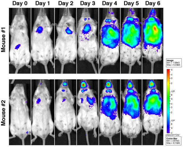
We documented the utility of pXB173-lux by testing whether it could be used as a reporter to follow F. tulanensis growth in a murine model of tularemia in real-time. We therefore challenged two BALB/c mice with ~105 cfu of LVS-pXB173-lux by the intranasal route (Fig. 3). In the first mouse, the majority of the LVS inoculum was detected in the stomach at three hours post challenge, suggesting that at least a portion of the intranasal challenge dose failed to reach the lungs and was swallowed. Twenty four-hours later, the bioluminescence production in the stomach of mouse 1 had resolved and LVS was clearly visualized in the lungs of both animals and in the upper respiratory tract of mouse two. The upper airway infection intensified over the six day course of the experiment in mouse two. It is unclear whether this upper respiratory tract infection occurs in natural inhalation infections or is an artifact of the intranasal inoculation method that is widely used by the tularemia research community. The images also showed that LVS disseminated to the cervical lymph nodes of mouse one (day 3) and mouse two (day 2) and that colonization of the lymph nodes intensified throughout the study period. Beginning on day 3 post-challenge LVS was observed in the liver of both mice, and by day 4 post-challenge, the livers of both mice were heavily colonized. On day 6 the both mice exhibited extensive bacterial dissemination which correlated with other signs of severe tularemic disease (i.e. significant weight loss, ruffled fur and reduced physical activity) and the experiment was terminated. These results validate that pXB173-lux can be used as a reporter to follow F. tularensis dissemination in mice.
Fig. 3.
Visualization of F. tularensis LVS-pXB173-lux in mice by bioluminescence imaging. Two twelve week-old BALB/c mice were challenged with 5 × 10⁵ CFU F. tularensis LVS-pXB173-lux in a total volume of 50 μl of PBS via the intranasal route. Bioluminescence production in the mice was then visualized using an IVIS Spectrum Imaging system at 24-hour intervals beginning 3 hours after administration of the challenge dose. Exposure times varied based on bioluminescent signal intensities in an effort to collect between 600 and 60,000 counts, and image scaling was normalized by converting total counts to photons/second. Results shown here are representative of several experiments of similar design.
The stability of pXB173-lux in F. tularensis Schu S4 was assessed to validate the use of this plasmid in a Type A strain background. The plasmid was well maintained in vitro with 85% of the bacteria retaining the plasmid following growth for four successive subcultures in the absence of antibiotic selection. In vivo stability was similar to the in vitro results with 82% and 75% and of the bacteria retaining the plasmid on days 5 and 6 post-challenge, respectively. This demonstrates that pXB173-lux is stable in F. tularensis Schu S4, while the data presented in figure 2 demonstrate that the lux reporter works in F. tularensis Schu S4. Collectively these results strongly suggest that pXB173-lux will be useful for in vivo studies with Type A F. tularensis strains as we have documented with F. tularensis LVS.
The results presented above show that bioluminescence is a highly sensitive reporter that can be used to follow F. tularensis growth in mice in real time. Bioluminescence represents a new tool for the tularemia research community that has not been previously available. In particular, the use of pXB173-lux can greatly facilitate animal and cell culture studies with virulent type A F. tularensis strains. As most analysis previously depended on terminal end point assays, the use of bioluminescence should greatly reduce both the labor cost and number of animals that are required for these assays while limiting the potential for occupational exposure of researchers to a potentially fatal pathogen.
Acknowledgments
This work was supported by NIH grant #U54 AI057157 from Southeastern Regional Center of Excellence for Emerging Infections and Biodefense, by NIH grants AI074582 and AI079482 (to JEB), AI061260 (to MAM), and by DOD grant W81XHW-05-1-0227. Its contents are solely the responsibility of the authors and do not necessarily represent the official views of the NIH.
Footnotes
Publisher's Disclaimer: This is a PDF file of an unedited manuscript that has been accepted for publication. As a service to our customers we are providing this early version of the manuscript. The manuscript will undergo copyediting, typesetting, and review of the resulting proof before it is published in its final citable form. Please note that during the production process errors may be discovered which could affect the content, and all legal disclaimers that apply to the journal pertain.
References
- Alexeyev MF, Shokolenko IN. RP4 oriT and RP4 oriT-R6K oriV DNA cassettes for construction of specialized vectors. Biotechniques. 1995;19:22–4. 26. [PubMed] [Google Scholar]
- Anda P, et al. Waterborne outbreak of tularemia associated with crayfish fishing. Emerging Infectious Diseases. 2001;7:575–82. doi: 10.3201/eid0707.010740. [DOI] [PMC free article] [PubMed] [Google Scholar]
- Bina XR, et al. The Bla2 beta-lactamase from the live-vaccine strain of Francisella tularensis encodes a functional protein that is only active against penicillin-class beta-lactam antibiotics. Arch Microbiol. 2006;186:219–28. doi: 10.1007/s00203-006-0140-6. [DOI] [PubMed] [Google Scholar]
- Cross TJaRLP. Francisella tularensis (tularemia) In: Mandell GL, Bennett JE, Dolin R, editors. Principles and Practice of Infectious Diseases. Churchill Livingston; Philadelphia: 2000. [Google Scholar]
- Evans ME. Francisella tularensis. Infection Control. 1985;6:381–3. [PubMed] [Google Scholar]
- Francis E. Sources of infection and seasonal incidence of tularemia in man. Public Health Reports. 1937;52:103. [Google Scholar]
- Greco D, et al. A waterborne tularemia outbreak. European Journal of Epidemiology. 1987;3:35–8. doi: 10.1007/BF00145070. [DOI] [PubMed] [Google Scholar]
- Karpoff SP, Antononoff NI. The spread of tularemia through water as a new factor in its epidemiology. Journal of Bacteriology. 1936;32:243. doi: 10.1128/jb.32.3.243-258.1936. [DOI] [PMC free article] [PubMed] [Google Scholar]
- Lavine CL, et al. Immunization with heat-killed Francisella tularensis LVS elicits protective antibody-mediated immunity. Eur J Immunol. 2007;37:3007–20. doi: 10.1002/eji.200737620. [DOI] [PubMed] [Google Scholar]
- LoVullo ED, et al. Genetic tools for highly pathogenic Francisella tularensis subsp. tularensis. Microbiology. 2006;152:3425–35. doi: 10.1099/mic.0.29121-0. [DOI] [PubMed] [Google Scholar]
- Maier TM, et al. Construction and characterization of a highly efficient Francisella shuttle plasmid. Appl Environ Microbiol. 2004;70:7511–9. doi: 10.1128/AEM.70.12.7511-7519.2004. [DOI] [PMC free article] [PubMed] [Google Scholar]
- Pavlov VM, et al. Cryptic plasmid pFNL10 from Francisella novicida-like F6168: the base of plasmid vectors for Francisella tularensis. FEMS Immunol Med Microbiol. 1996;13:253–56. doi: 10.1111/j.1574-695X.1996.tb00247.x. [DOI] [PubMed] [Google Scholar]
- Sjostedt A. Tularemia: history, epidemiology, pathogen physiology, and clinical manifestations. Ann N Y Acad Sci. 2007;1105:1–29. doi: 10.1196/annals.1409.009. [DOI] [PubMed] [Google Scholar]
- Syrjala H, et al. Airborne transmission of tularemia in farmers. Scandinavian Journal of Infectious Diseases. 1985;17:371–5. doi: 10.3109/13813458509058777. [DOI] [PubMed] [Google Scholar]
- Tarnvik A. Nature of protective immunity to Francisella tularensis. Reviews of Infectious Diseases. 1989;11:440–51. [PubMed] [Google Scholar]
- Teutsch SM, et al. Pneumonic tularemia on Martha’s Vineyard. New England Journal of Medicine. 1979;301:826–8. doi: 10.1056/NEJM197910113011507. [DOI] [PubMed] [Google Scholar]


