Abstract
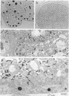
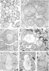
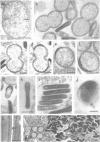
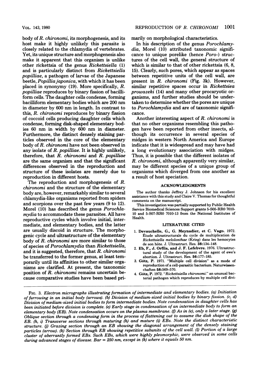
The reproduction and morphogenesis of Rickettsiella chironomi, an unusual procaryotic parasite of midge larvae, was studied with electron microscopy. The morphogenic cycle was similar to that found in chlamydia and consisted of four major cell types: (i) medium-sized spherical initial bodies (ca. 1 micrometers in diameter), (ii) large spherical initial bodies (1.5-2 micrometers), (iii) spherical intermediate bodies (600 to 700 nm), and (iv) disk-shaped elementary bodies (60 X 600 nm). The primary mode of reproduction involved binary fission of initial bodies to form other initial bodies or intermediate bodies. Each intermediate body condensed, forming an elementary body. The morphogenesis of R. chironomi is compared with that of several other organisms to which it is possibly related, including vertebrate chlamydia and invertebrate pathogens of the genera Rickettsiella and Porochlamydia, and its taxonomic position in regard to these is discussed. Additionally, a brief description of the pathology caused by the development of R. chironomi in larvae of Chironomus decorus and Chironomus frommeri is given.
Full text
PDF
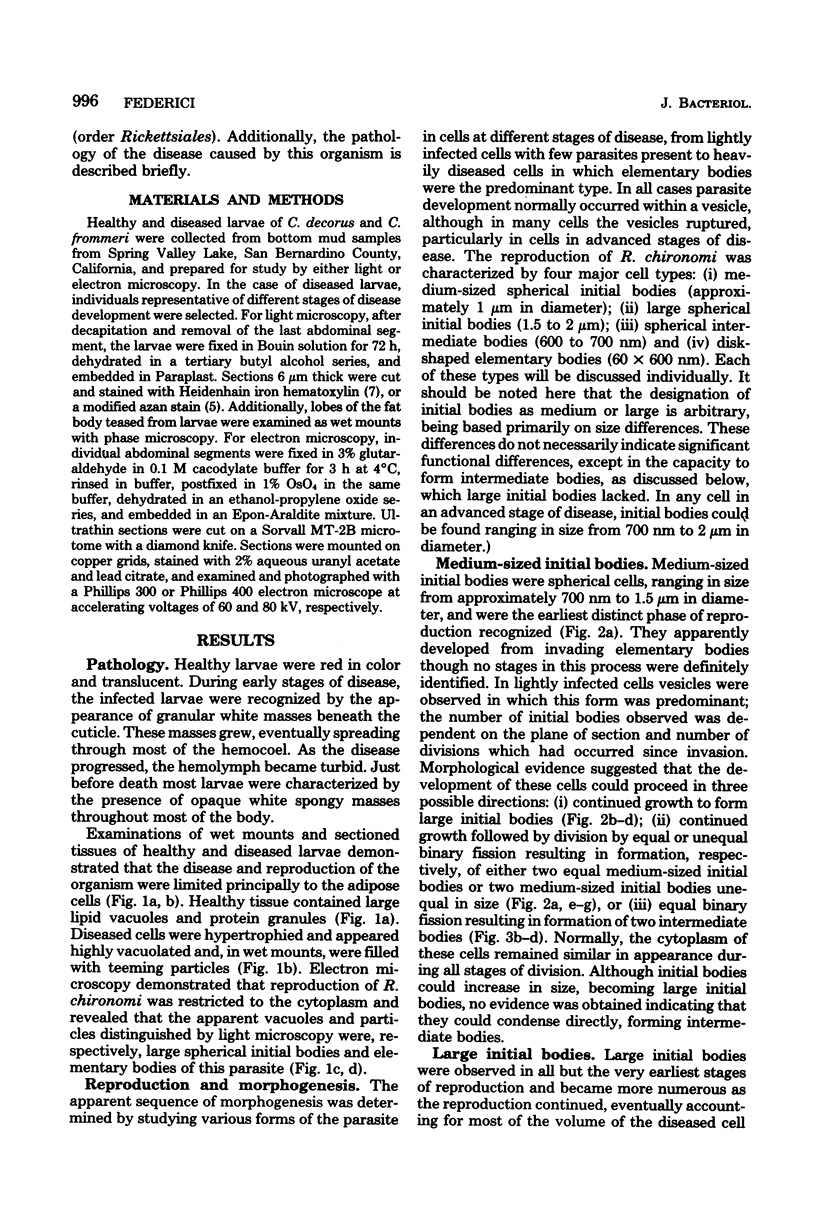
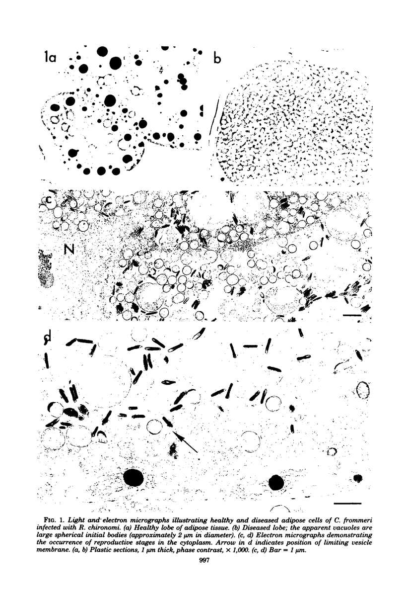
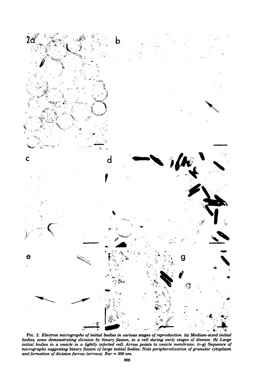
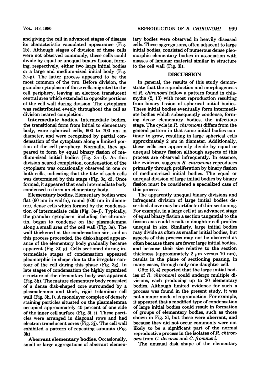

Images in this article
Selected References
These references are in PubMed. This may not be the complete list of references from this article.
- Devauchelle G., Meynadier G., Vago C. Etude ultrastructurale du cycle de multiplication de Rickettsiella melolonthae (Krieg), Philip, dans les hémocytes de son hôte. J Ultrastruct Res. 1972 Jan;38(1):134–148. doi: 10.1016/s0022-5320(72)90088-3. [DOI] [PubMed] [Google Scholar]
- Eb F., Orfila J., Lefebvre J. F. Ultrastructural study of the development of the agent of ewe's abortion. J Ultrastruct Res. 1976 Aug;56(2):177–185. doi: 10.1016/s0022-5320(76)80164-5. [DOI] [PubMed] [Google Scholar]
- Götz P. "Multiple cell division" as mode of reproduction of a cell-parasitic bacterium. Naturwissenschaften. 1971 Nov;58(11):569–570. doi: 10.1007/BF00598727. [DOI] [PubMed] [Google Scholar]
- Hamm J. J. A modified azan staining technique for inclusion body viruses. J Invertebr Pathol. 1966 Mar;8(1):125–126. doi: 10.1016/0022-2011(66)90113-3. [DOI] [PubMed] [Google Scholar]
- Hayes S. F., Burgdorfer W. Ultrastructure of Rickettsia rhipicephali, a new member of the spotted fever group rickettsiae in tissues of the host vector Rhipicephalus sanguineus. J Bacteriol. 1979 Jan;137(1):605–613. doi: 10.1128/jb.137.1.605-613.1979. [DOI] [PMC free article] [PubMed] [Google Scholar]
- ITO S., VINSON J. W. FINE STRUCTURE OF RICKETTSIA QUINTANA CULTIVATED IN VITRO AND IN THE LOUSE. J Bacteriol. 1965 Feb;89:481–495. doi: 10.1128/jb.89.2.481-495.1965. [DOI] [PMC free article] [PubMed] [Google Scholar]
- Palmer E. L., Mallavia L. P., Tzianabos T., Obijeski J. F. Electron microscopy of the cell wall of Rickettsia prowazeki. J Bacteriol. 1974 Jun;118(3):1158–1166. doi: 10.1128/jb.118.3.1158-1166.1974. [DOI] [PMC free article] [PubMed] [Google Scholar]
- Silverman D. J., Wisseman C. L., Jr Comparative ultrastructural study on the cell envelopes of Rickettsia prowazekii, Rickettsia rickettsii, and Rickettsia tsutsugamushi. Infect Immun. 1978 Sep;21(3):1020–1023. doi: 10.1128/iai.21.3.1020-1023.1978. [DOI] [PMC free article] [PubMed] [Google Scholar]


