Abstract
Birds have excellent visual abilities that are comparable or superior to those of primates, but how the bird brain solves complex visual problems is poorly understood. More specifically, we lack knowledge about how such superb abilities are used in nature and how the brain, especially the telencephalon, is organized to process visual information. Here we review the results of several studies that examine the organization of the avian telencephalon and the relevance of visual abilities to avian social and reproductive behavior. Video playback and photographic stimuli show that birds can detect and evaluate subtle differences in local facial features of potential mates in a fashion similar to that of primates. These techniques have also revealed that birds do not attend well to global configural changes in the face, suggesting a fundamental difference between birds and primates in face perception. The telencephalon plays a major role in the visual and visuo-cognitive abilities of birds and primates, and anatomical data suggest that these animals may share similar organizational characteristics in the visual telencephalon. As is true in the primate cerebral cortex, different visual features are processed separately in the avian telencephalon where separate channels are organized in the anterior-posterior axis roughly parallel to the major laminae. Furthermore, the efferent projections from the primary visual telencephalon form an extensive column-like continuum involving the dorsolateral pallium and the lateral basal ganglia. Such a column-like organization may exist not only for vision, but for other sensory modalities and even for a continuum that links sensory and limbic areas of the avian brain. Behavioral and neural studies must be integrated in order to understand how birds have developed their amazing visual systems through 150 million years of evolution.
Key Words: Birds, Columnar organization, Courtship, Parallel processing, Pigeons
Introduction
The visual abilities of birds are exceptional [Hodos, 1993; Frost and Sun, 1997; Shimizu et al., 2008; Güntürkün, 2000]. Their superb color perception, spatial and temporal resolving power, and visual learning and memory capabilities have been thoroughly investigated and documented. For example, birds have four types of cone photopigments with which they can see ultraviolet or near ultraviolet light in addition to the part of the spectrum that humans can see [Bowmaker et al., 1997]. In addition, many birds, and in particular predatory birds such as eagles and hawks, have high resolving power. Their globose-shaped eyes and high density of retinal cells enable them to see fine details much better than humans [Hodos, 1993]. For example, humans can detect only 30 cycles/degree while American kestrels (Falco sparverius) can detect 46 cycles/degree [Gaffney and Hodos, 2003]. Birds also have a superb temporal sensitivity for fast-changing images. In pigeons (Columba livia), the temporal resolution threshold reaches up to 100 Hz whereas human observers can detect just about 60 Hz [Hodos et al., 2003]. Finally, avian visuo-cognitive abilities are impressive too [Waldvogel, 1990; Cook, 2001; Wasserman et al., 2006]. For instance, in their classic concept formation study, Herrnstein and Loveland [1964] used operant-conditioning techniques to train pigeons to discriminate a number of stimulus photographs into two categories: photographs with people and those without people. Pigeons not only learned to discriminate them successfully, but they could also generalize this classification rule to novel photographs they had not previously seen. The results were interpreted to demonstrate that birds can form a visual concept of people. Using similar methods, Watanabe et al. [1995] trained pigeons to discriminate the stylistically different paintings of Monet (Impressionism) and Picasso (Cubism). When birds were then tested with additional paintings by these artists that had not been used during the training, they were also able to generalize the classification rule to successfully discriminate the two styles of paintings.
In this paper, three issues associated with the impressive visual abilities of birds are presented. The first issue concerns the adaptational value of these visual capabilities. Why do birds have such excellent visual abilities? How do they use such abilities in nature? Undoubtedly, the demand for fast and accurate aerial maneuvering during flight is one of the major reasons that birds have a highly developed visual system. Visual cues are also abundant and important among conspecifics for complex social maneuvering. They most likely use these cues to select mates, identify rivals, locate young, and differentiate members of higher and lower ranks. These are not easy tasks, considering that such visual stimuli are constantly viewed from different visual angles, under different levels of luminance, and from different distances. A series of behavioral studies are discussed that reveal that birds use their visual abilities to detect and evaluate the faces of potential mates.
The second and third issues concern the neural substrates underlying the outstanding visual performance of birds. There are two anatomical characteristics of the avian visual telencephalon that have not been well recognized and fully appreciated: (1) parallel processing and (2) columnar organization. As for the former, visual processing in the avian brain is highly distributed, as in mammals. Different visual features are processed and analyzed in physically distinct regions in the avian forebrain. This parallel processing in the telencephalon is similar to the functional segregation found in the dorsal and ventral streams of the mammalian cortices, each component of which is associated with specific visual functions. Such similarities suggest the existence of basic neural principles for visual processing shared by highly visual birds and primates.
Finally, a columnar organization in the visual telencephalon is discussed. The avian telencephalon is known to consist of multiple nuclear cell groups. For the visual system, relevant nuclei are ostensibly anatomically separate and spatially distant within the avian telencephalon. These nuclei are actually organized in a more systematic fashion than was previously understood. Thus, there is an extensive columnar continuum involving different telencephalic structures separated by several laminae. Such a column-like organization may exist not only for vision, but other sensory modalities and even non-sensory systems, suggesting that a columnar organization is another neural principle shared by avian and mammalian telencephalons. In particular, we suggest that there is a column-like continuum that links sensory and limbic areas of the avian brain. The expansion of this continuum may be relevant to the enlargement of the telencephalon in some species, such as corvids, that are known for their complex behavior.
Recognition of a Potential Mate
Video Playback Techniques
Pigeons are social animals. They often communicate with each other by exhibiting distinct behaviors, such as courtship displays in front of potential mates. Accordingly, the occurrence of a courtship display indicates that a bird ‘recognizes’ the visual object as a potential mate [Shimizu, 1998; Partan et al., 2005; Patton et al., 2010]. In studies in our laboratory, as well as that of Barrie J. Frost and Nikolaus F. Troje [Frost et al., 1998], subject pigeons were exposed to live stimulus pigeons, videotaped pigeons, and photographs of pigeons to examine which visual features can trigger spontaneous courtship displays.
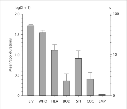
In order to compare the effects of various visual stimuli, a metric for measuring courtship responses needed to be developed. In our laboratory, subjects’ naturally occurring behaviors toward a real live potential mate were investigated for this purpose. In a study by Shimizu [1998], subject males were individually placed in an open-field apparatus with opaque walls. In one wall there was an embedded Plexiglas window, through which males could view a caged live stimulus female. The top of the open-field apparatus and the stimulus cage were covered only by wire-mesh ceilings, and thus the subject and stimulus birds could see, hear, smell and interact with each other. All responses of male subjects were video- and audio-recorded and several robust behavioral categories were identified, including bowing, tail-dragging and vocalization. In bowing, a male lowers its head while turning in full or half circles. In tail-dragging, a bird runs a short distance while dragging its spread tail along the ground. In vocalization, a bird produces a ‘coo’ sound, usually while bowing. The majority of male subjects exhibited these responses vigorously to a live stimulus. The frequency and duration of these responses were collected by analyzing video- and audio-recordings. The live female pigeon (LIV) condition in figure 1 shows the duration of ‘coo’ vocalizations. The response patterns were very similar for bowing and tail-dragging.
Fig. 1.
A histogram showing the duration of ‘coo’ vocalizations elicited in front of a live female pigeon (LIV) and six types of video-playback stimuli. The visual stimuli included a whole dynamic female pigeon (WHO), the head region (HEA), the body region (BOD), still images of a whole female pigeon (STI), a female cockatoo (COC), and the empty cage (EMP). Values are expressed as group means ± SE log display durations.
Once the baseline responses to a live stimulus were established, then video-playback stimuli were presented on a monitor screen in place of a live stimulus. One of these video stimuli was a life-sized, video-playback stimulus of a female pigeon (WHO in fig. 1). No auditory cues were included. The results showed that male pigeons exhibited stereotyped responses quite vigorously in front of the video playback. The results were consistent with many previous studies, in which taxidermic models of female birds effectively elicited courtship responses from males [Fisher and Hale, 1957; Carbaugh et al., 1962; Domjan et al., 1989; Crawford and Akins, 1993]. Thus, the video-playback study confirmed two conclusions from these previous studies. First, because only visual cues were included in video-playbacks and taxidermic models, a visual signal alone is sufficient to trigger courtship responses even without any other sensory signals. Second, reactions from a female or interactions between males and females are not necessarily essential for eliciting males’ responses, although such an interaction plays a significant role in actual courtship in nature [Lehrman, 1964; Burley, 1977; Patricelli et al., 2006].
The significant results of video-playback stimuli further emphasize the potency of visual signals in courtship, considering the fact that the quality of a video-playback stimulus is much poorer compared to both real live stimulus birds and taxidermic models [D'Eath, 1998]. The video-playbacks were presented on a 2-dimensional monitor screen that was designed for the eyes of human observers. Video stimuli cannot provide depth information nor accurate color information for the avian visual system. However, the fact that video-playbacks can trigger courtship responses does not imply that the stimulus deficiencies are irrelevant for the expression of courtship responses. In one of our studies [Shimizu, 1998], each trial in the study lasted only a short period of time (2 min). Subsequently, we found that subject birds reacted longer to live stimuli than to video-playback stimuli as the duration of a trial was extended in similar experiments.
In addition to the whole dynamic female pigeon (WHO), other visual stimuli were presented on the monitor screen [Shimizu, 1998]. These stimuli included only the head portion (HEA) or body portion (BOD) of a video-taped female pigeon, motionless images of the whole female (STI), and a video-taped female cockatoo (COC). The results showed that two visual features of a female are particularly effective in triggering reactions from males. First, the head region of a female pigeon was important. In the head (HEA) and body (BOD) conditions, only parts of the female pigeon were visible since the rest was occluded. Male subjects exhibited courtship displays for a much longer period to the head (HEA) stimulus than to the body (BOD) stimulus. Second, the stimulus was more effective when it was in motion. The motionless images (STI) resulted in shorter response durations compared to the dynamic view of the female (WHO). However, it was not solely the presence of movement that elicited responses from male subjects. Males showed almost no responses to a heterospecific stimulus (COC), even though it too was constantly in motion. Thus, species-specific patterns of movement may be more effective than other kinds of movement.
Significance of the Face
The results showing the efficacy of the head region of pigeons in eliciting courtship responses were in accordance with previous studies involving Japanese quail (Coturnix coturnix japonica). Domjan and Nash [1988] showed that males spent almost the same amount of time looking through a window at models of just the female head region compared to a full taxidermic model of a female quail. Domjan et al. [1989] further showed that female quail models of only the head region elicited as much copulatory behaviors from male subjects as a live female bird. Altogether, these studies suggest that the head region of potential mates is visually more salient, as well as socially important, compared to the body region. It is possible that males pay attention to the head region simply because it is situated in the upper part of a female body. In both pigeons and quails, males and females are about the same size and height. When males detect a potential mate, the head of a female is salient because the upper part of the body happens to be at the eye level of males. However, a more likely reason that the female head region is important is because it includes the face, characterized by a number of local elements – the eyes, beak, cere (the exposed skin at the base of the upper beak) and plumage. The manner in which these elements are spatially arranged is likely also important as a global feature for defining the face.
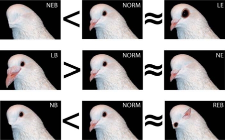
In order to examine these two possibilities, Patton et al. [2010] conducted a preference test, in which male pigeons were exposed simultaneously to pairs of photographic images of females. These stimuli were presented on the screens of two monitors, each of which was seen through a window in the opposing wall of an open-field apparatus. On one monitor screen, images of a female pigeon with a normal head were presented, whereas images of the same female with digitally and systematically altered facial features were shown on the opposite monitor. Males’ courtship responses displayed near each monitor were measured and compared to determine the significance of specific facial features. Instead of video-playback stimuli, static photographs had to be used in this study in order to make precise alterations to the facial features. They facilitated males’ responses by presenting successively different photographs of a female every five seconds. The results showed that male subjects reacted quite vigorously to these static stimuli. Figure 2 shows some examples of the normal (NORM) and altered stimulus categories used in the study. These stimuli included the following alterations: (1) no eyes or beak condition (NEB; both eyes and beak were removed); (2) large beak condition (LB; beak size was enlarged); (3) no beak condition (NB; the beak was removed); (4) large eyes condition (LE; the eye size was enlarged); (5) no eyes condition (NE; the eyes were removed), and (6) rearranged eyes and beak condition (REB).
Fig. 2.
Photographic images of altered and unaltered facial features of a female pigeon. NORM = Normal unaltered image; NEB = no eyes or beak; LB = large beak; NB = no beak; LE = large eyes; NE = no eyes; REB = rearranged eyes and beak. Significant preferences are represented by the symbols > and <, whereas no clear preferences are represented by the symbol ≈.
The results of the preference test between the intact original and these alterations are summarized in figure 2, in which the preferences based on ‘coo’ vocalization durations were depicted by three symbols, <, >, and ≈. As seen in figure 2, males preferred (i.e. vocalized longer near) the intact female images compared to those missing the eyes and beak (NEB condition in fig. 2). This means that male pigeons prefer the female with the facial features, and that pigeons must naturally pay attention to the face of a potential mate. When the effects of the beak and eyes were compared, the results showed that enlarging or removing the beak had a significant impact on preference (LB and NB conditions), whereas manipulating the eyes had a much weaker effect (LE and NE conditions). The morphology of the avian beak is associated with feeding [Weiner, 1995; Grant and Grant, 2002] and preening behavior to remove harmful ectoparasites [Clayton et al., 2005]. The presence and size of the beak of a female may be a useful visual feature to evaluate mates in terms of the efficacy of feeding and parasite control. The finding that the manipulation of the eyes did not have as strong an effect as the beak appears to be counterintuitive because previous studies showed that avian eye models triggered robust reactions [Blest, 1957; Scaife, 1976; Emery, 2000; Watve et al., 2002]. The main difference between these studies and the study by Patton et al. [2010] is whether stimulus eyes belong to dangerous predators or harmless conspecifics. Because the appearance of eyes by themselves is not necessarily different between a predator and prey, the location of stimulus eyes in the head may convey important cues to trigger different reactions. The eyes of predatory birds, like owls, are situated in the front of the head to widen the binocular vision for hunting, whereas the majority of birds, including pigeons, have laterally situated eyes to increase monocular visual fields for vigilance. The study by Patton et al. [2010] also showed that male pigeons did not discriminate between intact normal faces and globally altered faces in which local features were spatially rearranged in the REB condition. The results suggested that when birds see the face of another bird, they are predisposed to attend to local facial features, rather than the configuration of these features. This does not mean that birds are incapable of paying attention to global configuration. Many previous studies have shown that global aspects of visual stimuli can be used as discriminative stimuli [Wasserman et al., 1993; Kirkpatrick-Steger et al., 1996, 1998; Fremouw et al., 1998, 2002; Cavoto and Cook, 2001; Aust and Huber, 2003]. Finally, Patton et al. [2010] used only the faces of female birds as stimuli. Even if they used the faces of male pigeons as stimuli, the results would most likely be similar because previous studies suggest that the static visual cues of pigeons are not greatly sexually dimorphic. For example, Burley [1981] observed that when male pigeons encountered conspecific strangers, they initially started courting indiscriminately both female and male pigeons. Nakamura et al. [2006] also showed that when they trained pigeons to discriminate between the photographs of males and females, only some of the subjects were able to do so.
In humans and other primates, the faces of conspecifics are socially vital and visually salient stimuli. Numerous primate studies show that there are certain brain regions specifically associated with the processing of facial information (fusiform face area) [Grill-Spector et al., 2004]. The observation that some bird species also pay special attention to subtle differences in facial features of conspecifics is intriguing since it suggests that the avian visual system may also be equipped with such a neural mechanism that processes facial features. At the same time, there are also significant differences between birds and primates in terms of face perception. For instance, humans and other primates naturally attend to the eyes of conspecifics [Kano and Tomonaga, 2009, 2010]. As seen above, this appears not to be true for pigeons. Similarly, the global configuration of facial features is not important for pigeons whereas it provides an essential cue for individual recognition in humans.
Parallel Processing in the Avian Visual System
Functional Segregation in the Collothalamic Pathway
Underlying their superb visual abilities, birds have a highly developed and differentiated neural system [Zeigler and Bischof, 1993; Güntürkün, 2000; Shimizu, 2001; Shimizu et al., 2008]. Especially in species with lateral eyes, the majority of retinal fibers terminate in the optic tectum of the midbrain, which in turn projects to the nucleus rotundus, the largest nucleus of the dorsal thalamus. Neurons of the nucleus rotundus further send prominent projections to the entopallium in the telencephalon [Karten and Hodos, 1970; Benowitz and Karten, 1976; Husband and Shimizu, 1999; Laverghetta and Shimizu, 2003; Krützfeldt and Wild, 2004, 2005]. This collothalamic visual pathway plays a pivotal role in visual information processing in birds. Damage to different centers of this pathway causes devastating effects on performance in various visual tasks, including color, brightness and pattern discriminations [Hodos, 1993].
The avian optic tectum is a large, highly laminated structure, where the visual input projects retinotopically to the superficial layers. The retinotopic organization disappears as neurons of the deeper layers of the tectum send projections to the nucleus rotundus in a non-topographic fashion. Instead, different attributes of visual information are processed in separate and parallel channels in the nucleus rotundus and entopallium. The nucleus rotundus consists of anatomically distinct subdivisions, such as the anterior, central and posterior parts [Benowitz and Karten, 1976; Mpodozis et al., 1996]. They can be differentiated on the basis of cell type, density and neurochemical contents [Martinez-de-la-Torre et al., 1990; Laverghetta and Shimizu, 2003]. For instance, the rotundal subdivisions can be visualized using the distribution pattern of a calcium-binding-protein (calbindin)-like immunoreactivity [Husband and Shimizu, 1999]. These different parts receive projections from different classes of neurons in the deeper layers of the optic tectum [Hellman and Güntürkün, 2001; Marín et al., 2003].
Using electrophysiological techniques, Frost and his colleagues showed that neurons in the anatomically different regions in the nucleus rotundus have physiologically different response characteristics [Wang et al., 1993]. Their results showed that changes in color and luminance features of visual stimuli increased responses in neurons of the anterior rotundus whereas moving stimuli increased responses in the central and posterior rotundus. The results suggest that static and dynamic attributes of visual stimuli are processed in parallel channels within the nucleus rotundus. When selective lesion studies were conducted, the results were consistent with the physiological data. Lesions in the anterior part, but not in the central and posterior parts of the rotundus, resulted in deficits in color discrimination tasks [Laverghetta and Shimizu, 1999]. Together, these data suggest that the nucleus rotundus can be divided into anatomically, physiologically and functionally segregated divisions.
Parallel Processing in the Telencephalon
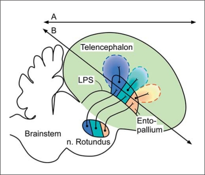
The parallel channels of the collothalamic pathway extend from the thalamic level to higher visual structures in the telencephalon. Previous connection studies [Benowitz and Karten, 1976; Husband and Shimizu, 1999] suggested that there are some topographical relationships between subdivisions of the nucleus rotundus and different parts of the entopallium. Anterograde tracers have been injected into different subdivisions of the nucleus rotundus in zebra finches (Taeniopygia guttata) [Laverghetta and Shimizu, 2003]. The results showed that the projection sites of the anterior, central and posterior rotundus appeared to be topographically organized in the entopallium along the anterior-posterior axis. Thus, as the source of the ascending projection moved caudally in the nucleus rotundus, the target of the projection in the entopallium also shifted caudally. This projection pattern was later confirmed in finches and pigeons [Krützfeldt and Wild, 2004, 2005]. These results suggested that the primary visual area in the telencephalon maintains separate and parallel channels (fig. 3).
Fig. 3.
Schematic diagram showing the parallel system within the collothalamic visual pathway in a sagittal section of a bird brain. Different subdivisions of nucleus rotundus send projections to different subdivisions of the entopallium. Areas defined by dotted lines represent the higher visual areas receiving projections from the entopallium. Line A represents the axis according to the standard coordinate system for bird brains, whereas line B represents the axis roughly parallel to the major laminae in the telencephalon. LPS = Pallium-subpallium lamina.
Several studies have shown that the functional segregation of the parallel channels extends from the thalamus to the telencephalon. For instance, in collaboration with Robert Cook, we made lesions selectively in different portions of the entopallium to study the behavioral effects on various visual tasks [Patton et al., 2004]. All birds were tested simultaneously with two different tasks: (1) discrimination of different textures of color and form (i.e. static-feature analysis), and (2) discrimination of different types of motion (dynamic-feature analysis). The results suggest the possibility of a double dissociation in structure and function. Birds with anterior entopallium lesions showed significant deficits in the color/form task and very little decline in the motion task. In contrast, birds with posterior entopallial lesions showed greater deficits in the motion task than in the color/form task. Nguyen et al. [2004] also carried out a similar, but more systematic, lesion study using different visual discrimination tasks and reached basically the same conclusion regarding this functional segregation in the telencephalon. Finally, physiological data suggest that the response characteristics of neurons in the posterior entopallium are similar to those of the posterior rotundus [Xiao et al., 2006; Xiao and Frost, 2009].
The parallel visual channels extend into even higher areas than the entopallium of the telencephalon. Small injections of tracers were made to clarify the efferent connections of different portions of the entopallium [Husband and Shimizu, 1999]. The results showed that different parts of the entopallium send projections to different telencephalic regions, which are also organized roughly along the anterior-posterior axis. These telencephalic regions include part of the lateral portion of the frontal nidopallium (NFL), the temporo-parieto-occipital area (TPO), the lateral portion of the intermediate nidopallium (NIL) as well as the lateral portion of the caudal nidopallium (NCL). These results suggest that different features of visual stimuli (e.g. static and dynamic features) are processed separately in anatomically different parallel channels in the thalamic nucleus (rotundus), primary telencephalic area (entopallium) and higher telencephalic areas (e.g. NFL, TPO, NIL and NCL).
Still, comparatively little is known about anatomical, physiological and functional segregation among these secondary visual areas. Among these higher visual areas, NIL receives major projections from the posterior entopallium. The NIL is difficult to distinguish from the surrounding region in terms of cytoarchitecture and neurochemical contents. We found out that this region can be visualized by analyzing the expression of an immediate early gene zenk. This immediate early gene (also known as zif-268, Egr-1, NGFI-A and Krox-24) encodes transcriptional regulators and is believed to be a crucial step in the cellular process underlying long-term memory formation. It has been used extensively in birds to specify various cell groups characterized by gene expression following an exposure to specific stimuli or behaviors. These studies include the analysis of the song control system in songbirds [Mello et al., 1992; Jarvis et al., 1995; Mello and Ribeiro, 1998; Jarvis et al., 2000], sexual behavior in quail and starlings (Sturnus vulgaris) [Ball and Balthazart, 2001; Can et al., 2007], sexual imprinting in finches [Lieshoff et al., 2004; Huchzermeyer et al., 2006] and homing behavior in pigeons [Shimizu et al., 2004]. In general, high levels of ZENK protein have been observed especially in the avian telencephalon compared to the lower brain regions, and, within the telencephalon, higher sensory and/or association regions compared to the primary sensory regions. Studies in our laboratory have shown that the NIL region contained more ZENK-positive cells when pigeons were exposed to a potential mate than an empty chamber [Patton et al., 2009]. Similar findings have been reported in the finch brain area LNH, which likely corresponds to part of the NIL region in pigeons. After their ‘first courtship’ experience, neurons of LNH showed increased levels of ZENK [Lieshoff et al., 2004] and another immediate early gene protein (FOS) [Sadananda and Bischof, 2002, 2006].
Comparison with the Primate Visual System
In primates, the primary visual route from the retina to the telencephalon is the lemnothalamic pathway via the lateral geniculate nucleus [Livingstone and Hubel, 1988]. Lesions in the lemnothalmic pathway cause severe effects on vision, including deteriorated color perception, acuity, and blindness, which are comparable to lesion effects in the avian collothalamic pathway. For primates, the collothalamic visual pathway (retina-superior colliculus-extrastriate cortex) is mainly involved in visuomotor behavior, such as orienting and attending to a visual stimulus. The avian lemnothlamic pathway runs from the retina to the dorsal thalamus to the visual Wulst in the telencephalon. This pathway is relatively minor for many birds (especially those with lateral eyes), and lesions here cause relatively minor deficits in many visual discrimination tasks [Hodos, 1993, but see Shimizu and Hodos, 1989; Güntürkün and Hahmann, 1999; Budzynski et al., 2002; Mouritsen et al., 2005]. Therefore, the avian collothalamic pathway and the primate lemnothalamic pathway are functionally comparable in that both play essential and comparable roles in visual processing [Bischof and Watanabe, 1997; Shimizu and Bowers, 1999].
Despite the aforementioned similarities, caution is clearly warranted when comparing the avian collothalamic pathway and the primate lemnothalamic pathway, which are anatomically different and evolutionarily distinct [Nguyen et al., 2004]. Nevertheless, the parallel processing found in the avian collothalamic pathway is reminiscent of the functional segregation found in the separate channels of the primate lemnothalamic visual pathway. In the primate retina, parvocellular neurons are responsive to fine details and color of stimuli whereas magnocellular neurons respond strongly to moving stimuli. The functional segregation found at the retinal level is maintained at the lateral geniculate and the primary visual cortex as well. Within the cerebral cortex, visual information from the primary visual cortex travels further to multiple higher visual areas, where there are at least two distinct pathways: the dorsal and ventral streams [Ungerleider and Mishkin, 1982]. With mostly parvocellular input, neurons in the ventral stream are sensitive to details of shape and color. In contrast, neurons in the dorsal stream received mostly magnocellular input, sensitive to movements. These observations of the avian and primate visual pathways suggest the possibility that these similar principles of functional segregation are the neural foundation for the exceptional visual and vision-associated cognitive abilities for each animal group. At the same time, there are several significant differences between the pathways of birds and primates. For example, there are extensive interactions between the two visual channels and among the primary and higher visual areas in primates [DeYoe and Van Essen, 1988; Merigan and Maunsell, 1993; Nassi and Callaway, 2009], whereas reciprocal connections between the visual areas in the avian forebrain appear to be limited [Husband and Shimizu, 1999]. These anatomical differences may be related to visual behavioral differences between birds and primates. As described above, one clear difference revealed by ethological studies is that birds are predisposed to attend to local facial features, but not to global features. The perception of a global configuration may require the presence of such interactive circuitries within higher visual areas.
Columnar Organization in the Avian Telencephalon
Organization in the Visual Telencephalon
The majority of areas in the avian telencephalon consist of a voluminous nuclear mass called the dorsal ventricular ridge (DVR). It is comprised of several pallial regions, including the mesopallium, nidopallium and arcopallium [Reiner et al., 2004; Jarvis et al., 2005]. While the exact mammalian counterpart of the avian DVR is still debated [Karten, 1969, 1991; Karten and Shimizu, 1989; Bruce and Neary, 1995; Northcutt and Kaas, 1995; Puelles et al., 2000; Butler and Molnár, 2002; Striedter, 2004; Reiner et al., 2005], the avian DVR clearly plays a major role in various sensory, motor, and cognitive functions, as is the case with the mammalian neocortex. However, neurons of DVR are aggregated as multiple nuclear clusters and nuclei in a non-laminar fashion, instead of a cortex-like laminar fashion. For different functions, a specific neural circuitry is formed by a group of nuclei that are closely interconnected, but spatially dispersed in DVR. The apparent lack of a familiar laminar arrangement often prevents us from recognizing a coherent organization or architecture in these neural circuitries, giving the erroneous impression that neural computation in the avian telencephalon is carried out not as efficiently as we assume a more orderly, laminar system would.
A close examination of these circuitries in DVR suggests that a more systematic organization exists in the avian telencephalon than previously considered. In particular, many of these circuits appear to form an orderly columnar organization, although it is sometimes severely distorted. One main reason that such an organization is difficult to identify in standard brain sections is due to the ‘rotated’ nature of commonly available coordinate systems for bird brains. For many systems, the beak (i.e. the anterior fixation point) is placed 45 degrees below the horizontal plane defined by the ear bar (i.e. the posterior fixation point) of the stereotaxic instrument. This rotation is useful especially when the brainstem structures are compared to the mammalian equivalents in transverse sections [e.g. Karten and Hodos, 1967; Bischof, 1981], but, at the same time, it can cause confuse examinations of telencephalic organization.
In the case of the visual system, sensory information through the collothalamic pathway reaches the entopallium, which is a part of the DVR. While, as described above, this rotundus-entopallium connection is organized along the antero-posterior axis, the axis is actually rotated relative to the axis of a standard coordinate system. On transverse sections based on a standard coordinate system, injections of anterograde tracers into the anterior rotundus produced terminal fields in the anterior and also ventral portions of the entopallium, whereas injections into the caudal rotundus showed terminals in caudal and dorsal portions of the entopallium. These results could mistakenly imply that the rotundus-entopallial correspondence is more complicated and convoluted than it really is. However, as seen in figure 3, the antero-posterior axis of the entopallium is not matched to the horizontal plane of a coordinate system, but parallel to major telencephalic laminae, such as the pallial-subpallial lamina (LPS). Accordingly, each subdivision of the entopallium forms a column-like organization almost perpendicular to these laminae.
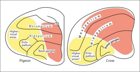
In addition to their afferents, the main efferent projections of different parts of the entopallium are also arranged in a column-like fashion. The anterior, intermediate and posterior entopallium all send dorsolateral projections which eventually reach anterior (NFL), intermediate (TPO) and posterior regions (NIL and NCL), respectively near the edge of the telencephalon. Veenman et al. [1995] designated these regions, as well as other somatic regions associated with different modalities, as the pallium externum. The pallium externum further gives rise to a projection to the lateral striatum [Veenman et al., 1995]. The entopallium also has reciprocal connections with a nucleus in the lateral mesopallium. This nucleus resides dorsal to the entopallium [Husband and Shimizu, 1999; Krützfeldt and Wild, 2004, 2005]. These findings suggest that the visual columns extend from the entopallium to higher pallial areas in the dorsolateral telencephalon, which in turn have close connections with the lateral portion of the basal ganglia (fig. 4). Thus, these visual columns form extensive dorsolateral continuums across DVR and basal ganglia. Studies using zenk expression show that such a columnar organization is not unique to vision, but is also present in other sensory modalities and movement-associated areas [Feenders et al., 2008].
Fig. 4.
Schematic diagrams showing transverse sections of the telencephalon of the pigeon (left) and the crow (right). The vision-related continuum is situated in the lateral region, whereas the limbic-associated continuum is located in the medial region.
Organization of the Limbic-Associated Telencephalon
A columnar organization may not be unique to the somatic telencephalon. It may also exist in the limbic-associated regions, the medial DVR and basal ganglia in particular. In birds, the medial basal ganglia are located just lateral to the ventral portion of the lateral ventricle. The rostral and intermediate portions of the medial basal ganglia in particular are considered to be part of the limbic system, including the nucleus accumbens and ventral pallidum. When Husband and Shimizu [submitted] injected anterograde and retrograde tracers into the medial basal ganglia at rostral and intermediate levels, the results showed that there are prominent reciprocal connections between the medial basal ganglia and the dorsally located medial DVR regions (i.e. medial nidopallium and mesopallium). The injections into the medial basal ganglia also revealed connections with the medial hyperpallium and hippocampus, forming an extensive medial continuum almost parallel to the lateral ventricle. The results are consistent with a previous study in chicks, in which tracer injections in the intermediate medial DVR resulted in extensive anterograde labeling in the medial basal ganglia [Metzger et al., 1998]. The results are also consistent with the many retrogradely labeled cells in the medial DVR after injections in the medial basal ganglia in chicks [Székely et al., 1994] and pigeons [Veenman et al., 1995; Kröner and Güntürkün, 1999] and the connections with the hippocampus [Veenman et al., 1995; Székely and Krebs, 1996; Atoji et al., 2002].
The exact functional roles of the medial pallium, and its potential mammalian equivalent, are not clear. In galliforms, this area is involved in learning and memory processes, rather than sensory processing or motor control. More specifically, it has been associated with visual, acoustic, and sexual imprinting, whereby preference is developed for a certain stimulus in the formation of filial behavior [Maier and Scheich, 1987; Gruss and Braun, 1996; Horn, 1998; Bolhuis, 1999; Thode et al., 2005] or mate choice [Bischof and Herrmann, 1986; Scheich et al., 1992]. Metzger et al. [1998] proposed that part of the avian medial pallium corresponds to the prefrontal cortex of mammals. As the possible avian equivalent of the prefrontal cortex, the NCL in particular is more commonly suggested in terms of chemical, hodological and functional evidence [Mogensen and Divac, 1982; Waldmann and Güntürkün, 1993; Kröner and Güntürkün, 1999; Güntürkün and Durstewitz, 2001]. However, part of the medial pallium also has several characteristics similar to the mammalian prefrontal cortex. In terms of connections, the close association with the limbic regions, such as hippocampus and medial basal ganglia is similar to the prefrontal neocortex [Heimer et al., 1985, 1997]. Like the prefrontal cortex, the medial pallium receives input from higher sensory areas, such as NCL, which in turn receives secondary and tertiary input from the visual, auditory, and trigeminal systems [Bonke et al., 1979; Wild et al., 1993; Leutgeb et al., 1996; Metzger et al., 1996; Kröner and Güntürkün, 1999]. Furthermore, the mammalian mediodorsal nucleus sends a major projection to the prefrontal cortex [Divac et al., 1978; Ray and Price, 1993; Kuroda et al., 1998]. The possible avian counterpart of these nuclei (part of the dorsomedial thalamic nuclei) also sends a projection to the medial pallium, but not to NCL [Kitt and Brauth, 1982; Wild, 1987; Metzger et al., 1996; Kröner and Güntürkün, 1999; Montagnese et al., 2003]. Regardless of the functions and mammalian correspondence, this DVR region is a part of the extensive medial column, in which highly processed sensory input is linked with the pallial and subpallial limbic regions.
Enlargement of a Column
According to volumetric measurements, the size of the avian telencephalon is proportionally larger in some species (e.g. parrots, passerines) than others (e.g. pigeons, galliforms) [Burish et al., 2004; Lefebvre et al., 2004; Iwaniuk and Hurd, 2005]. These studies further show that the size increase is not necessarily due to the overall telencephalic expansion, but because specific regions within it are comparatively enlarged in some species. For example, the expansion of the Wulst is associated with more frontally oriented eyes [Iwaniuk et al., 2008]. If a neural circuit associated with a certain function is indeed organized in a columnar fashion, as described above, it is reasonable to assume that development and sophistication of the function may be reflected in the complexity and expansion of the relevant column [Jerison, 1973]. Some preliminary evidence suggests that this may be the case at least for the medial limbic-associated column.
Jungle crows (Corvus macrorhynchos) are corvids commonly found in cities in Japan, where their ‘intelligence’ is well known [Izawa and Watanabe, 2008a], as is suggested to be true for other corvids as well [Hunt, 1996; Clayton et al., 2003; Emery, 2006; Prior et al., 2008]. Izawa and Watanabe [2008b] have created an atlas of the jungle crow brain, in which it is clear that the crow's telencephalon is much larger than that of a pigeon, despite their similar total body weight. Within the telencephalon, several regions are proportionally larger in the crow than those same regions in the pigeon, such as the anterior Wulst and caudal nidopallium. Among these obvious expansions, of particular note in the crow is that the intermediate telencephalon has expanded extensively in the lateral direction. This lateral expansion appears not to be due to enlargement of the dorsolateral column (including the entopallium), but likely due to the enlarged medial continuum discussed above, as seen in figure 4. Further examination of the significance of this enlarged medial continuum is warranted and suggests the fascinating possibility that, given its likely association with learning and memory in corvids, this medial continuum may, at least in part, correspond to the prefrontal cortex.
Conclusion
Pioneering neuroscientists like Barrie J. Frost used behavioral, physiological and anatomical techniques to study the avian visual system decades ago. Since then, research has demonstrated the exceptional behavioral abilities of this comparatively miniscule neural system and the complex and sophisticated circuitries in the avian brain underlying such abilities. However, more studies are clearly necessary to paint a more comprehensive picture of avian vision and its neural substrates.
In terms of visual behavior, extensive studies have been conducted, especially those using operant conditioning techniques to clarify the capabilities and limits of the visual abilities of birds. The traditional learning research with pigeons has produced an enormous amount of literature about their visual and visuo-cognitive abilities. However, the interdisciplinary exchange of such crucial information between researchers performing these studies and those who are interested in vision and bird behaviors has not been widespread. It is a shame that the voluminous basic behavioral data are not as often or broadly shared by investigators outside the field of learning as they should be. Studies from an ecological and ethological point of view on the avian visual system are also limited compared to traditional learning research. These studies should be encouraged to investigate exactly how the avian visual system solves complicated biological problems in the real world. For instance, Partan et al. [2005] modified the ethological methods described above [Shimizu, 1998] to investigate how visual and auditory channels interact and are integrated. While birds in nature often combine visual and auditory (e.g. vocal) signals to communicate with each other, little is known about multisensory interactions and effects.
For the neural aspects of the avian visual system, studies on the telencephalon have just begun. While ample investigations have focused on the retina and well developed midbrain of the avian visual system, more physiological and anatomical data on the visual telencephalon are imperative, as it clearly plays an essential role in various visual tasks. Response characteristics of different visual areas need to be documented; in addition, the expression of immediate early genes in these areas following different visual experiences needs to be collected. Studies on more diverse birds are also necessary in order to understand the effects of various selective pressures on the visual telencephalon over the course of avian evolution. While only a limited number of avian species (e.g. chickens and pigeons) have been used for the majority of vision research, there are about 10,000 living species of birds in various environments. Ultimately, these behavioral and neuroethological studies must be and will be integrated in order to understand how birds have developed their amazing visual systems through 150 million years of evolution.
Acknowledgements
The authors thank Dr. Andrew N. Iwaniuk and Dr. Douglas R.W. Wylie for their efforts to organize the 21st Annual Karger Workshop, ‘Vision with an Eye to Ecology’. Without their hard work and patience, this special issue would not have been possible. The workshop was a tribute to Dr. Barrie J. Frost, whom the authors also thank for his thoughtful and constructive guidance for our own work. This research was supported by an Established Researcher Grant from the University of South Florida and a research grant from the National Science Foundation to T.S. (IBN-0091869). All methods used in these experiments comply with the IACUC of the University of South Florida.
References
- Atoji Y, Wild JM, Yamamoto Y, Suzuki Y. Intratelencephalic connections of the hippocampus in pigeons (Columba livia) J Comp Neurol. 2002;447:177–199. doi: 10.1002/cne.10239. [DOI] [PubMed] [Google Scholar]
- Aust U, Huber L. Elemental versus configural perception in a people-present/people-absent discrimination task by pigeons. Learn Behav. 2003;31:213–224. doi: 10.3758/bf03195984. [DOI] [PubMed] [Google Scholar]
- Ball GF, Balthazart J. Ethological concepts revisited: Immediate early gene induction in response to sexual stimuli in birds. Brain Behav Evol. 2001;57:252–270. doi: 10.1159/000047244. [DOI] [PubMed] [Google Scholar]
- Benowitz LI, Karten HJ. Organization of the tectofugal pathway in the pigeon: A retrograde transport study. J Comp Neurol. 1976;167:503–520. doi: 10.1002/cne.901670407. [DOI] [PubMed] [Google Scholar]
- Bischof HJ. Stereotaxic headholder for small birds. Brain Res Bull. 1981;7:435–436. doi: 10.1016/0361-9230(81)90042-3. [DOI] [PubMed] [Google Scholar]
- Bischof HJ, Herrmann K. Arousal enhances [14C]2-deoxyglucose uptake in four forebrain areas of the zebra finch. Behav Brain Res. 1986;21:215–221. doi: 10.1016/0166-4328(86)90239-1. [DOI] [PubMed] [Google Scholar]
- Bischof HJ, Watanabe S. On the structure and function of the tectofugal visual pathway in laterally eyed birds. Eur J Morphol. 1997;35:246–254. doi: 10.1076/ejom.35.4.246.13080. [DOI] [PubMed] [Google Scholar]
- Blest AD. The function of eyespot patterns in the Lepidoptera. Behaviour. 1957;11:209–256. [Google Scholar]
- Bolhuis JJ. Early learning and the development of filial preferences in the chick. Behav Brain Res. 1999;98:245–252. doi: 10.1016/s0166-4328(98)00090-4. [DOI] [PubMed] [Google Scholar]
- Bonke BA, Bonke D, Scheich H. Connectivity of the auditory forebrain nuclei in the guinea fowl (Numida meleagris) Cell Tissue Res. 1979;200:101–121. doi: 10.1007/BF00236891. [DOI] [PubMed] [Google Scholar]
- Bowmaker JK, Heath LA, Wilkie SE, Hunt DM. Visual pigments and oil droplets from six classes of photoreceptor in the retinas of birds. Vision Res. 1997;37:2183–2194. doi: 10.1016/s0042-6989(97)00026-6. [DOI] [PubMed] [Google Scholar]
- Bruce LL, Neary TJ. The limbic system of tetrapods: a comparative analysis of cortical and amygdalar populations. Brain Behav Evol. 1995;46:224–234. doi: 10.1159/000113276. [DOI] [PubMed] [Google Scholar]
- Budzynski CA, Gagliardo A, Ioalé P, Bingman VP. Participation of the homing pigeon thalamofugal visual pathway in sun-compass associative learning. Eur J Neurosci. 2002;15:197–210. doi: 10.1046/j.0953-816x.2001.01833.x. [DOI] [PubMed] [Google Scholar]
- Burish MJ, Kueh HY, Wang SSH. Brain architecture and social complexity in modern and ancient birds. Brain Behav Evol. 2004;63:107–124. doi: 10.1159/000075674. [DOI] [PubMed] [Google Scholar]
- Burley N. Parental investment, mate choice, and mate quality. Proc Natl Acad Sci. 1977;74:3476–3479. doi: 10.1073/pnas.74.8.3476. [DOI] [PMC free article] [PubMed] [Google Scholar]
- Burley N. The evolution of sexual indistinguishability. In: Alexander RD, Tinkle DW, editors. Natural Selection and Social Behaviour: Recent Research and New Theory. New York: Chiron Press; 1981. pp. 121–137. [Google Scholar]
- Butler AB, Molnár Z. Development and evolution of the collopallium in amniotes: a new hypothesis of field homology. Brain Res Bull. 2002;57:475–479. doi: 10.1016/s0361-9230(01)00679-7. [DOI] [PubMed] [Google Scholar]
- Carbaugh BT, Schein MW, Hale EB. Effects of morphological variations of chicken models on sexual responses of cocks. Anim Behav. 1962;10:235–238. [Google Scholar]
- Can A, Domjan M, Delville Y. Sexual experience modulates neuronal activity in male Japanese quail. Horm Behav. 2007;52:590–599. doi: 10.1016/j.yhbeh.2007.07.011. [DOI] [PMC free article] [PubMed] [Google Scholar]
- Cavoto K, Cook RG. Cognitive precedence for local information in hierarchical stimulus processing by pigeons. J Exp Psychol Anim Behav Process. 2001;27:3–16. [PubMed] [Google Scholar]
- Clayton DH, Moyer BR, Bush SE, Jones TG, Gardiner DW, Rhodes BB, Goller F. Adaptive significance of avian beak morphology for ectoparasite control. Proc Roy Soc Lond B. 2005;272:811–817. doi: 10.1098/rspb.2004.3036. [DOI] [PMC free article] [PubMed] [Google Scholar]
- Clayton NS, Bussey TJ, Dickinson A. Can animals recall the past and plan for the future? Nature Rev Neurosci. 2003;4:685–691. doi: 10.1038/nrn1180. [DOI] [PubMed] [Google Scholar]
- Cook RG (2001): Avian Visual Cognition. www.pigeon.psy.tufts.edu/avc/
- Crawford LL, Akins CK. Stimulus control of copulatory behavior in male Japanese quail. Poultry Sci. 1993;72:722–727. doi: 10.3382/ps.0720722. [DOI] [PubMed] [Google Scholar]
- D'Eath RB. Can video images initiate real stimuli in animal behaviour experiments? Biol Rev. 1998;73:267–292. [Google Scholar]
- DeYoe EA, Van Essen DC. Concurrent processing streams in monkey visual cortex. Trends Neurosci. 1988;11:219–226. doi: 10.1016/0166-2236(88)90130-0. [DOI] [PubMed] [Google Scholar]
- Divac I, Björklund A, Lindvall O, Passingham RE. Converging projections from the medial thalamic nucleus and mesencephalic dopaminergic neurons to the neocortex in three species. J Comp Neurol. 1978;180:59–72. doi: 10.1002/cne.901800105. [DOI] [PubMed] [Google Scholar]
- Domjan M, Nash S. Stimulus control of social behavior in male Japanese quail (Coturnix coturnix japonica) Anim Behav. 1988;36:1006–1015. [Google Scholar]
- Domjan M, Greene P, North NC. Contextual conditioning and the control of copulatory behavior by species-specific sign stimuli in male Japanese quail. J Exp Psychol Anim Behav Process. 1989;15:147–153. [PubMed] [Google Scholar]
- Emery NJ. The eyes have it: the neuroethology, function and evolution of social gaze. Neurosci Biobehav Rev. 2000;24:581–604. doi: 10.1016/s0149-7634(00)00025-7. [DOI] [PubMed] [Google Scholar]
- Emery NJ. Cognitive ornithology: the evolution of avian intelligence. Philosophic Trans R Soc Lond B. 2006;361:23–43. doi: 10.1098/rstb.2005.1736. [DOI] [PMC free article] [PubMed] [Google Scholar]
- Feenders G, Liedvogel M, Rivas M, Zapka M, Horita H, Hara E, Wada K, Mouritsen H, Jarvis ED (2008): Molecular mapping of movement-associated areas in the avian brain: a motor theory for vocal learning origin. PLoS ONE 3: e1768. DOI: 10.1371/journal.pone. 0001768 [DOI] [PMC free article] [PubMed]
- Fisher AE, Hale EB. Stimulus determinants of sexual and aggressive behavior in male domestic fowl. Behaviour. 1957;10:309–323. [Google Scholar]
- Fremouw T, Herbranson WT, Shimp CP. Priming of attention to local or global levels of visual analysis. J Exp Psychol Anim Behav Process. 1998;24:278–290. doi: 10.1037//0097-7403.24.3.278. [DOI] [PubMed] [Google Scholar]
- Fremouw T, Herbranson WT, Shimp CP. Dynamic shifts of pigeon local/global attention. Anim Cogn. 2002;5:233–243. doi: 10.1007/s10071-002-0152-9. [DOI] [PubMed] [Google Scholar]
- Frost BJ, Sun H. Visual motion processing for figure/ground segregation, collision avoidance, and optic flow analysis in the pigeon. In: Srinivasan MV, Venkatesh S, editors. From Living Eyes to Seeing Machines. New York: Oxford University Press; 1997. pp. 80–103. [Google Scholar]
- Frost BJ, Troje NF, David S (1998): Pigeon courtship behaviour in response to live birds and video presentations. 5th International Conference of Neuroethology, Ontario, Canada.
- Gaffney MF, Hodos W. The visual acuity and refractive state of the American kestrel (Falco sparveerius). Vis Res. 2003;43:2053–2059. doi: 10.1016/s0042-6989(03)00304-3. [DOI] [PubMed] [Google Scholar]
- Grant PR, Grant BR. Unpredictable evolution in a 30-year study of Darwin's finches. Science. 2002;296:707–711. doi: 10.1126/science.1070315. [DOI] [PubMed] [Google Scholar]
- Grill-Spector K, Knouf N, Kanwisher N. The fusiform face area subserves face perception, not generic within-category identification. Nature Neurosci. 2004;7:555–561. doi: 10.1038/nn1224. [DOI] [PubMed] [Google Scholar]
- Gruss M, Braun K. Stimulus-evoked increase of glutamate in the mediorostral neostriatum/hyperstriatum ventrale of domestic chick after auditory filial imprinting: an in vivo microdialysis study. J Neurochem. 1996;66:1167–1173. doi: 10.1046/j.1471-4159.1996.66031167.x. [DOI] [PubMed] [Google Scholar]
- Güntürkün O. Sensory physiology: vision. In: Whittow GC, editor. Sturkie's Avian Physiology. ed 5. New York: Academic Press; 2000. pp. 1–19. [Google Scholar]
- Güntürkün O, Durstewitz D. Multimodal areas of the avian forebrain: blueprints for cognition? In: Roth G, Wulliman MF, editors. Brain Evolution and Cognition. New York: Wiley/Spektrum; 2001. pp. 431–450. [Google Scholar]
- Güntürkün O, Hahmann U. Functional subdivisions of the ascending visual pathways in the pigeon. Behav Brain Res. 1999;98:193–201. doi: 10.1016/s0166-4328(98)00084-9. [DOI] [PubMed] [Google Scholar]
- Heimer L, Alheid GF, de Olmos JS, Groenewegen HJ, Haber SN, Harlan RE, Zahm DS. The accumbens: beyond the core-shell dichotomy. J Neuropsychol Clin Neurosci. 1997;9:354–381. doi: 10.1176/jnp.9.3.354. [DOI] [PubMed] [Google Scholar]
- Heimer L, Alheid GF, Zaborszky L. Basal ganglia. In: Paxinos G, editor. The Rat Nervous System. New York: Academic Press; 1985. pp. 37–86. [Google Scholar]
- Hellmann B, Güntürkün O. Structural organization of parallel information processing within the tectofugal visual system of the pigeon. J Comp Neurol. 2001;429:94–112. doi: 10.1002/1096-9861(20000101)429:1<94::aid-cne8>3.0.co;2-5. [DOI] [PubMed] [Google Scholar]
- Herrnstein RJ, Loveland DH. Complex visual concept in the pigeon. Science. 1964;146:549–551. doi: 10.1126/science.146.3643.549. [DOI] [PubMed] [Google Scholar]
- Hodos W. Visual capabilities of birds. In: Zeigler HP, Bischof HJ, editors. Vision, Brain and Behavior in Birds. Cambridge: MIT Press; 1993. pp. 63–76. [Google Scholar]
- Hodos W, Potocki A, Ghim MM, Gaffney M. Temporal modulation of spatial contrast vision in pigeons (Columba livia) Vis Res. 2003;43:761–767. doi: 10.1016/s0042-6989(02)00417-0. [DOI] [PubMed] [Google Scholar]
- Horn G. Visual imprinting and the neural mechanisms of recognition memory. Trends Neurosci. 1998;21:300–305. doi: 10.1016/s0166-2236(97)01219-8. [DOI] [PubMed] [Google Scholar]
- Huchzermeyer C, Husemann P, Lieshoff C, Bischof HJ. ZENK expression in a restricted forebrain area correlates negatively with preference for an imprinted stimulus. Behav Brain Res. 2006;171:154–161. doi: 10.1016/j.bbr.2006.03.034. [DOI] [PubMed] [Google Scholar]
- Hunt GR. Manufacture and use of hook-tools by New Caledonian crows. Nature. 1996;379:249–251. [Google Scholar]
- Husband SA, Shimizu T. Efferent projections of the ectostriatum in the pigeon (Columba livia) J Comp Neurol. 1999;406:329–345. [PubMed] [Google Scholar]
- Husband SA, Shimizu T: Calcium-binding protein distributions and fiber connections of the nucleus accumbens in the pigeon (Columba livia) Submitted. [DOI] [PubMed]
- Iwaniuk AN, Hurd PL. The evolution of cerebrotypes in birds. Brain Behav Evol. 2005;65:215–230. doi: 10.1159/000084313. [DOI] [PubMed] [Google Scholar]
- Iwaniuk AN, Heesy CP, Hall MI, Wylie DRW. Relative Wulst volume is correlated with orbit orientation and binocular visual field in birds. J Comp Physiol A. 2008;194:267–282. doi: 10.1007/s00359-007-0304-0. [DOI] [PubMed] [Google Scholar]
- Izawa E, Watanabe S. Formation of linear dominance relationship in captive jungle crows (Corvus macrorhynchos): implications for individual recognition. Behav Process. 2008a;78:44–52. doi: 10.1016/j.beproc.2007.12.010. [DOI] [PubMed] [Google Scholar]
- Izawa E, Watanabe S. A stereotaxic atlas of the brain of the jungle crow (Corvus macrorhynchos); In: Watanabe S, editor. Integration of Comparative Neuroanatomy and Cognition. Tokyo: Keio Univesity Press; 2008b. pp. 212–273. [Google Scholar]
- Jarvis ED, Güntürkün O, Bruce L, Csillag A, Karten HJ, Kuenzel W, Medina L, Paxinos G, Perkel DJ, Shimizu T, Striedter GF, Wild JM, Ball GF, Douglas-Ford J, Durand S, Hough G, Husband S, Kubikova L, Lee DW, Mello CV, Powers A, Siang C, Smulders TV, Wada K, White SA, Yamamoto K, Yu J, Reiner A, Butler AB. Avian brains and a new understanding of vertebrate brain evolution. Nature Rev Neurosci. 2005;6:151–159. doi: 10.1038/nrn1606. [DOI] [PMC free article] [PubMed] [Google Scholar]
- Jarvis ED, Mello CV, Nottebohm F. Associative learning and stimulus novelty influence the song-induced expression of an immediate early gene in the canary forebrain. Learn Mem. 1995;2:62–80. doi: 10.1101/lm.2.2.62. [DOI] [PubMed] [Google Scholar]
- Jarvis ED, Ribeiro S, da Silva ML, Ventura D, Vielliard J, Mello CV. Behaviourally driven gene expression reveals song nuclei in hummingbird brain. Nature. 2000;406:628–632. doi: 10.1038/35020570. [DOI] [PMC free article] [PubMed] [Google Scholar]
- Jerison HJ. Evolution of the Brain and Intelligence. New York: Academic Press; 1973. [Google Scholar]
- Kano F, Tomonaga M. How chimpanzees look at pictures: a comparative eye-tracking study. Proc Natl Acad Sci. 2009;276:1949–1955. doi: 10.1098/rspb.2008.1811. [DOI] [PMC free article] [PubMed] [Google Scholar]
- Kano F, Tomonaga M. Face scanning in chimpanzees and humans: continuity and discontinuity. Anim Behav. 2010;79:227–235. [Google Scholar]
- Karten HJ. The organization of the avian telencephalon and some speculations on the phylogeny of the amniote telencephalon. Ann NY Acad Sci. 1969;167:164–179. [Google Scholar]
- Karten HJ. Homology and evolutionary origins of the ‘neocortex’. Brain Behav Evol. 1991;38:264–272. doi: 10.1159/000114393. [DOI] [PubMed] [Google Scholar]
- Karten HJ, Hodos W. A Stereotaxic Atlas of the Brain of the Pigeon (Columba livia) Baltimore: Johns Hopkins University Press; 1967. [Google Scholar]
- Karten HJ, Hodos W. Telencephalic projections of the nucleus rotundus in the pigeon (Columba livia) J Comp Neurol. 1970;140:35–51. doi: 10.1002/cne.901400103. [DOI] [PubMed] [Google Scholar]
- Karten HJ, Shimizu T. The origins of neocortex: connections and lamination as distinct events in evolution. J Cogn Neurosci. 1989;1:291–301. doi: 10.1162/jocn.1989.1.4.291. [DOI] [PubMed] [Google Scholar]
- Kirkpatrick-Steger K, Wasserman EA, Biederman I. Effects of spatial rearrangement of object components on picture recognition in pigeons. J Exp Anal Behav. 1996;65:465–475. doi: 10.1901/jeab.1996.65-465. [DOI] [PMC free article] [PubMed] [Google Scholar]
- Kirkpatrick-Steger K, Wasserman EA, Biederman I. Effects of geon deletion, scrambling, and movement on picture recognition in pigeons. J Exp Psychol Anim Behav Process. 1998;24:34–36. doi: 10.1037//0097-7403.24.1.34. [DOI] [PubMed] [Google Scholar]
- Kitt CA, Brauth SE. A paleostriatal-thalamic-telencephalic path in pigeons. Neuroscience. 1982;7:2735–2751. doi: 10.1016/0306-4522(82)90097-5. [DOI] [PubMed] [Google Scholar]
- Kröner S, Güntürkün O. Afferent and efferent connections of the caudolateral neostriatum in the pigeon (Columba livia): a retro- and anterograde pathway tracing study. J Comp Neurol. 1999;407:228–260. doi: 10.1002/(sici)1096-9861(19990503)407:2<228::aid-cne6>3.0.co;2-2. [DOI] [PubMed] [Google Scholar]
- Krützfeldt NOE, Wild JM. Definition and connections of the entopallium in the zebra finch (Taeniopygia guttata) J Comp Neurol. 2004;468:452–465. doi: 10.1002/cne.10972. [DOI] [PubMed] [Google Scholar]
- Krützfeldt NOE, Wild JM. Definition and novel connections of the entopallium in the pigeon (Columba livia) J Comp Neurol. 2005;490:40–56. doi: 10.1002/cne.20627. [DOI] [PubMed] [Google Scholar]
- Kuroda M, Yokofujita J, Murakami K. An ultrastructural study of the neural circuit between the prefrontal cortex and the mediodorsal nucleus of the thalamus. Prog Neurobiol. 1998;54:417–458. doi: 10.1016/s0301-0082(97)00070-1. [DOI] [PubMed] [Google Scholar]
- Laverghetta AV, Shimizu T. Visual discrimination in the pigeon (Columba livia): effects of selective lesions of the nucleus rotundus. NeuroReport. 1999;10:981–985. doi: 10.1097/00001756-199904060-00016. [DOI] [PubMed] [Google Scholar]
- Laverghetta AV, Shimizu T. Organization of the ectostriatum based on afferent connections in the zebra finch (Taeniopygia guttata) Brain Res. 2003;963:101–112. doi: 10.1016/s0006-8993(02)03949-5. [DOI] [PubMed] [Google Scholar]
- Lefebvre L, Reader SM, Sol D. Brains, innovations and evolution in birds and primates. Brain Behav Evol. 2004;63:233–246. doi: 10.1159/000076784. [DOI] [PubMed] [Google Scholar]
- Lehrman DS. The reproductive behavior of ring doves. Sci Am. 1964;211:48–54. doi: 10.1038/scientificamerican1164-48. [DOI] [PubMed] [Google Scholar]
- Leutgeb S, Husband S, Riters LV, Shimizu T, Bingman VP. Telencephalic afferents to the caudolateral neostriatum of the pigeon. Brain Res. 1996;730:173–181. doi: 10.1016/0006-8993(96)00444-1. [DOI] [PubMed] [Google Scholar]
- Lieshoff C, Grosse-Ophoff J, Bischof HJ. Sexual imprinting leads to lateralized and non-lateralized expression of the immediate early gene zenk in the zebra finch brain. Behav Brain Res. 2004;148:145–155. doi: 10.1016/s0166-4328(03)00189-x. [DOI] [PubMed] [Google Scholar]
- Livingstone M, Hubel D. Segregation of form, color, movement, and depth: anatomy, physiology and perception. Science. 1988;240:740–749. doi: 10.1126/science.3283936. [DOI] [PubMed] [Google Scholar]
- Maier V, Scheich H. Acoustic imprinting in guinea fowl chicks: age dependence of 2-deoxyglucose uptake in relevant forebrain areas. Brain Res. 1987;428:15–27. doi: 10.1016/0165-3806(87)90079-4. [DOI] [PubMed] [Google Scholar]
- Marín G, Letelier JC, Henny P, Sentis E, Farfán G, Fredes F, Pohl N, Karten H, Mpodozis J. Spatial organization of the pigeon tectorotundal pathway: an interdigitating topographic arrangement. J Comp Neurol. 2003;458:361–380. doi: 10.1002/cne.10591. [DOI] [PubMed] [Google Scholar]
- Martinez-de-la-Torre M, Martinez S, Puelles L. Acetylcholinesterase-histochemical differential staining of subdivisions within the nucleus rotundus of the chick. Anat Embryol. 1990;181:129–135. doi: 10.1007/BF00198952. [DOI] [PubMed] [Google Scholar]
- Mello CV, Ribeiro S. ZENK protein regulation by song in the brain of songbirds. J Comp Neurol. 1998;393:426–438. doi: 10.1002/(sici)1096-9861(19980420)393:4<426::aid-cne3>3.0.co;2-2. [DOI] [PubMed] [Google Scholar]
- Mello CV, Vicario DS, Clayton DF. Song presentation induces gene expression in the songbird forebrain. Neurobiol. 1992;89:6818–6822. doi: 10.1073/pnas.89.15.6818. [DOI] [PMC free article] [PubMed] [Google Scholar]
- Merigan WH, Maunsell JHR. How parallel are the primate visual pathways? Annu Rev Neurosci. 1993;16:369–402. doi: 10.1146/annurev.ne.16.030193.002101. [DOI] [PubMed] [Google Scholar]
- Metzger M, Jiang S, Braun K. Organization of the dorsocaudal neostriatal complex: a retrograde and anterograde tracing study in the domestic chick with special emphasis on pathways relevant to imprinting. J Comp Neurol. 1998;395:380–404. [PubMed] [Google Scholar]
- Metzger M, Jiang S, Wang J, Braun K. Organization of the dopaminergic innervation of forebrain areas relevant to learning: a combined immunohistochemical/retrograde tracing study in the domestic chick. J Comp Neurol. 1996;376:1–27. doi: 10.1002/(SICI)1096-9861(19961202)376:1<1::AID-CNE1>3.0.CO;2-7. [DOI] [PubMed] [Google Scholar]
- Mogensen J, Divac I. The prefrontal ‘cortex’ in the pigeon: behavioral evidence. Brain Behav Evol. 1982;21:60–66. doi: 10.1159/000121617. [DOI] [PubMed] [Google Scholar]
- Montagnese CM, Mezey SE, Csillag A. Efferent connections of the dorsomedial thalamic nuclei of the domestic chick (Gallus domesticus) J Comp Neurol. 2003;459:301–326. doi: 10.1002/cne.10612. [DOI] [PubMed] [Google Scholar]
- Mouritsen H, Feenders G, Liedvogel M, Wada K, Jarvis ED. Night-vision brain area in migratory songbirds. Proc Natl Acad Sci USA. 2005;102:8339–8344. doi: 10.1073/pnas.0409575102. [DOI] [PMC free article] [PubMed] [Google Scholar]
- Mpodozis J, Cox K, Shimizu T, Bischof HJ, Woodson W, Karten HJ. GABAergic inputs to the nucleus rotundus (pulvinar inferior) of the pigeon (Columba livia) J Comp Neurol. 1996;374:204–222. doi: 10.1002/(SICI)1096-9861(19961014)374:2<204::AID-CNE4>3.0.CO;2-6. [DOI] [PubMed] [Google Scholar]
- Nakamura T, Ito M, Croft DB, Westbrook RF. Domestic pigeons (Columba livia) discriminate between photographs of male and female pigeons. Learn Behav. 2006;34:327–339. doi: 10.3758/bf03193196. [DOI] [PubMed] [Google Scholar]
- Nassi JJ, Callaway EM. Parallel processing strategies of the primate visual system. Nature Rev Neurosci. 2009;10:360–372. doi: 10.1038/nrn2619. [DOI] [PMC free article] [PubMed] [Google Scholar]
- Nguyen AP, Spetch ML, Crowder NA, Winship IR, Hurd PL, Wylie DRW. A dissociation of motion and spatial-pattern vision in the avian telencephalon: implication for the evolution of ‘visual stream’. J Neurosci. 2004;24:4962–4970. doi: 10.1523/JNEUROSCI.0146-04.2004. [DOI] [PMC free article] [PubMed] [Google Scholar]
- Northcutt RG, Kaas JH. The emergence and evolution of mammalian neocortex. Trends Neurosci. 1995;18:373–379. doi: 10.1016/0166-2236(95)93932-n. [DOI] [PubMed] [Google Scholar]
- Partan S, Yelda S, Price V, Shimizu T. Female pigeons, Columba livia, respond to multisensory audio/video playbacks of male courtship behavior. Anim Behav. 2005;70:957–966. [Google Scholar]
- Patricelli GL, Coleman SW, Borgia G. Male satin bowerbirds, Ptilonorhynchus violaceus, adjust their display intensity in response to female starling: an experiment with robotic females. Anim Behav. 2006;71:49–59. [Google Scholar]
- Patton TB, Husband SA, Shimizu T. Female stimuli trigger gene expression in male pigeons. Soc Neurosci. 2009;4:28–39. doi: 10.1080/17470910801936803. [DOI] [PubMed] [Google Scholar]
- Patton TB, Szafranski G, Shimizu T (2010): Male pigeons react differentially to altered facial features of female pigeons. Behaviour, in press.
- Patton TB, VandenBosche J, Koban AC, Cook RG, Shimizu T. Functional segregation within the entopallium in pigeons (Columba livia) Soc Neurosci Abst. 2004;30:89.13. [Google Scholar]
- Prior H, Schwarz A, Güntürkün O. Mirror-induced behavior in the magpie (Pica pica): evidence of self-recognition. PLoS Biol. 2008;6:1642–1650. doi: 10.1371/journal.pbio.0060202. [DOI] [PMC free article] [PubMed] [Google Scholar]
- Puelles L, Kuwana E, Puelles E, Bulfone A, Shimamura K, Keleher J, Smiga S, Rubenstein JL. Pallial and subpallial derivatives in the embryonic chick and mouse telencephalon, traced by the expression of the genes Dlx-2, Emx-1, Nkx-2.1, Pax-6, and Tbr-1. J Comp Neurol. 2000;424:409–438. doi: 10.1002/1096-9861(20000828)424:3<409::aid-cne3>3.0.co;2-7. [DOI] [PubMed] [Google Scholar]
- Ray JP, Price JL. The organization of projections from the mediodorsal nucleus of the thalamus to orbital and medial prefrontal cortex in macaque monkeys. J Comp Neurol. 1993;337:1–31. doi: 10.1002/cne.903370102. [DOI] [PubMed] [Google Scholar]
- Reiner A, Perkel DJ, Bruce LL, Butler AB, Csillag A, Kuenzel W, Medina L, Paxinos G, Shimizu T, Striedter G, Wild M, Ball GF, Durand S, Güntürkün O, Lee DW, Mello CV, Powers A, White SA, Hough G, Kubikova L, Smulders TV, Wada K, Dugas-Ford J, Husband S, Yamamoto K, Yu J, Siang C, Jarvis ED. Revised nomenclature for avian telencephalon and some related brainstem nuclei. J Comp Neurol. 2004;473:377–414. doi: 10.1002/cne.20118. [DOI] [PMC free article] [PubMed] [Google Scholar]
- Reiner A, Yamamoto K, Karten HJ. Organization and evolution of the avian forebrain. Anat Rec. 2005;287A:1080–1102. doi: 10.1002/ar.a.20253. [DOI] [PubMed] [Google Scholar]
- Sadananda M, Bischof HJ. Enhanced fos expression in the zebra finch (Taeniopygia guttata) brain following first courtship. J Comp Neurol. 2002;448:150–164. doi: 10.1002/cne.10232. [DOI] [PubMed] [Google Scholar]
- Sadananda M, Bischof HJ. C-fos induction in forebrain areas of two different visual pathways during consolidation of sexual imprinting in the zebra finch (Taeniopygia guttata) Behav Brain Res. 2006;173:262–267. doi: 10.1016/j.bbr.2006.06.033. [DOI] [PubMed] [Google Scholar]
- Scaife M. The response to eye-like shapes by birds. I. The effect of context: a predator and a strange bird. Anim Behav. 1976;24:195–199. [Google Scholar]
- Scheich H, Wallhäusser-Franke E, Braun K. Does synaptic selection explain auditory imprinting? In: Squire LR, Weinberger NM, Lynch G, McGaugh JL, editors. Memory: Organization and Locus of Change. New York: Oxford University Press; 1992. pp. 114–159. [Google Scholar]
- Shimizu T. Conspecific recognition in pigeons (Columba livia) using dynamic video images. Behaviour. 1998;135:43–53. [Google Scholar]
- Shimizu T. Evolution of the forebrain in tetrapods. In: Roth G, Wulliman MF, editors. Brain Evolution and Cognition. New York: Wiley/Spektrum; 2001. pp. 135–184. [Google Scholar]
- Shimizu T, Bowers AN. Visual pathways in the avian telencephalon: evolutionary implications. Behav Brain Res. 1999;98:183–191. doi: 10.1016/s0166-4328(98)00083-7. [DOI] [PubMed] [Google Scholar]
- Shimizu T, Hodos W. Reversal learning in pigeons: effects of selective lesions of the Wulst. Behav Neurosci. 1989;103:262–273. doi: 10.1037//0735-7044.103.2.262. [DOI] [PubMed] [Google Scholar]
- Shimizu T, Bowers AN, Budzynski CA, Kahn MC, Bingman VP. What does a pigeon brain look like during homing? Selective examination of ZENK expression. Behav Neurosci. 2004;118:845–851. doi: 10.1037/0735-7044.118.4.845. [DOI] [PubMed] [Google Scholar]
- Shimizu T, Patton TB, Szafranski G. Evolution of the visual system in birds. In: Binder MD, Hirokawa N, Windhorst U, Hirsch MC, editors. Encyclopedic Reference of Neuroscience. Heidelberg: Springer; 2008. [Google Scholar]
- Striedter GF. Principles of Brain Evolution. Sunderland: Sinuaer Associates; 2004. [Google Scholar]
- Székely AD, Krebs JR. Efferent connectivity of the hippocampal formation of the zebra finch (Taenopygia guttata): an anterograde pathway tracing study using Phaseolus vulgaris leucoagglutinin. J Comp Neurol. 1996;368:198–214. doi: 10.1002/(SICI)1096-9861(19960429)368:2<198::AID-CNE3>3.0.CO;2-Z. [DOI] [PubMed] [Google Scholar]
- Székely AD, Boxer MI, Steward MG, Csillag A. Connectivity of the lobus parolfactorius of the domestic chicken (Gallus domesticus): an anterograde and retrograde pathway tracing study. J Comp Neurol. 1994;348:374–393. doi: 10.1002/cne.903480305. [DOI] [PubMed] [Google Scholar]
- Thode C, Bock J, Braun K, Darlison MG. The chicken immediate-early gene ZENK is expressed in the medio-rostral neostriatum/hyperstriatum ventrale, a brain region involved in acoustic imprinting, and is up-regulated after exposure to an auditory stimulus. Neuroscience. 2005;130:611–617. doi: 10.1016/j.neuroscience.2004.10.015. [DOI] [PubMed] [Google Scholar]
- Ungerleider LG, Mishkin M. Two cortical visual systems. In: Ingle DJ, Goodale MA, Mansfield RJW, editors. Analysis of Visual Behavior. Cambridge: MIT Press; 1982. pp. 549–586. [Google Scholar]
- Veenman CL, Wild JM, Reiner A. Organization of the avian ‘corticostriatal’ projection system: a retrograde and anterograde pathway tracing study in pigeons. J Comp Neurol. 1995;354:87–126. doi: 10.1002/cne.903540108. [DOI] [PubMed] [Google Scholar]
- Waldmann C, Güntürkün O. The dopaminergic innervation of the pigeon caudolateral forebrain: immunocytochemical evidence for a ‘prefrontal cortex’ in birds? Brain Res. 1993;600:225–234. doi: 10.1016/0006-8993(93)91377-5. [DOI] [PubMed] [Google Scholar]
- Waldvogel JA. The bird's eye view. Am Sci. 1990;78:342–353. [Google Scholar]
- Wang YC, Jiang S, Frost BJ. Visual processing in pigeon nucleus rotundus: luminance, color, motion, and looming subdivisions. Vis Neurosci. 1993;10:21–30. doi: 10.1017/s0952523800003199. [DOI] [PubMed] [Google Scholar]
- Wasserman EA, Zentall TR. Comparative Cognition: Experimental Explorations of Animal Intelligence. New York: Oxford University Press; 2006. [Google Scholar]
- Wasserman EA, Kirkpatrick-Steger K, Van Hamme LJ, Biederman I. Pigeons are sensitive to the spatial organization of complex visual stimuli. Psychol Sci. 1993;4:336–341. [Google Scholar]
- Watanabe S, Sakamoto J, Wakita M. Pigeons' discrimination of paintings by Monet and Picasso. J Exp Anal Behav. 1995;63:165–174. doi: 10.1901/jeab.1995.63-165. [DOI] [PMC free article] [PubMed] [Google Scholar]
- Watve M, Thakar J, Kale A, Puntambekar S, Shaikh I, Vaze K, Jog M, Paranjape S. Bee-eaters (Merops orientalis) respond to what a predator can see. Anim Cogn. 2002;5:253–259. doi: 10.1007/s10071-002-0155-6. [DOI] [PubMed] [Google Scholar]
- Weiner J. The Beak of the Finch: A Story of Evolution in Our Time. New York: Vintage Books; 1995. [Google Scholar]
- Wild JM. Thalamic projections to the paleostriatum and neostriatum in the pigeon (Columba livia) Neuroscience. 1987;20:305–327. doi: 10.1016/0306-4522(87)90022-4. [DOI] [PubMed] [Google Scholar]
- Wild JM, Karten HJ, Frost BJ. Connections of the auditory forebrain in the pigeon (Columba livia) J Comp Neurol. 1993;337:32–62. doi: 10.1002/cne.903370103. [DOI] [PubMed] [Google Scholar]
- Xiao Q, Frost BJ. Looming responses of telencephalic neurons in the pigeon are modulated by optic flow. Brain Res. 2009;1035:40–46. doi: 10.1016/j.brainres.2009.10.008. [DOI] [PubMed] [Google Scholar]
- Xiao Q, Li DP, Wang SR. Looming-sensitive responses and receptive field organization of telencephalic neurons in the pigeon. Brain Res Bull. 2006;68:322–328. doi: 10.1016/j.brainresbull.2005.09.003. [DOI] [PubMed] [Google Scholar]
- Zeigler HP, Bischof HJ. Vision, Brain, and Behavior in Birds. Cambridge: MIT Press; 1993. [Google Scholar]


