Abstract
Dystrophia myotonia type 1 (DM1; Steinert's disease; myotonic dystrophy) is an autosomal dominant disorder due to a large CTG expansion in the 3′-untranslated region (UTR) of the DM protein kinase (DMPK) gene. Transcription of this gene yields a long CUGn-containing mutant (mut) RNA, in which clinical disease is associated with repeats of n=100–5000. Phenomenologically, the expression of mut RNA is correlated with the morphologic observation of ribonucleoprotein precipitates (‘foci') in the nuclei of DMPK-expressing cells. The prevailing view is that the identification of proteins in these foci is the sine qua non of protein–mut RNA interactions. In this viewpoint, I contend that this is an unwarranted inference that falls short in explaining published data. A new model of mut RNA–protein interactions is proposed with distinct binding properties for soluble and insoluble (focus) mut RNA that accommodate these data without exclusions.
Keywords: mutant RNA, RNA configuration, protein binding, CUGBP, MBNL, transcription factors
Mechanisms in dystrophia myotonia
Dystrophia myotonia (DM) includes an array of seemingly unrelated clinical phenotypes, and a variety of mechanisms have been posited to account for its diverse features. The CUGn-expanded DMPK mRNA (mutant (mut) RNA) is not transported to the cytoplasm for translation but retained in the nucleus.1, 2 Early research examined the effects of the resultant haploinsufficiency of DMPK, and then that of the transcriptionally disrupted flanking genes, SIX5 and DMAHP, but all of these cis effects together did not have explanatory value for more than a minor portion of the phenotypes.3, 4
Ensuing work then focused on trans gain-of-function effects of the mut RNA itself. Initially implicated in this group was the splicing factor CUG-binding protein (CUGBP).5, 6 In the presence of mut RNA, CUGBP was bound into a ribonucleoprotein (RNP) complex that delayed its normally rapid catabolism, leading to CUGBP accumulation and hyperactivity in the nucleus. This hyperactivity led to improper mRNA splicing of various RNAs and aberrent isoforms of the CIC-1 chloride channel molecules that were implicated in myotonia.7, 8, 9, 10 DM splicing and cardiac and skeletal muscle abnormalities were recapitulated in transgenic models of CUGBP overexpression.11
Subsequently, another RNA-splicing protein, muscleblind-like (MBNL), was shown to be bound by mut RNA, but it was inhibited in its activity.12 As MBNL works contrarily to CUGBP, its suppression provided similar effects as CUGBP overexpression, creating analogous splicing abnormalities. Correspondingly, MBNL knockout mice had features of eye, muscle and splicing abnormalities seen in DM.13 MBNL was suggested by RNAi experiments to be the more central factor in splicing abnormalities for one mRNA species in affected myocytes,14 but other mRNAs were not studied nor were other tissues.
More recently, a further potential mechanism was described in DM-affected cells. In this, mut RNA binds and sequesters nuclear transcription factors (TFs), leading to the depletion of TFs (‘leaching') from active chromatin.15 This in turn leads to the depressed expression of a wide array of genes (∼50%) in DMPK-expressing cells, among which select TF restorations were shown to reverse transcriptional loss.
Morphologically, mut RNA appears in vivo as insoluble foci in the nuclei of affected cells.2, 16 Data with in vivo immunofluorescence show an association of MBNL with nuclear foci.17 RNAi studies suggested a central role for MBNL in creating foci,14 a conclusion that is tempered by results in Drosophila where non-MBNL proteins can also induce mut RNA foci.18 In contrast, focus localization was not seen for CUGBP19, 20, 21, 22, 23 or for TFs.22 These latter observations of non-focus localization in morphologic studies were seen to juxtapose against biochemical data for mut RNA binding by CUGBP and TFs alike. These observations have been interpreted by some to be discrepant. However, I will contend that there are no logical grounds for this interpretation, that all data are mutually compatible and accurate.
Foci: cause or corollary?
There has been a prevailing notion that (i) mut RNA in DM cells exists solely as foci. A less restrictive model would be that (ii) mut RNA in foci is representative of all mut RNA for protein binding – if it is shown that mut RNA also exists outside the foci. Model (i) ‘foci mut RNA being all mut RNA' can be seen to be a special case of model (ii) ‘foci mut RNA representing all mut RNA'. For the purpose of this essay, the consequences are the same for either representation (i and ii) of mut RNA properties, and we group them collectively under model A.
The identification of proteins in foci became the de facto sine qua non of protein–mut RNA interactions: it has been variously stated that binding of CUGBP to mut RNA in vivo is controversial because experiments do not detect CUGBP in foci,14 CUGBP has been questioned for having any interaction with DMPK in vivo,19 CUGBP binding by mut RNA was unlikely to be responsible for splicing defects,22 in general challenging any role of CUGBP binding in DM1 pathophysiology.19 Similarly, once TFs were shown not to be in foci, the in vivo data on binding of TFs by mut RNA and leaching from chromatin, on depressed mRNA and proteins in DM1 cells and on their normalization with select TF restorations were discounted as an indirect evidence.22 The foci-causal view is summarized as follows: ‘evidence suggests that RNA inclusions (foci) are directly involved in disease pathogenesis, through a mechanism that involves sequestration of muscleblind proteins and misregulation of alternative splicing'.22
An alternate view to the above is that it is mut RNA binding rather than focus formation that is the important feature in DM, irrespective of the fraction of a protein that is bound into mut RNA foci. In this view, the appearance of foci may reflect the disease but is not causative or essential to the disease process. It was conceded earlier that there could be a non-focus form of mut RNA with a contributing role in DM,21 and the role of morphologic foci in splicing dysregulation has been questioned by others.24 Yet even when high levels of short CUGn-containing RNAs yielded DM-like findings without foci,25 attention remained entirely on unknown MBNL interactions in this experiment26 with no mention of the alternative possibility of contributions from CUGBP–mut RNA binding.
In this viewpoint, I propose that foci are only one of the different forms of mut RNA in the cells and that the different forms have distinct binding properties. The evidence derives from published data on CUGBP–mut RNA interactions, with more limited corroborating data on TF binding. From this, a new and testable model evolves that more broadly accommodates the data and resolves apparent discrepancies.
The example of CUGBP
Available data on CUGBP–mut RNA binding are detailed in the following and summarized in Table 1. We separately consider biochemical and morphologic data and distinguish whether they are derived in vivo or in vitro.
Table 1. Data on CUGBP binding to mut RNA (see text for citations).
| Data on CUGBP binding | Observation supports? | Comment | |
|---|---|---|---|
| Biochemical data on CUGBP In vivo | |||
| Coimmunoprecipitation with mut RNA | Yes | Ab to CUGBP recovers mut RNA | |
| HMW complexes by SEC | Yes | All CUGBP in mut RNA complex | |
| Increased effect with longer repeat | Yes | Prolonged CUGBP half-life, accumulation, increased splicing abnormalities | |
| Increased CUGBP (pharmacodynamics) | Yes | Level as predicted by mut RNA binding | |
| Increased effect with high levels of short repeat | Yes | CUGBP accumulation, splice abnormalities, phenotypic DM | |
| In vitro | |||
| Increased binding with longer repeat | Yes | No | For yes, max n=123; RNA-free |
| For no, max n=90; RNA-blocked | |||
| Morphological data on CUGBP | |||
| In vitro | |||
| Binding to mut RNA | Yes | By EM, binds to ss CUGn regions | |
| In vivo | |||
| Colocalization with mut RNA foci | No | Diffuse nuclear distribution for CUGBP | |
Biochemical studies
In vivo
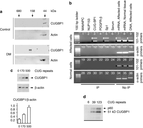
By a number of in vivo biochemical tests (using intact cells), CUGBP binds selectively to the CUGn expansions in mut RNA (Table 1). Extracts from DM (but not wild-type) cells showed increased molecular weight (MW) for CUGBP on size-exclusion chromatography, in accord with its binding to a large RNA (Figure 1a).8 Further, and importantly, Timchenko et al8 showed that CUGBP binding by mut RNA was not a minor fraction of the protein: virtually all of the CUGBP was shifted to higher MW (Figure 1a).a (See Technical note a in Supplementary online materials. All letter citations in this article refer to Technical notes located on this site.) In an independent study, immunoprecipitation of CUGBP from nuclear extracts of DM-affected cells recovered mutant but not wild-type DMPK RNA15 (Figure 1b), confirming the selectivity of this binding in vivo for RNA with large CUGn expansions. Earlier in vitro reconstructions showed binding of CUGBP to purified wild-type DMPK 3′-untranslated region (UTR) RNA with short CUGn (n=5–37), but not to actin RNA, consistent with the ability to detect CUGBP in vitro with small expansions, for example, labeled CUG8.5 However, the just-mentioned in vivo studies15 did not confirm binding of CUGBP to wild-type DMPK RNA as seen in in vitro, perhaps due to the array of small RNAs (not well characterized) that interact and compete27 or due to other conditions in vivo. When wt CUG5-containing mRNA is expressed at extremely high levels, however, then wt RNA can apparently compete with small RNAs for CUGBP binding as evidenced by elevated cellular CUGBP,25 paralleling what is seen in mut RNA-expressing cells. At normal RNA levels, only the larger expansion of the CUGn in mut RNA appears to have the affinity or molarity of binding sites to manifest major binding of CUGBP in vivob.28
Figure 1.
Demonstration that mut RNA binds CUGBP. (a) CUGBP in vivo is completely complexed with mut RNA. Protein from cardiac muscle was fractionated on size-exclusion chromatography and detected by antibody against CUGBP or actin (negative control). In normal hearts, CUGBP runs at 44 kDa, at its native protein MW, but in DM hearts runs at a high-MW position of 150 kDa that is RNase sensitive, corresponding to CUGn position (not shown) (from Timchenko et al8). (b) CUGBP binds only the mutant form of DMPK mRNA in vivo. Nuclear extracts underwent RT–PCR after immunoprecipitation (‘IP') or not (‘no IP'), with antibody to CUGBP, nuclear pore proteins, TF Sp1 and PDGF receptor. Anti-CUGBP coprecipitated DMPK RNA only from DM-affected, and not control, cells, using primers (101–102) spanning the CUGn repeat (n=100). Mut RNA was also precipitated with transcription factor Sp1 but not any other protein and actin RNA was not present in any IP. (Wild-type DMPK mRNA generates single band (150 nt) and mutant DMPK dual bands (150 and 450 nt), with the smaller band arising by template sliding across expanded CUGn during PCR.) (from Ebralidze et al15) (c) Binding of CUGBP to longer mut RNAs in vivo leads to increased CUGBP in cells that correlates with longer protein half-life. Upper: western blot. Lower: graph of data from three experiments (from Timchenko et al8). (d) Increased binding of CUGBP in vitro with longer repeat length. ‘RNA-free' CUGBP was mixed with equal molar quantities of end-labeled RNAs containing repeat lengths of 6, 39 and 123 CUG and assessed for binding (from Timchenko et al27).
As mentioned above under Mechanisms, binding by mut RNA in vivo has the effect of prolonging the normally short half-life of CUGBP.8 This longer survival translates into higher in vivo concentrations of CUGBP8, 25, 29 and increased net splicing activity that recapitulates the splice variants seen in DM cells.7, 8, 9, 10, 11, 25 It is clinically observed that disease severity parallels increased repeat length. Phillips et al7 similarly showed that increased length of CUGn led to increased splicing in their model system. Using relative CUGBP levels as a readout, Timchenko et al8 then showed that expression of longer mut RNAs protected CUGBP from its normally rapid catabolism and led to higher accumulations of CUGBP (Figure 1c) to result in these elevated in vivo splicing activities. Thus, all in vivo observations are consistent with increased CUGBP effect with longer CUGn by biochemical criteria.
This observation leads to a further – pharmacodynamic – argument that has not previously been considered. By first principles of interacting decaying systems, when a short-lived component binds to a longer lived, the ratio of increase in steady-state concentration of the first approaches the ratio of the half-lives.30 That is, with the normal CUGBP half-life of 3 h8 and mut RNA half-life of ∼15 h (same as wt),2 the half-life ratio of 5 directly predicts up to fivefold increase in steady-state CUGBP levels due to complexing. Up to fivefold increase in CUGBP levels is what was observed in the quantitation of Figure 1c8 (see also reference Savkur et al29), with similar values for human DM1 tissues,25 lending weight to the plausibility of CUGBP binding to mut RNA by this independent measure.
A further consequence of the pharmacodynamic argument is that protein levels at steady state cannot increase by more than the excess of the second and longer surviving component to which it is bound.30 That is, an observed fivefold increase in CUGBP levels in DM tissue requires at least a fivefold excess in available mut RNA-binding sites – and the excess could be much greater. It also implies that CUGBP is virtually totally bound, as supported by the observations of Figure 1a. Correspondingly, CAGn expansions do not bind CUGBP, do not lead to elevated CUGBP levels and do not induce splicing abnormalities, although CAGn binds and sequesters MBNL into foci.24 This indicates that CUGBP elevation is a secondary consequence neither of focus formation nor of MBNL depletion, consistent with the direct binding role of CUGn in prolonging CUGBP survival and increasing cellular levels.
In vitro
The results of in vitro biochemical tests merit close examination. In vitro tests showed CUGBP binding that does not31 or does27 increase with longer repeat length. This discrepancy may be explained by the shorter maximum CUGn in the earlier study (n=90 vs n=123) that may not exceed a sequestration threshold; mut RNA is not thought to cause disease for expansions of less than 100. It could also derive from differences in binding conditions that affect CUGBP binding (discussed below under Morphological studies). But a more likely explanation has been advanced (L Timchenko, personal communication) as follows. CUGBP extracted from normal, mut RNA-negative cells in the earlier study (Michalowski et al,31 employing methods of Timchenko et al5) was later shown to be in complex with small RNAs that block efficient CUGn binding under the kinetic conditions of the in vitro test.27 c In separate tests with such ‘RNA-blocked' CUGBP, Timchenko et al27 confirmed mut RNA binding to only a minor fraction of CUGBP that does not change with more CUGs per mole of RNA (L Timchenko, personal communication), a situation that may not be overcome by varying extract-to-probe ratios. Using newer methods with CUGBP stripped of bound RNAs, all CUGBP was made ‘available' and binding increased with CUG length for constant moles of mut RNA (Figure 1d). This in vitro observation with ‘RNA-free' CUGBP then parallels the in vivo length effects of Figure 1c.32d
Morphological studies
In vitro
Earlier in vitro biochemical tests had shown that purified CUGn forms double-stranded (duplex) hairpins in solution.33, 34 In vitro examinations by electron microscopy (EM) confirmed duplex regions to which added purified CUGBP did not bind, attaching only to the CUGn single-stranded tails at hairpin bases.31 As longer CUGn would be absorbed into longer CUG:GUC hairpins, this observation has been interpreted to imply that the CUGn length effect noted clinically could not be explained by CUGn binding of CUGBP: there would always be a constant molar binding ratio. However, this interpretation from in vitro data is inconsistent with in vivo observations of Figure 1c, where a length effect is evident.
This apparent conflict is plausibly resolved by the following considerations. First, the protein used in these EM tests was the same ‘RNA-blocked' CUGBP (see above) that failed to show increased binding with increased CUGn length in vitro,31 in contrast to the ‘RNA-free' CUGBP that bound more to longer CUGn (Figure 1d).27 With the small active fraction of CUGBP in the Michalowski et al31 preparation, preference for binding to single-stranded (ss) CUGn tails under low available CUGBP could be expected thermodynamically because there is less opportunity at the molecular ends to form double-stranded (ds) domains that could compete with and inhibit CUGBP binding. This EM test has never been performed with suitably prepared ‘RNA-free' CUGBP. Second, the real in vivo setting is likely to be different for RNA configuration, as allowed by the authors who first described CUGn hairpins.33 In the EM studies showing binding solely to the base of hairpins,31 incubations were at low temperature (30 and 20°C) and in the presence of spermidine, both of which favor duplex hairpin formation.35, 36 e With higher temperatures in vivo (37°C) and with proteins present in nuclei such as CUGBP and others that preferentially bind ss RNA, the stability of ss vs ds regions is predicted to be more favored in vivo than in vitro. Thus, this in vitro reconstruction may under-represent the ss regions in vivo to which CUGBP might bind in proportion to CUGn length.f
In vivo
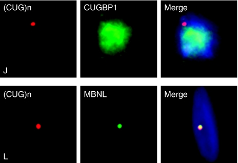
Finally, and the key reason for the current proposal, in vivo morphologic studies showed that CUGBP does not colocalize with mut RNA in foci (Figure 2).21, 22 Instead, CUGBP is dispersed throughout the nucleoplasm. Following the evidence that all or most CUGBP is bound into a mut RNA complex in vivo (Figure 1a, Table 1), observation of dispersed CUGBP implies that mut RNA, bound to CUGBP, is also dispersed in the nucleoplasm, that is, in a soluble, or at least microdisperse form – in addition to the fraction that is in foci. This inference is corroborated by analogous observations of TF binding: mut RNA that leaches Sp1 and RAR out of chromatin and into mut RNA-containing RNP15 is revealed by TF staining in vivo as a dispersed complex in the nucleoplasm of DM-affected cells.22
Figure 2.
CUGBP distribution. CUGBP is dispersed throughout DM nuclei (J), whereas MBNL colocalizes with DM foci (CUGn) (L) (from Mankodi et al, 200321). Ribonuclear inclusions in skeletal muscle in myotonic dystrophy types 1 and 2, Vol 54, No. 6, 2003, page 764. Copyright John Wiley & Sons, 2003. Reprinted with permission of John Wiley & Sons, Inc.
The weight of diverse biochemical and functional measures seems to support a biologically relevant association of CUGBP and TFs with mut RNA. Under the currently prevailing concepts, however, these factors have been undervalued as potential contributors to DM pathology.
Logic for a new model
This situation has been suggested to confront us with a disjunction between the biochemical binding data for CUGBP and TFs and their morphologic non-observation in foci. Yet both the biochemical and morphological data were separately judged as credible by peer-reviewers when they were published. To then downgrade the biochemical data, however, it is fair to ask: what logic impels us to their exclusion? Model A provides one such logic, with the key component highlighted. From model A:
(1) Proteins are bound to mut RNA. (2) Bound proteins are found in foci. (3) Proteins not bound to mut RNA are not found in foci. (4) Proteins not found in foci are not bound to mut RNA.
Elements 1–3 are entirely rational and acceptable. However, statement 4, although superficially also acceptable, provokes the current examination. It is this element of model A that brings about an apparent conflict between the biochemical and morphological data sets. Statement 4 is in truth a hypothesis, and were it so stated, it could attract an experimental test.
Yet, if we reconsider the above formulation to modify the last statement (4) to an alternative hypothesis, another, less restrictive model (model B) emerges:
(1) Proteins are bound to mut RNA. (2) Bound proteins are found in foci. (3) Proteins not bound to mut RNA are not found in foci. (4) Some proteins not found in foci are bound to mut RNA. (5) Some mut RNA is not in foci (ie, soluble). (6) Some proteins are selectively bound to soluble mut RNA. (7) Soluble and focus mut RNA have different binding properties.
We note that there is nothing in the logic of statements 1–3 that is inherently in conflict with the new statement 4. However, this formulation, inescapably implying (7), begs the question: How can a soluble form of mutant RNA differ from a focus form? The RNAs are the same chemically.
A new model of mut RNA conformation and binding
Here, I propose a model (B) that is consistent with the broader range of data, in which focus localization is sufficient, but not necessary for, a conclusion of protein binding by mut RNA.
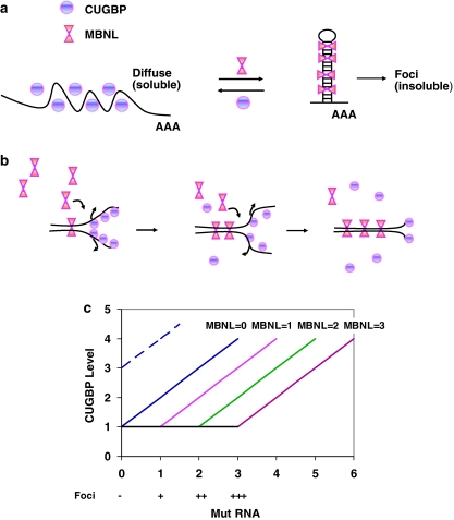
In vitro, CUGn self-pairs as CUG:GUC, in which non-standard U:U pairing has been shown to be permissive, creating ‘metastable slippery hairpins'.33 In vivo, RNA, including mut RNA, exists not as naked RNA but in complex with RNA-binding proteins as RNP with distinct properties.15, 37, 38 Proteins that bind to ss RNA (we call ‘class I') will tend to melt out ds duplex regions and favor a linear single-stranded state. CUGBP would be a class I protein, as would influenza ssRNA-binding protein,39 in parallel with class I proteins first described for DNA. There is ample precedent for polynucleotide melting by ss-binding proteins: for example, fd gene 5 protein causes a 30°C drop in melting temperature (Tm) for poly(dA.dT) in 0.1 Na+40 and T4 gene 32 I* fragment causes a 50°C drop in the Tm of natural DNA.41 Although RNA–protein interactions are less studied, a similar effect on the Tms for the patient CUG repeats could be predicted for any protein that preferentially binds ss CUGn regions, including CUGBP and what other undefined nuclear proteins may perform this function in vivo. Proteins that preferentially bind ds RNA (we call ‘class II') will naturally promote ds regions. MBNL would be a class II protein. The balance between linear, mixed and duplex structures in vivo will be determined by salt and temperature and by the relative abundance of class I and II proteins in a tissue and their relative energetics of binding (Figure 3a).
Figure 3.
New model (model B) for mut RNA binding. (a) CUGBP binds to ‘melted' CUGn single-stranded regions that remain soluble and MBNL binds to double-stranded ‘hairpin' regions that lead to insoluble foci. (Mixed ds/ss complexes are likely to be soluble.) (b) Model of how MBNL binding promotes double-stranded regions that successively extrude CUGBP, with the net result of CUGBP being excluded from insoluble foci. On account of the relative absence of CUGBP from foci, the energetics of the class II-type binding of MBNL are inferred to dominate over that of class I-type binding by CUGBP until free MBNL is substantially depleted. (c) Predicted effects of mut RNA and MBNL binding on CUGBP levels under model B. Abscissa Mut RNA refers to total CUGn-binding sites, whether by overexpression or longer CUGn. MBNL binds first. For a given level of mut RNA, increasing MBNL predicts more foci until MBNL or mut RNA is exhausted (− → +++). On the ordinate, CUGBP level is represented, with 1 being normal muscle. If mut RNA exceeds MBNL, then CUGBP binding proceeds with increased CUGBP until mut RNA is exhausted or the half-life ratio has been reached (nominally estimated as 5; see text). MBNL knockout corresponds to MBNL=0. Higher levels of MBNL ‘protect' cells from CUGBP binding and elevation, depending on cell type and developmental stage or on whether MBNL is artificially overexpressed. Where CUGBP starts high normally, as in heart (eg, at level 3), the absolute increment in CUGBP level is the same for a given CUGn RNA excess above MBNL, but the fractional change in CUGBP is smaller. (The example of Mahadevan et al25 is modeled with MBNL=0 for not interacting with CUG5 RNA; dashed line.)
When MBNL levels are suppressed experimentally by RNAi, foci are reduced14 and, because the quantity of mut RNA is the same, a substantial fraction of mut RNA therefore cannot but be in the non-focus (ie, soluble or disperse) fraction. This, per se, is evidence that focus and soluble mut RNA fractions can coexist in vivo. This result indicates that mut RNA is soluble in vivo in the absence of MBNL and that the complex of MBNL with mut RNA to stabilize ds regions renders a portion of the RNA insoluble.g Inasmuch as both ss and ds nucleic acids are soluble (including purified CUGn), it is some property of the MBNL protein–RNA complex, perhaps involving charge neutralization and/or secondary protein–protein interactions, that renders mut RNA non-soluble to result in foci. In general, ds nucleic acids have lower solubility and more readily aggregate or precipitate in complex.42 Where RNAs are MBNL bound but interspersed with ss or unbound ds domains, these complexes are predicted not to be in foci, but to remain soluble (or microdisperse), as recently shown with the troponin T pre-mRNA–MBNL complex.26 A dense coalescence of protein onto the RNA is likely to be the mechanism for insolubility, as will occur for MBNL on extended CUG hairpins26 (Figure 3a, right).
The act of stabilizing hairpins by MBNL binding is predictably a cooperative phenomenon (Figure 3b). This means that binding of each MBNL clamps the adjacent duplex to facilitate the binding of the next MBNL, with each bound MBNL rendering the duplex more stable, closing like a zipper. Our supposition of cooperative binding is buttressed by the recent demonstration of MBNL homotypic protein–protein interactions on the CUGn duplex.26 Logically, this zippering process also progressively excludes CUGBP that is bound to the ss CUGn, leading ultimately to essentially pure cocrystals of mut RNA and MBNL, in direct analogy to the exclusion of impurities during a crystallization. This mechanism could explain why CUGBP is absent from foci (Figure 2) yet highly bound to non-focus soluble mut RNA (Figure 1a).
As foci form, the deeper portion is removed from the exchangeable solution. This is seen in photobleaching experiments by the large immobile fraction of MBNL in foci,h whereas MBNL not in foci is fully mobile and recovers fluorescence completely.24 In turn, as MBNL coprecipitates with mut RNA into insoluble foci, the residual free MBNL in the solution is progressively depleted (Figure 3b) and thus less able to exclude CUGBP from mut RNA binding. With CUGBP now predominating in solution, the remaining binding of mut RNA to CUGBP results in a soluble mut RNA form that is substantially single stranded. Other data suggest that not all MBNL is concentrated into DM foci, with up to 30–40% dispersed in nucleoplasm.22, 43 In this case, non-focus MBNL at depleted concentrations may also be bound to mut RNA that is still soluble, that is, mixed ds/ss in configuration as discussed above, to which CUGBP is concurrently bound.
Mut RNA with accessible ss sequence binds and protects CUGBP from its normally rapid catabolism, prolonging CUGBP survival and accumulating to high levels.8 As a still-active soluble form in complex, bound CUGBP mediates splicing to higher net activity.i,j In contrast, binding of MBNL by mut RNA in ds form suppresses its splicing activity by active site blockade or by its sequestration into insoluble foci, which removes it from solution.k
CUGBP binding occurs only after MBNL is essentially fully absorbed into complex, with the extent of CUGBP binding, prolongation of survival and increased cellular levels being dictated by the excess of mut RNA-binding sites. This is represented conceptually in Figure 3c. Longer mut RNAs will have more CUGBP-binding sites after MBNL saturation. If MBNL is always fully bound first due to its higher avidity complex, then it is likely that it is this variable binding of CUGBP that mediates the length effect of CUGn in DM1 through the resulting progressive increase in CUGBP levels. For example, in Figure 3c with fixed MBNL=1 and a mut RNA=0, CUGBP=1 (normal); for mut RNA=2, CUGBP=3; and for mut RNA=4, CUGBP=5. This parallels observations in Figure 1c, with longer RNAs supplying more moles of mut RNA-binding sites. By extension, the binding of TFs to mut RNA15 would occur in the same sequence and at the same time as CUGBP: after the MBNL absorption into mut RNA is complete, the residual soluble mut RNA binds TFs, which in turn leads to TF leaching from chromatin and decreased transcription of selected genes.
Explanatory value
The key features of the revised model are (i) the presence of a second, non-focus form of mut RNA and (ii) distinct characteristics of protein binding for the two forms. The focus form is mainly a duplex of mutant RNA bound with MBNL and other class II proteins that excludes CUGBP and TFs, whereas the soluble form is substantially single stranded, with binding of CUGBP, TFs and other class I proteins. This new model is compatible with a number of observations on which the more restrictive model A fails (Table 2).
Table 2. Explanatory value of models of CUGn properties.
| CUGn model | ||
|---|---|---|
| Data explained? | A | B |
| 1 Mut RNA binds MBNL | Ok | Ok |
| 2 MBNL in foci | Ok | Ok |
| 3 Mut RNA binds CUGBP | Fails | Ok |
| 4 CUGBP levels increase | Fails | Ok |
| 5 CUGBP not in foci | Fails | Ok |
| 6 Mut RNA binds TFs | Fails | Ok |
| 7 TFs not in foci | Fails | Ok |
| 8 CAGn make foci not DM | Fails | Ok |
| 9 High CUG5 causes DM | Fails?* | Ok |
| 10 Low CUG200 not cause DM | Ok | Ok |
See Technical note l and associated text.
From all considerations, one view evolves for DM: ‘loss of alternative splicing regulation results directly from… unopposed [CUGBP] activity'.13 In this, MBNL serves as a ‘brake' by binding to stem-loop structures and blocking splicing activity, as recently and elegantly demonstrated by Swanson and colleagues.26 As MBNL opposes CUGBP action, DM can be achieved experimentally by the suppression of MBNL to leave CUGBP unopposed, as in MBNL knockouts, and yield DM-splicing defects. Where MBNL is undisturbed, CUGBP overexpression can overcome the MBNL ‘brake'. In natural DM, CUGBP is elevated and MBNL is suppressed. Still, it is said: ‘it is unclear how (CUG)n … expansion RNAs upregulate [CUGBP] protein levels'13 and ‘the mechanism of this increase has not been determined'.44 Model B provides a mechanism to explain increased CUGBP levels.
By this, we would say that the concept of Mahadevan et al25 is right: both MBNL and CUGBP are in the same pathway and the balance of their activities will determine net splicing in vivo. We would also agree with the accompanying editorial of Timchenko45 that proposed that MBNL binds first, and then CUGBP. Model B provides a physicochemical basis for this sequence. Finally, any effect of TF leaching by soluble mut RNA would modify these effects by suppressing mRNA levels for specific genes as well.
The following are additional points to be considered for model B:
Until now, the observation that CUG5 overexpression caused DM was considered to pose a potential ‘conundrum'.25 However, the data can be understood under model B as follows: CUGBP binds short ss CUGn RNAs.5 The large overexpression of CUG5 provides excess binding sites to compete with endogenous small ‘blocking' RNAs27 to absorb all of the CUGBP, delaying CUGBP catabolism to result in increased in vivo concentrations with CUGBP hyperactivity and increased splicing. As CUG5 RNA is too short to form ds hairpins,33 there is no competing type II protein (MBNL) binding or focus formation and all binding is therefore ss (Figure 3a-left) (for caveat, see note l).
Also of interest in these CUG5 experiments is the absence of CUGBP elevation or splicing abnormalities in the heart muscle. This is understood by model B as follows. The CUG5 RNA transgene was much less expressed in the heart than in the skeletal muscle (three- to fivefold induced in cardiac vs 10- to 20-fold in skeletal25). In addition, the baseline levels of CUGBP are much higher in the heart, and therefore requiring more RNA binding to increase CUGBP levels. This is seen from Figure 3c: for skeletal muscle, if the overexpressed CUG5 is equivalent to level 3 on the mut RNA scale with a starting level CUGBP=1 and MBNL=0 (for no ‘protection' from MBNL), CUGBP increases from 1 to 4. In the case of heart, with CUGBP=3 at baseline (Figure 3, dashed line) and a CUG5 level one-third as high, or 1.0 equivalent of mut RNA, the increase would only be to CUGBP=4, not readily distinguishable vs CUGBP=3 at baseline. If there is also MBNL bindingl, this would be particularly protective with this lower DMPK RNA expression in heart muscle, possibly blocking any effect on CUGBP level. This says nothing, however, about the cells of the conduction system where the DMPK gene induction and baseline CUGBP may be very different, resulting in splicing and/or transcription abnormalities in those cells to explain the cardiac arrhythmias observed.
In separate experiments with CUG200 heterozygotes that had very low mut RNA expression, MBNL binding yielded mut RNA precipitation and focus formation. However, the mut RNA expression was too low to deplete MBNL (eg, Figure 3c with mut RNA=0.5 and MBNL=1). With excess MBNL, all mut RNA was absorbed into insoluble complex (foci) and stabilized into a ds state (Figure 3a-right) to which CUGBP could not bind. With still adequate MBNL and with CUGBP levels undisturbed, animals were without DM phenotype.
In summary, model B for mut RNA–protein interactions is seen to have a broad explanatory value for DM-associated phenomena.
Future directions
Model B is a hypothesis. I believe that it has merits over the until-now prevailing model A, but tests will be important to contrast their elements. Several predictions of the new model are testable, as outlined in the following.
What is the proportion of mut RNA in foci (insoluble) vs that not in foci (soluble)? This has not been measured; indeed, the coexistence of a soluble form of mut RNA in DM1 has not formally been acknowledged. The actual balance of these forms in a given tissue will depend upon the particular stoichiometries of the interacting and competing chemical components in that tissue. Although the issue of detection of soluble mut RNA vs background is not trivialm, it should be doable in expert hands with current imaging and molecular techniques.
What is the proportion of CUGBP in complex in DM? From studies of Timchenko8 cited above (Figure 1a), nearly all of it is, and pharmacodynamic considerations indicate that it should be. The same experiments with nuclear fractions and added controls for cross-contamination to rule out post-extraction association28 will support (or falsify) model B. UV cross-linking and extraction under denaturing conditions may be useful as an adjunctive study, with normalization for recovery vs control MBNL cross-linking, where binding is uncontroversial.
Does CUGBP phosphorylation contribute to mut RNA binding and/or CUGBP half-life prolongation? PKC induction by CUG960 RNA mediates CUGBP hyperphosphorylation. A PKC activator (PMA) was shown to induce hyperphosphorylation and to prolong CUGBP survival to >4 h in normal cells.46 Still needed are half-life studies in DM cells, where inhibitors could test the relevance of phosphorylation to the observed CUGBP elevations in such cells. Binding to mut RNA should yield CUGBP half-lives up to ∼15 h as noted above, consistent with one earlier measure.8 Rigorous half-life measurements and CUGBP quantitation in DM-affected cells, without and with PKC inhibitors, will be very informative to this question. If PKC inhibitors reverse the prolonged CUGBP t1/2, it will be of interest to examine their effects on mut RNA binding (if reconfirmed) as in 2. above. Mut RNA half-life should be redetermined in the same system, which sets the upper limit for the t1/2 of complexed CUGBP.
How depleted is MBNL functionally in DM? The absence of findings where MBNL is bound but CUGBP is not disturbed (eg, CAGn expansions,24 perhaps also in low level CUG20025) contrasts with the MBNL knockouts where the DM syndrome is recapitulated. If a residual 10% activity of MBNL is sufficient to mediate normal in vivo functions, for example, then the splicing abnormalities may be primarily due to CUGBP increase that occurs with binding to excess mut RNA after MBNL is maximally bound. This would make foci, in one sense, ‘protective' against DM, as supposed by Mahadevan25 and Timchenko45, but by reason of blocking sites for CUGBP binding under model B.
Does MBNL binding to mut RNA ‘protect' against CUGBP binding and elevation? The chart of Figure 3c predicts that more CUGBP will accumulate in DM cells if MBNL protein is also artificially suppressed by RNAi.14 The same is predicted in DM mice47, 48 if they are crossed with MBNL knockouts.13 MBNL knockout mice have CUGBP levels that are normal and DM mice are elevated for CUGBP. The cross is predicted to be more elevated.m,n Correspondingly, MBNL overexpression is predicted to normalize (reduce) CUGBP in DM cells (eg, with mut RNA=3, move from MBNL=1 to MBNL=3 in Figure 3c); in the single relevant report, CUGBP was not measured.49
Does MBNL binding to mut RNA protect against TF leaching from chromatin15? Similar tests using MBNL suppression in DM cells as above could be performed. The readouts in this case are the RNP/chromatin ratio of TFs and mRNA levels in DM cells after suppressing MBNL. If MBNL is protective, then MBNL suppression will exaggerate the leaching and mRNA suppression in DM cells. Conversely, MBNL overexpression should reduce TF leaching.
Does MBNL bind to wt DMPK 3′-UTR RNA? This question follows from the speculation in note l on possible MBNL-binding sites with CUG5 in the 3′-UTR. If MBNL in fact does not bind, as originally supposed, it leaves only non-MBNL components (eg, CUGBP and TFs) to induce DM in the Mahadevan model.25 If MBNL does bind, this ‘high-CUG5' situation likely represents a model setting of CUGBP and MBNL both binding to soluble RNA; their bound ratio could then be determined.
Conclusion
This proposal aims to reconcile several aspects of DM pathobiology not satisfactorily explained by current concepts. These concepts have centered around the observation of foci and an inference that proteins should be found in such foci to be considered mut RNA bound. As CUGBP and TFs do not colocalize with foci, there has been a disposition to exclude roles for CUGBP and TF binding in DM pathology. I present evidence for the existence of mut RNA in a non-focus, soluble fraction in vivo that selectively binds CUGBP and, by extension, nuclear TFs as well. I describe a model that accommodates these data, and experiments are proposed for its test.
With this, the three major models of trans dominant effects from mut RNA binding should be assessed, without prejudgment, to sort out the relative contribution of mut RNA-mediated biochemical events for the individual genes and tissues affected, while remaining open to what other mediators may yet be discovered. Plausibly, one gene that is critical in one tissue may be more affected by one mechanism, and another gene in another tissue more affected by a different mechanism.
Acknowledgments
I acknowledge personal communications, thoughtful comments and generous sharing of unpublished data by Drs Thomas Cooper, Yoshihiro Kino, Wlodzimierz Krzyzosiak, Mani Mahadevan, Maurice Swanson and Lubov Timchenko. However, to absolve all of any responsibility for the views expressed in this article, I state that they are solely my own. This paper was presented at the New England Chapter Muscular Dystrophy Association Annual Conference, Rhode Island Hospital, Providence on 30 November 2007. Preparation of this article was supported in part by the Association Française contre les Myopathies.
Footnotes
Supplementary Information accompanies the paper on European Journal of Human Genetics website (http://www.nature.com/ejhg)
Supplementary Material
References
- Hamshere MG, Newman EE, Alwazzan M, Athwal BS, Brook JD. Transcriptional abnormality in myotonic dystrophy affects DMPK but not neighboring genes. Proc Natl Acad Sci USA. 1997;94:7394–7399. doi: 10.1073/pnas.94.14.7394. [DOI] [PMC free article] [PubMed] [Google Scholar]
- Davis BM, McCurrach ME, Taneja KL, Singer RH, Housman DE. Expansion of a CUG trinucleotide repeat in the 3′ untranslated region of myotonic dystrophy protein kinase transcripts results in nuclear retention of transcripts. Proc Natl Acad Sci USA. 1997;94:7388–7393. doi: 10.1073/pnas.94.14.7388. [DOI] [PMC free article] [PubMed] [Google Scholar]
- Klesert TR, Cho DH, Clark JI, et al. Mice deficient in Six5 develop cataracts: implications for myotonic dystrophy. Nat Genet. 2000;25:105–109. doi: 10.1038/75490. [DOI] [PubMed] [Google Scholar]
- Sarkar PS, Appukuttan B, Han J, et al. Heterozygous loss of Six5 in mice is sufficient to cause ocular cataracts. Nat Genet. 2000;25:110–114. doi: 10.1038/75500. [DOI] [PubMed] [Google Scholar]
- Timchenko LT, Miller JW, Timchenko NA, et al. Identification of a (CUG)n triplet repeat RNA-binding protein and its expression in myotonic dystrophy. Nucleic Acids Res. 1996;24:4407–4414. doi: 10.1093/nar/24.22.4407. [DOI] [PMC free article] [PubMed] [Google Scholar]
- Timchenko LT, Timchenko NA, Caskey CT, Roberts R. Novel proteins with binding specificity for DNA CTG repeats and RNA CUG repeats: implications for myotonic dystrophy. Hum Mol Genet. 1996;5:115–121. doi: 10.1093/hmg/5.1.115. [DOI] [PubMed] [Google Scholar]
- Philips AV, Timchenko LT, Cooper TA. Disruption of splicing regulated by a CUG-binding protein in myotonic dystrophy. Science. 1998;280:737–741. doi: 10.1126/science.280.5364.737. [DOI] [PubMed] [Google Scholar]
- Timchenko NA, Cai ZJ, Welm AL, Reddy S, Ashizawa T, Timchenko LT. RNA CUG repeats sequester CUGBP1 and alter protein levels and activity of CUGBP1. J Biol Chem. 2001;276:7820–7826. doi: 10.1074/jbc.M005960200. [DOI] [PubMed] [Google Scholar]
- Mankodi A, Takahashi MP, Jiang H, et al. Expanded CUG repeats trigger aberrant splicing of ClC-1 chloride channel pre-mRNA and hyperexcitability of skeletal muscle in myotonic dystrophy. Mol Cell. 2002;10:35–44. doi: 10.1016/s1097-2765(02)00563-4. [DOI] [PubMed] [Google Scholar]
- Charlet-B N, Savkur RS, Singh G, Philips AV, Grice EA, Cooper TA. Loss of the muscle-specific chloride channel in type 1 myotonic dystrophy due to misregulated alternative splicing. Mol Cell. 2002;10:45–53. doi: 10.1016/s1097-2765(02)00572-5. [DOI] [PubMed] [Google Scholar]
- Ho TH, Bundman D, Armstrong DL, Cooper TA. Transgenic mice expressing CUG-BP1 reproduce splicing mis-regulation observed in myotonic dystrophy. Hum Mol Genet. 2005;14:1539–1547. doi: 10.1093/hmg/ddi162. [DOI] [PubMed] [Google Scholar]
- Miller JW, Urbinati CR, Teng-Umnuay P, et al. Recruitment of human muscleblind proteins to (CUG)(n) expansions associated with myotonic dystrophy. EMBO J. 2000;19:4439–4448. doi: 10.1093/emboj/19.17.4439. [DOI] [PMC free article] [PubMed] [Google Scholar]
- Kanadia RN, Johnstone KA, Mankodi A, et al. A muscleblind knockout model for myotonic dystrophy. Science. 2003;302:1978–1980. doi: 10.1126/science.1088583. [DOI] [PubMed] [Google Scholar]
- Dansithong W, Paul S, Comai L, Reddy S. MBNL1 is the primary determinant of focus formation and aberrant insulin receptor splicing in DM1. J Biol Chem. 2005;280:5773–5780. doi: 10.1074/jbc.M410781200. [DOI] [PubMed] [Google Scholar]
- Ebralidze A, Wang Y, Petkova V, Ebralidse K, Junghans RP. RNA leaching of transcription factors disrupts transcription in myotonic dystrophy. Science. 2004;303:383–387. doi: 10.1126/science.1088679. [DOI] [PubMed] [Google Scholar]
- Taneja KL, McCurrach M, Schalling M, Housman D, Singer RH. Foci of trinucleotide repeat transcripts in nuclei of myotonic dystrophy cells and tissues. J Cell Biol. 1995;128:995–1002. doi: 10.1083/jcb.128.6.995. [DOI] [PMC free article] [PubMed] [Google Scholar]
- Mankodi A, Urbinati CR, Yuan QP, et al. Muscleblind localizes to nuclear foci of aberrant RNA in myotonic dystrophy types 1 and 2. Hum Mol Genet. 2001;10:2165–2170. doi: 10.1093/hmg/10.19.2165. [DOI] [PubMed] [Google Scholar]
- Houseley JM, Wang Z, Borck GJR, et al. Myotonic dystrophy associated expanded CUG repeat muscleblind positive ribonuclear foci are not toxic to Drosophila. Hum Mol Genet. 2005;14:873–883. doi: 10.1093/hmg/ddi080. [DOI] [PubMed] [Google Scholar]
- Fardaei M, Larkin K, Brook JD, Hamshere MG. In vivo co-localisation of MBNL protein with DMPK expanded-repeat transcripts. Nucleic Acids Res. 2001;29:2766–2771. doi: 10.1093/nar/29.13.2766. [DOI] [PMC free article] [PubMed] [Google Scholar]
- Fardaei M, Rogers MT, Thorpe HM, et al. Three proteins, MBNL, MBLL and MBXL, co-localize in vivo with nuclear foci of expanded-repeat transcripts in DM1 and DM2 cells. Hum Mol Genet. 2002;11:805–814. doi: 10.1093/hmg/11.7.805. [DOI] [PubMed] [Google Scholar]
- Mankodi A, Teng-Umnuay P, Krym M, Henderson D, Swanson M, Thornton CA. Ribonuclear inclusions in skeletal muscle in myotonic dystrophy types 1 and 2. Ann Neurol. 2003;54:760–768. doi: 10.1002/ana.10763. [DOI] [PubMed] [Google Scholar]
- Jiang H, Mankodi A, Swanson MS, Moxley RT, Thornton CA. Myotonic dystrophy type 1 is associated with nuclear foci of mutant RNA, sequestration of muscleblind proteins and deregulated alternative splicing in neurons. Hum Mol Genet. 2004;13:3079–3088. doi: 10.1093/hmg/ddh327. [DOI] [PubMed] [Google Scholar]
- de Haro M, Al-Ramahi I, De Gouyon B, et al. MBNL1 and CUGBP1 modify expanded CUG-induced toxicity in a Drosophila model of myotonic dystrophy type 1. Hum Mol Genet. 2006;15:2138–2145. doi: 10.1093/hmg/ddl137. [DOI] [PubMed] [Google Scholar]
- Ho TH, Savkur RS, Poulos MG, Mancini MA, Swanson MS, Cooper TA. Colocalization of muscleblind with RNA foci is separable from mis-regulation of alternative splicing in myotonic dystrophy. J Cell Sci. 2005;118:2923–2933. doi: 10.1242/jcs.02404. [DOI] [PubMed] [Google Scholar]
- Mahadevan MS, Yadava RS, Yu Q, et al. Reversible model of RNA toxicity and cardiac conduction defects in myotonic dystrophy. Nat Genet. 2006;38:1066–1070. doi: 10.1038/ng1857. [DOI] [PMC free article] [PubMed] [Google Scholar]
- Yuan Y, Compton SA, Sobczak K, et al. Muscleblind-like 1 interacts with RNA hairpins in splicing target and pathogenic RNAs. Nucleic Acids Res. 2007;35:5474–5486. doi: 10.1093/nar/gkm601. [DOI] [PMC free article] [PubMed] [Google Scholar]
- Timchenko LT, Iakova P, Welm AL, Cai ZJ, Timchenko NA. Calreticulin interacts with C/EBPalpha and C/EBPbeta mRNAs and represses translation of C/EBP proteins. Mol Cell Biol. 2002;22:7242–7257. doi: 10.1128/MCB.22.20.7242-7257.2002. [DOI] [PMC free article] [PubMed] [Google Scholar]
- Mili S, Steitz JA. Evidence for reassociation of RNA-binding proteins after cell lysis: implications for the interpretation of immunoprecipitation analyses. RNA. 2004;10:1692–1694. doi: 10.1261/rna.7151404. [DOI] [PMC free article] [PubMed] [Google Scholar]
- Savkur RS, Philips AV, Cooper TA. Aberrant regulation of insulin receptor alternative splicing is associated with insulin resistance in myotonic dystrophy. Nat Genet. 2001;29:40–47. doi: 10.1038/ng704. [DOI] [PubMed] [Google Scholar]
- Junghans RP, Waldmann TA. Metabolism of Tac (IL2Rα): physiology of cell surface shedding and renal elimination, and suppression of catabolism by antibody binding. J Exp Med. 1996;183:1587–1602. doi: 10.1084/jem.183.4.1587. [DOI] [PMC free article] [PubMed] [Google Scholar]
- Michalowski S, Miller JW, Urbinati CR, Paliouras M, Swanson MS, Griffith J. Visualization of double-stranded RNAs from the myotonic dystrophy protein kinase gene and interactions with CUG-binding protein. Nucleic Acids Res. 1999;27:3534–3542. doi: 10.1093/nar/27.17.3534. [DOI] [PMC free article] [PubMed] [Google Scholar]
- Kino Y, Mori C, Oma Y, Takeshita Y, Sasagawa N, Ishiura S. Muscleblind protein, MBNL1/EXP, binds specificallyto CHHG repeats. Hum Mol Genet. 2004;3:495–507. doi: 10.1093/hmg/ddh056. [DOI] [PubMed] [Google Scholar]
- Napierela M, Krzyzosiak WJ. CUG repeats present in myotonin kinase RNA form metastable ‘slippery' hairpins. J Biol Chem. 1997;272:31079–31085. doi: 10.1074/jbc.272.49.31079. [DOI] [PubMed] [Google Scholar]
- Tian B, White RJ, Xia T, et al. Expanded CUG repeat RNAs form hairpins that activate the double-stranded RNA-dependent protein kinase PKR. RNA. 2000;6:79–87. doi: 10.1017/s1355838200991544. [DOI] [PMC free article] [PubMed] [Google Scholar]
- Hou MH, Lin SB, Yuann JM, Lin WC, Wang AH, Kan LL. Effects of polyamines on the thermal stability and formation kinetics of DNA duplexes with abnormal structure. Nucleic Acids Res. 2001;29:5121–5128. doi: 10.1093/nar/29.24.5121. [DOI] [PMC free article] [PubMed] [Google Scholar]
- Venkiteswaran S, Vijayanathan V, Shirahata A, Thomas T, Thomas TJ. Antisense recognition of the HER-2 mRNA: effects of phosphorothioate substitution and polyamines on DNA.RNA, RNA.RNA, and DNA.DNA duplex stability. Biochemistry. 2005;44:303–312. doi: 10.1021/bi0485272. [DOI] [PubMed] [Google Scholar]
- Hieronymus H, Silver PA. A systems view of mRNP biology. Genes Dev. 2004;18:2845–2860. doi: 10.1101/gad.1256904. [DOI] [PubMed] [Google Scholar]
- Moore MJ. From birth to death: the complex lives of eukaryotic mRNAs. Science. 2005;309:1514–1518. doi: 10.1126/science.1111443. [DOI] [PubMed] [Google Scholar]
- Portela A, Digard P. The influenza virus nucleoprotein: a multifunctional RNA-binding protein pivotal to virus replication. J Gen Virol. 2002;83:723–734. doi: 10.1099/0022-1317-83-4-723. [DOI] [PubMed] [Google Scholar]
- Sang BC, Gray DM. Fd gene 5 protein binds to double-stranded polydeoxyribonucleotides poly(dA.dT) and poly[d(A-T).d(A-T)] Biochemistry. 1987;26:7210–7214. doi: 10.1021/bi00397a002. [DOI] [PubMed] [Google Scholar]
- Pant K, Karpel RL, Rouzina I, Williams MC. Salt dependent binding of T4 gene 32 protein to single and double-stranded DNA: single molecule force spectroscopy measurements. J Mol Biol. 2005;349:317–330. doi: 10.1016/j.jmb.2005.03.065. [DOI] [PubMed] [Google Scholar]
- Saminathan M, Antony T, Shirahata A, Sigal LH, Thomas T, Thomas TJ. Ionic and structural specificity effects of natural and synthetic polyamines on the aggregation and resolubilization of single-, double-, and triple-stranded DNA. Biochemistry. 1999;38:3821–3830. doi: 10.1021/bi9825753. [DOI] [PubMed] [Google Scholar]
- Finkelman FD, Madden KB, Morris SC, et al. Anti-cytokine antibodies as carrier proteins. Prolongation of in vivo effects of exogenous cytokines by injection of cytokine–anti-cytokine antibody complexes. J Immunol. 1993;151:1235–1244. [PubMed] [Google Scholar]
- Lin X, Miller JW, Mankodi A, et al. Failure of MBNL1-dependent post-natal splicing transitions in myotonic dystrophy. Hum Mol Genet. 2006;15:2087–2097. doi: 10.1093/hmg/ddl132. [DOI] [PubMed] [Google Scholar]
- Timchenko LT. Reversal of fortune. Nat Genet. 2006;38:976–977. doi: 10.1038/ng0906-976. [DOI] [PubMed] [Google Scholar]
- Kuyumcu-Martinez NM, Wang G-S, Cooper TA. Increased steady-state levels of CUGBP1 in myotonic dystrophy are due to PKC-mediated hyperphosphorylation. Mol Cell. 2007;28:68–78. doi: 10.1016/j.molcel.2007.07.027. [DOI] [PMC free article] [PubMed] [Google Scholar]
- Mankodi A, Logigian E, Callahan L, et al. Myotonic dystrophy in transgenic mice expressing an expanded CUG repeat. Science. 2000;289:1769–1773. doi: 10.1126/science.289.5485.1769. [DOI] [PubMed] [Google Scholar]
- Seznec H, Agbulut O, Sergeant N, et al. Mice transgenic for the human myotonic dystrophy region with expanded CTG repeats display muscular and brain abnormalities. Hum Mol Genet. 2001;10:2717–2726. doi: 10.1093/hmg/10.23.2717. [DOI] [PubMed] [Google Scholar]
- Kanadia RN, Shin J, Yuan Y, et al. Reversal of RNA missplicing and myotonia after muscleblind overexpression in a mouse poly(CUG) model for myotonic dystrophy. Proc Natl Acad Sci USA. 2006;103:11748–11753. doi: 10.1073/pnas.0604970103. [DOI] [PMC free article] [PubMed] [Google Scholar]
Associated Data
This section collects any data citations, data availability statements, or supplementary materials included in this article.


