Abstract
Background and Objective:
Peritoneal dialysis (PD) remains the generally accepted method for management of renal failure in chronic and acute renal failure. Despite the rapidly increasing use of continuous ambulatory peritoneal dialysis (CAPD) since its introduction, controversy persists as to the efficacy and exact role of the modality in the treatment of end stage renal failure. The aim of this paper is to present the experience with laparoscopic placement of a peritoneal dialysis catheter and starting the peritoneal dialysis on the same day.
Methods:
The laparoscopic placement of a peritoneal dialysis catheter was performed on 11 patients (10 males and 1 female) with an average age of 35 years, over a 12-month period. The procedure was done using two 5 mm abdominal trocars. The precise position of the catheter on the pelvis was ensured laparoscopically. One to two liters exchange dialysis was used for every patient, and no leakage was recorded.
Results:
The patients tolerated the procedure well. The peritoneal dialysis was started immediately. Patients were discharged after an overnight stay, and PD was carried out routinely.
Conclusion:
The results of laparoscopic placement of a peritoneal dialysis catheter show the following advantages: minimal incision; less surgical trauma; the procedure hastens the early start of peritoneal dialysis and has no complications.
Keywords: Lkaparoscopy, Peritoneal dialysis, Catheter
INTRODUCTION
Continuous ambulatory peritoneal dialysis is an effective treatment for acute or chronic renal failure. Popovich et al first introduced the continuous ambulatory peritoneal dialysis (CAPD) technique in 1976.1 CAPD is widely used throughout the world. As of 1989, about half of all American pediatric patients who suffered from chronic renal failure were managed by CAPD.2 Advantages of peritoneal dialysis over hemodialysis include its relative simplicity and lack of life-threatening condition. Peritoneal dialysis is preferred for patients with cardiovascular instability, for patients for whom vascular access is difficult and for patients for whom anticoagulants would be hazardous.3 There are four options for placement of chronic peritoneal catheters: surgical placement; blind placement, using the Tenckhoff trocar; blind placement, using a guidewire; and minitrocar placement using peritoneoscopy.4 Kimmelstiel et al introduced laparoscopy in the management of peritoneal dialysis catheters (PDC).5 The initial experience of laparoscopic placement of PDC has been presented in the literature.6 Furthermore, the experience of surgical placement of CAPD has progressed to the point that it can be done laparoscopically in almost all patients who need peritoneal dialysis. Our follow-up of all patients is presented.
MATERIALS AND METHODS
In this series of 11 patients, all underwent laparoscopic placement of a peritoneal dialysis catheter for chronic renal failure. The procedure was performed between February 1998 and December 1998 at King Khalid University Hospital, Riyadh, Saudi Arabia.
OPERATIVE TECHNIQUE
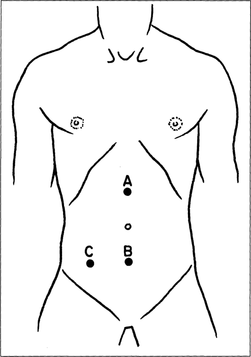
The patient is placed in the supine position, and general endotracheal anesthesia is induced. A preoperative prophylactic antibiotic is given routinely for all patients. The patient's abdomen is prepared and draped in the standard sterile fashion. The port sites and the exit site of the catheter are chosen (Figure 1). The first incision is 10 mm in length, 3 cm below the umbilicus. The pneumoperitoneum is established by using the Veress needle until the intra-abdominal pressure is 8 mm Hg. A 5 mm trocar is introduced and a 5 mm scope is used to visualize the abdominal cavity. The second 5 mm trocar is introduced 3 cm supraumbilically, and the first trocar is removed. Visualization of the abdominal cavity is done through the second port. The AlKhuwaiter-AlDohayan needle7 is used to close the port site and to close the entrance of the peritoneal catheter using synthetic absorbable sutures. A double cuff catheter is utilized for dialysis. The needle is mounted with the suture and passed through the fascia above the defect and retrieved by a 2 mm grasper through the fascia below the defect. The catheter is introduced, placing the inner cuff above the peritoneum and the fascia.
Figure 1.
The diagram shows the site of incision used for laparoscopic placement of a peritoneal dialysis catheter. A) 5 mm incision for camera; B) l cm incision for introduction of the peritoneal dialysis catheter; and C) 3 mm incision for exit of the peritoneal dialysis catheter.
The suture is tied around the cuff and checked to make sure there is no leakage of gas after positioning the catheter in the pelvis over the urinary bladder. The other tip of the catheter is tunneled and exit is 3-4 cm right to the midline at the same level of the entrance of the catheter, ensuring that the second cuff is placed subcutaneously. One to two liters dialysis exchange is performed immediately to test catheter functions.
RESULTS
Chronic renal failure requiring dialysis occurred in 11 patients (10 males and 1 female). Ages ranged from 20 to 45 years (mean age 35 years). All patients tolerated the procedure well. Immediate low volume (1L) peritoneal dialysis (PD) was initiated in all patients after laparoscopic surgery. No hernia or wound infections occurred in the following 6-12 months. The patients resumed full activity in seven days. To date, 11 patients have well-functioning catheters and remain on peritoneal dialysis.
DISCUSSION
Continuous ambulatory peritoneal dialysis has many advantages, both in terms of quality of life and in terms of fewer complications.8,9 Since the introduction of CAPD by Popovich and Moncrief in 1975, the controversy persists as to the efficacy and exact role of this modality in the treatment of end stage renal disease.1
This study shows the advantages of CAPD: safety, minimal incisions, faster recovery, early start of dialysis and no dialysis leakage. CAPD is a true alternative dialysis modality. It is easily applicable, and has a high degree of patient acceptance. The cost of CAPD is low, with dis-ease and death rates and clinical outcome comparable to those of continuous hemodialysis (CHD).4 There are other methods for performing peritoneal dialysis; however, these methods have many disadvantages. Even though surgical placement is done under direct vision, the larger incision and poor healing in uremie patients limits its success.10 In contrast, laparoscopic placement of PDC has a smaller incision and provides proper placement of the catheter under vision. Additionally, minimal incision promotes fast healing. On the other hand, percutaneous placement of dialysis drain is simple and can be done without a special preparation.
However, the procedure is done blindly, and there is a risk of early leakage and bowel perforation. Kimmelstiel introduced laparoscopy in the management of PD catheter in 1993.5 Laparoscopic placement of PDC is a minimally invasive procedure. It is suitable for obese patients, and complications can be managed laparoscopically. Furthermore, with laparoscopy, the surgeon can place the catheter in the proper position in the pelvis; omentumetectomy can also be done easily. In this technique, 5 mm incisions are used to decrease the risk of hernia. Ilhan reported hernia occurrence when he used a 1 cm incision.11 Fascial incisions should be closed since early leakage can occur in an open wound. Recent reports compare the laparoscopic placement of PDC and omentumetry showing its merits.12
In conclusion, laparoscopic placement of a PD catheter is easy and can be safely done under vision. Excision of omentum and complications can be managed laparoscopically. Laparoscopy should be the first method to be considered for the placement of a PD catheter.
Footnotes
The initial experience of this paper was presented as an abstract in the 7th International Meeting of the Society of Laparoendoscopic Surgeons, December 9-12, 1998, in San Diego, California, USA.
References:
- 1. Popovich RP, Moncrief JW, Decherd JF, Pyle WR. The definition of a novel portable/wearable equilibrium peritoneal technique (Abstract). ASAIO Trans. 1976;5:64 [Google Scholar]
- 2. Alexander SR, Hondra M. Continuous peritoneal dialysis for children: a decade of worldwide growth and development. Kidney Int. 1993;43:S65–S74 [PubMed] [Google Scholar]
- 3. Fleisher AG, Kimmelstiel FM, Lattes CG, Miller RE. Surgical complications of peritoneal dialysis catheters. Am J Surg. 1985;149:726–729 [DOI] [PubMed] [Google Scholar]
- 4. Ash SR, Dargirdas JT. Hand Book of Dialysis. Philadelphia: Little and Brown Company; 1994:274–300 [Google Scholar]
- 5. Kimmelstiel FM, Miller RE, Molinelli BM, Losch JA. Laparoscopic management of peritoneal dialysis catheters. Surg Gynecol Obstet. 1993;176:565–570 [PubMed] [Google Scholar]
- 6. Al-Dohayan A. Laparoscopic placement of peritoneal dialysis catheter. In Montori A., ed. The Procedure of the 6th World Congress of Endoscopie Surgery. Rome: The 6th World Congress; 1998:899–902 [Google Scholar]
- 7. Al-Dohayan AD, Al-Khuwaiter SA. Laparoscopic Bassini's repair. Surg Endose. 1998;12(5):774 [Google Scholar]
- 8. Charytan C, Spinowitz BS, Galler M. A comparative study of continuous ambulatory peritoneal dialysis and center hemodialysis. Arch Intern Med. 1986;146:1138–1143 [PubMed] [Google Scholar]
- 9. Simmons RG, Anderson BA, Kamstra BA. Comparison of quality of life of patients on continuous ambulatory peritoneal dialysis, hemodialysis, and after transplantation. Am J Kid Dis. 1984;4(3):253–255 [DOI] [PubMed] [Google Scholar]
- 10. Colin JF, Elliot P, Ellis H. The effect of uraemia upon wound healing: an experimental study. Br J Surg. 1979;66:793–797 [DOI] [PubMed] [Google Scholar]
- 11. Ilhan YS, Cifter C, Centinkay Z. Malfunction of peritoneal dialysis catheter repaired by laparoscopic surgery. In Montori A., ed. The Proceedings of the 6th World Congress of Endoscopie Surgery. Rome: The 6th World Congress; 1998:291–294 [Google Scholar]
- 12. Chasen A, Sawyer M, Popham S. Laparoscopic placement of peritoneal dialysis catheter with primary omentectomy reduced the omental catheter occlusion. In Montori A., ed. The Proceedings of the 6th World Congress of Endoscopie Surgery. Rome: The 6th World Congress; 1998:287–290 [Google Scholar]


