Abstract
Laparoscopists consider the umbilical and ventral midline area to be “vascular safe.” On occasion, however, the insertion of the first trocar at the umbilical port may result in severe abdominal wall hematoma.
Keywords: Laparoscopy, Abdominal wall hematoma
INTRODUCTION
Laparoscopic abdominal wall hemorrhage usually results from insertion of secondary trocars that injure branches of the epigastric vessels. The case reported herein shows that significant abdominal wall hemorrhage can result also from injury to paraumbilical vessels during the placement of the first trocar through the umbilical port.
CASE REPORT
A 76-year-old patient in good general health who had had an abdominal hysterectomy over 25 years previously was admitted to the hospital for removal of a persistent left adnexal mass of about 5-cm in diameter. Her serial cancer antigen (CA) 125 test was normal, her preoperative hemoglobin (Hgb) was 13.1G/dL, and her hematocrit (Hct) was 38.7%. The patient also had a third-degree symptomatic rectocele that needed correction. The surgery began with a diagnostic laparoscopy. With the patient in the Trendelenburg position, a 1-cm skin incision was made in the infraumbilical area with a scalpel.
A Veress needle was introduced into the peritoneal cavity while the saline drop test confirmed the intraperitoneal placement. After adequate pneumoperitoneum was obtained using a radially expanding access device, the umbilical port was dilated to accommodate a 10-mm trocar. Next, a 5-mm suprapubic trocar was inserted under direct visualization. The left adnexal mass was found to be extremely adherent to the pelvic wall, and for this reason the surgeon felt more comfortable proceeding with bilateral oophorectomy through laparotomy.
Prior to laparotomy, the 10-mm trocar was removed under direct laparoscopic visualization, no active bleeding was noticed, and the infraumbilical skin was closed with 3-0 Vicryl. Next, the rectocele was repaired. The total intraoperative blood loss was estimated to be less than 100 cc (most of which resulted from the repair of the rectocele).
On the first postoperative day, the patient was noted to be oliguric, with a pulse of 90 to 100 beats per minute. Her Hgb had dropped to 7.7 G/dL, and her Htc had dropped to 22.5%. The patient's coagulation system was normal, and she remained asymptomatic.
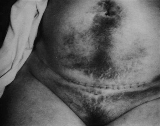
An ultrasound performed on the same day showed no free intraperitoneal fluid but a small anterior abdominal hematoma. The patient received 2 units of red blood cells. Her Hgb stabilized at around 10.3/G/dL, and her Htc stabilized at around 29.9%. The small periumbilical hematoma that appeared on the first postoperative day became obvious on the third postoperative day (Figure 1).
Figure 1.
Abdominal wall hematoma, postoperative day 3.
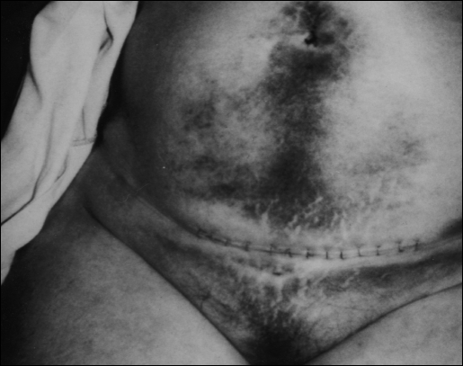
The abdominal hematoma was more pronounced on the next postoperative day, and a CT scan showed “soft tissue stranding in the anterior abdominal wall and mild thickening of the rectus muscle.” On discharge, on the sixth postoperative day, the patient's abdominal hematoma started showing discoloration due to the degradation in hemoglobin as verified by the yellow-greenish color at the periphery (Figure 2).
Figure 2.
Abdominal wall hematoma, postoperative day 6.
DISCUSSION
The umbilical area is the main insertion port for gyneco-logic laparoscopic procedures and together with the ventral midline area is considered in general “vascular safe.” More frequently, the secondary puncture sites are responsible for anterior abdominal wall vascular injuries. Most of the time, those injuries go unrecognized during laparoscopy because of the tamponade effect of the pneumoperitoneum and the reduction in venous return due to the steep Trendelenburg position in which the patient is placed.
Abdominal wall hematoma has been described as occurring in 6.25% of patients after laparoscopic cholecystectomy.1 They become evident in days 2 to 6 postoperatively, by visible bruises, excessive pain, or an asymptomatic drop in hematocrit. In a prospective, multicenter study,2 in an effort to reduce cannula-related laparoscopic complications, radial expanding devices were used. The results showed no major injuries, including no abdominal wall bleeding.
To minimize the danger of laparoscopic abdominal hematoma, some authors,3 based on an anatomical study of 21 human cadavers, are recommending the location of trocars in the ventral midline or in a zone of 5-cm width lateral to the lateral border of the rectus sheath. It is of interest that others4 who studied the vascular territories of the superior epigastric and the deep inferior epigastric systems described many perforating arteries that are emerging through the anterior rectus sheath, but the highest concentration of them is in the paraumbilical area. Those vessels are terminal branches of the deep inferior epigastric artery. They feed into a subcutaneus vascular network that radiates from the umbilicus like “the spokes of a wheel.”
CONCLUSION
With the rich periumbilical vascular network, it is a wonder that we do not see more frequent laparoscopic abdominal wall hematoma in the umbilical area. One possible explanation could be that, in general, laparoscopy is performed in women younger than our patient. If a trocar-related vascular injury occurs then it is possible that the reflex vasoconstriction associated with immediate vascular injury response and the trocar-related tamponade are sufficient to produce umbilical hemostasis. We speculate that our 76-year-old patient, with her more pronounced atherosclerosis, lacked an effective vasoconstriction reflex response to vascular injury. Therefore, adequate umbilical hemostasis could not be achieved and abdominal wall hematoma resulted.
References:
- 1. Bhattacharya S, Tate JJ, Davidson BR, Hobbs KE. Abdominal wall hematoma complicating laparoscopic cholecystectomy. HPB Surg. 1994; 7(4):291–296 [DOI] [PMC free article] [PubMed] [Google Scholar]
- 2. Galen DI, Jacobson I, Weckstein LN, Kaplan RA, DeNevi KL. Reduction of cannula-related laparoscopic complications using a radially expending access device. J Am Assoc Gynecol Laparosc. February 1999; 6: 79–84 [DOI] [PubMed] [Google Scholar]
- 3. Balzer KM, Witte H, Recknagel S, Kozianka J, Waleczek H. Anatomic guidelines for the prevention of abdominal wall hematoma induced by trocar placement. Surg Radiol Anat. 1999; 21: 87–89 [DOI] [PubMed] [Google Scholar]
- 4. Boydy JB, Taylor GI, Corlett R. The vascular territories of the superior epigastric and the deep inferior epigastric systems. Plast Reconstr Surg. 1984; 73: 1–16 [DOI] [PubMed] [Google Scholar]


