Abstract
Plasmodium falciparum parasites evade the host immune system by clonal expression of the variant antigen, P. falciparum erythrocyte membrane protein 1 (PfEMP1). Antibodies to PfEMP1 correlate with development of clinical immunity but are predominantly variant-specific. To overcome this major limitation for vaccine development, we set out to identify cross-reactive epitopes on the surface of parasitized erythrocytes (PEs). We prepared mAbs to the cysteine-rich interdomain region 1 (CIDR1) of PfEMP1 that is functionally conserved for binding to CD36. Two mAbs, targeting different regions of CIDR1, reacted with multiple P. falciparum strains expressing variant PfEMP1s. One of these mAbs, mAb 6A2-B1, recognized nine of 10 strains tested, failing to react with only one strain that does not bind CD36. Flow cytometry with Chinese hamster ovary cells expressing variant CIDR1s demonstrated that both mAbs recognized the CIDR1 of various CD36-binding PfEMP1s and are truly cross-reactive. The demonstration of cross-reactive epitopes on the PE surface provides further credence for development of effective vaccines against the variant antigen on the surface of P. falciparum-infected erythrocytes.
The variant surface protein Plasmodium falciparum erythrocyte membrane protein 1 (PfEMP1) plays a major role in the parasite–host interaction. Antibodies to PfEMP1 show significant correlation with development of clinical immunity, making PfEMP1 an important vaccine candidate (1–5). However, the variant nature of PfEMP1 remains the major obstacle for vaccine development, as the immune response to PfEMP1 is highly variant-specific. Individuals with relatively low exposure to P. falciparum parasites show very restricted recognition of the PE surface (1, 2, 4, 6). This is even more pronounced in sera from monkeys repeatedly exposed to a particular P. falciparum strain that recognize exclusively the PfEMP1 of that strain (7). Sera from adult residents of endemic areas can agglutinate parasitized erythrocytes (PEs) from various strains and isolates (1–3, 6, 8, 9). Yet, it is apparent that in most assays each variant is recognized by a different set of antibodies in the sera (6, 10). Immunization of various animals with recombinant fragments of PfEMP1 also tends to generate variant specific recognition at the PE surface (11, 12). Thus, it is apparent that PfEMP1 lacks cross-reactive antigenic epitopes and largely displays variant-specific immunodominant epitopes. Under strong, selective immune pressure, this results in the appearance of nonoverlapping antigenically distinct clones, as suggested by the mathematical model of Gupta et al. (13, 14). The lack of cross-reactive epitopes and the presence of immunodominant variant epitopes on PfEMP1 can explain how the immune response to PfEMP1 is kept variant-specific despite repeated exposure to the pathogen. These findings highlight the difficulties in using PfEMP1 for malaria vaccines. However, some structural conservation must exist in PfEMP1 to maintain its function as adhesion receptor (12, 15–18). Exposure to parasites that adhere in the human placenta or parasites that cause severe disease can elicit a response that is not variant-specific (19, 20). These results suggest that PfEMP1s associated with a particular adhesion or virulence properties may express conserved and immunogenic epitopes. Such epitopes are of great importance for development of anti-disease vaccines and therapeutics, but are yet to be identi-fied. Of particular interest is the cysteine-rich interdomain region 1 (CIDR1) of PfEMP1 that mediates PE adhesion to the major host receptor CD36 (12, 21). The domain is (relatively) conserved, mediates an important function, and does not induce high titer of antibodies during the infection (12, 15, 18). Immunization with this region gave, primarily, a strain-specific recognition at the PE surface (11, 12). Therefore, if conserved epitopes appear on this domain, they are predicted to be rare and not immunodominant.
To identify such epitopes, we generated mAbs and used various methods of screening to identify cross-reactive mAbs. We describe here two mAbs that show cross-reactivity with PfEMP1s of various strains. These mAbs may give rise to novel therapeutics and development of cross-reactive anti-PfEMP1 malaria vaccines.
Materials and Methods
Parasites and Cell Lines.
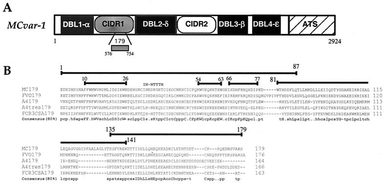
P. falciparum parasites were cultivated as described (11). In several cases, adherent PEs were enriched by selecting for binding to Chinese hamster ovary (CHO) K1 cells or CHO-CD36 cells (22). Parasite strains and isolates used were: Malayan Camp rosetting positive (MC R+), MC R−, FCR3-CD36, FCR3-ICAM1, FCR3-chondroitin sulfate A (CSA), ItA4, ItG 2F6, Santa Lucia (SL), and isolate RB8 R+. Parasites of the FVO strain in human erythrocytes were a gift from John Barnwell, Center for Disease Control and Prevention (Chamblee, GA). These parasites are known to express variant PfEMP1s (7, 11, 12, 21, 22). These parasites typed by microsatellites appear to be genotypically distinct from the Camp strain. CIDR1 domains of the expressed var gene from MC R+ (MCvar1395–852, AAB60251), FVO (FVOvar11–480, AF286005), ItA4 (A4var401–846, L42244), FCR3-CSA (FCR3varCSA418–824, AJ133811), and A4tres (A4Tvar375–724, AF193424) parasites were PCR-amplified and cloned into the expression vector pSRα5 (21). The MC R+ minimal CD36-binding region, MC 179 (MCvar1576–754), was also cloned and expressed as above. The amino acid boundaries of each clone and the accession number of the gene are provided. Site-directed mutagenesis replacing part of the MC CIDR1 with the FCR3varCSA CIDR1 sequence (Fig. 1B) was performed with the Quickchange site-directed mutagenesis kit (Stratagene). Each clone was verified by sequencing the entire insert. CHO-K1 cells were transfected by using Superfect reagent (Life Technologies, Gaithersburg, MD), and single-cell stable transfectants were cloned as described (21).
Figure 1.
Schematic representation and alignment of the amino acid sequences of the 179-aa minimal CD36-binding domain. (A) Schematic representation and amino acid boundaries of the MCvar1 gene (AAB60251) and the 179-aa CD36-binding domain. (B) Alignment of the amino acid sequences of the 179 region of PfEMP1 from various P. falciparum parasites. The consensus at 80% homology is also given. Alignment was performed by using the clustalw program (http://www.ebi.ac.uk/clustalw/), and the 80% consensus was performed by using the consensus program (http://www.bork.embl-heidelberg.de/cgi/consensus). Uppercase letters indicate conserved residues by the single-letter amino acid code. Lowercase letters indicate conserved classes of amino acids as follows: h, hydrophobic residues (A, C, F, G, H, I, K, L, M, T, T, V, W, Y); p, polar residues (C, D, E, H, K, N, Q, R, S, T); c, charged resides (D, E, H, K, R); a, aromatic residues (F, H, W, Y); s, small residues (A, C, D, G, N, P, S, T, V); +, positive residues (H, K, R); −, negative residues (D, E); l, aliphatic residues (I, L, V); and t, turnlike residues (A, C, D, E, G, H, K, N, Q, R, S, T). Peptides from the MC sequence used in various assays are indicated with their amino acid boundaries. The mutated site of the MC179 sequence (IN-NTTTH) also is indicated.
Recombinant Proteins and Peptides.
The production and purification of glutathione S-transferase (GST), GST-rC1–2 [1–179] (MCvar1576–754, AAB60251), maltose-binding protein (MBP) rC1–2 [1–233] (MCvar1576–808), and yeast rC1–2 [1–179] (y179) have been described (11, 12). An identical procedure was used to generate GST-rC1–2 [1–87], GST-rC1–2 [81–141], and GST-rC1–2 [135–179] (Fig. 1B). Peptides corresponding to the sequence rC1–2 [10–26], rC1–2 [54–63], and rC1–2 [66–77] (Fig. 1B) were synthesized with an additional cysteine residue at the amino terminal by SynPep (Dublin, CA) and conjugated to BSA by using the Imagect maleimide-activated conjugation kit (Pierce).
Immunization and Production of Hybridomas.
Sixteen NIH-Swiss mice were immunized (in groups of four) four times with either 5, 10, 25, or 50 μg of GST-rC1–2 [1–179] in Freund's complete adjuvant (first immunization) and incomplete adjuvant (immunizations 2–4). Mice were injected s.c. and i.p. with 200 μl of immunogen on days 0, 21, 35, and 49 and bled 7 days after each immunization. Hybridoma cells were prepared by Biocon (Rockville, MD). A single mouse, from the group immunized with 25 μg of protein, was chosen based on its reactivity with PEs of strains other than MC R+. The mouse was boosted with 50 μg of y179 and then killed 2 days later, and its spleen cells were fused with SP2O myeloma cells by using the polyethylene glycol fusion method (23). Cells from wells positive by ELISA (hybridoma lines) were cloned into three 96-well plates by limiting dilution at 0.5, 1, and 2 cells per well and grown in Hybri-max (Sigma) medium containing 10–20% FCS. Only wells that contained a single colony were followed. Culture supernatant was collected when cultures turned yellow and the cells started to die and tested as below. Ascites fluid from clones 4B3-A11, 6A2-B1, and TIB-191 (anti-trinitrophenyl IgG1 control mAb; American Type Culture Collection) was prepared in mice by Covance (Richmond, CA). Both mAbs also were produced as culture supernatant in a bioreactor cellmax artificial capillary module according to the manufacturer's protocol (Spectrum, Laguna Hills, CA).
Antibody Reactivity Assays.
ELISA and agglutination assays were as described (11, 12). Agglutination was performed at several dilutions to ensure optimal conditions for agglutination. A pool of human hyperimmune sera was used to establish agglutinability of PEs for each agglutination assay, and an unrelated mouse IgG1 was used as specificity control. Agglutination scoring was performed in a blind fashion by using only a number assigned to each tube. Agglutination scores (11) were modified to give more accurate measurements and were based primarily on the size of the agglutinate followed by a secondary scoring for the number of agglutinates. At least five agglutinates of similar size had to be present in five fields (×400) to validate a score: 0, no agglutination; 1+, agglutinates of 10–20 PEs; 2+, agglutinates of 20–50 PEs; 3+, agglutinates of 50–100 PEs; 4+, agglutinates of 100–250 PEs; 5+, agglutinates of >250 PEs. After scoring for size, the number of agglutinates was counted and added to the size score: 0, <10 agglutinates; 0.25, 10–25 agglutinates; 0.5, 25–50 agglutinates; 0.75, >50 agglutinates. Ig isotope was determined with mAbs isotyping kit II (Pierce).
Blockade of Adhesion.
Blockade of PE adhesion to CD36 was performed as described (12). In brief, mature-stage PEs at 3% parasitemia were incubated for 30 min at 37°C in the presence of 1:4 dilution of ascites fluid or with 1 mg/ml (final) appropriate mAb in binding medium, pH 6.7 (RPMI 1640/25 mM Hepes/0.5% BSA), before being added to immobilized CD36. After a 60-min incubation, the plate was washed, fixed, and stained as described (12). Each assay was performed in duplicate, and at least five fields were counted randomly to determine the number of adherent cells.
Flow Cytometry Assays.
For antibody binding to the surface of transfected CHO cells, cells were removed by PBS containing 2 mM EDTA, centrifuged down for 5 min at 780 × g, and resuspended to 1–5 × 10⁶/ml with PBS/0.5% BSA/0.1% sodium azide (RIA buffer). Samples (100 μl) were added to round-bottomed, 96-well plates and centrifuged as above, and the liquid was discarded by inversion and tapping. Samples (100 μl) containing 100 μg/ml mAb 4B3-A11 or mAb 6A2-B1 were added, and the cells were incubated for 1 h at room temperature with rotation. mAb 179 (Affymax Research Institute, Santa Clara, CA) that recognizes an epitope tag incorporated to the carboxyl terminus of each clone, was used at 6 μg/ml to determine the expression of each clone. Cells were centrifuged and washed twice with 125 μl of RIA buffer. One hundred microliters of fluorescein-labeled goat anti-mouse IgG (Jackson ImmunoResearch) diluted 1:100 in RIA buffer was added, and the cells were incubated for 45 min and washed as above. The cells were resuspended in 100 μl of RIA buffer and kept on ice until added to 0.3 ml of PBS for flow cytometry analysis. Flow cytometry was performed by using a Becton Dickinson FACScalibur and the flowjo 3.1 analysis software (Tri Star, San Carlos, CA).
Results
Preparation and Characterization of Anti-rC1–2 [1–179] mAbs.
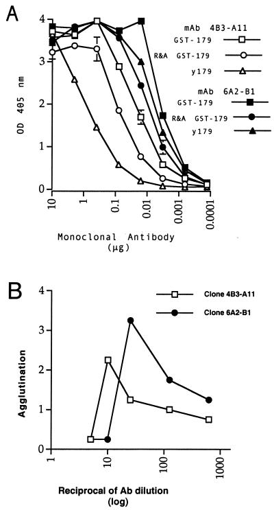
Sixteen mice were immunized with GST-rC1–2 [1–179], and sera from all 16 animals agglutinated MC R+ PEs. However, although there is apparent homology in the 179 region of CIDR1 among different PfEMP1s (Fig. 1B), only one mouse gave low but consistent agglutination with PEs of P. falciparum strains (ItG-ICAM and ItA4) other than MC R+ (data not shown). This mouse was boosted with a yeast recombinant protein, y179, the minimal protein from CIDR1 that binds to CD36. The spleen cells were fused 3 days later to produce hybridoma cell lines. Seven lines that were positive by ELISA to y179 and agglutinated MC R+ PEs were recloned by limiting dilution. The subclones that reacted by ELISA against y179 were tested against subregions of rC1–2 [1–179]. Subclones from six of seven lines reacted with amino acids 1–87 of rC1–2, one reacted with amino acids 81–141, and none reacted with the C-terminal region (amino acids 135–179). All clones were of the IgG1 subclass and reacted with the reduced and alkylated form of GST-rC1–2 [1–179] (Fig. 2A and data not shown). Subclones from all seven clones agglutinated MC R+. Two subclones, 4B3-A11 and 6A2-B1, also agglutinated PEs from other strains and, therefore, were analyzed further.
Figure 2.
Reactivity of mAbs 4B3-A11 and 6A2-B1. (A) The reactivity of mAb 4B3-A11 (open symbols) and mAb 6A2-B1 (solid symbols) with the GST-rC1–2 [1–179] (GST-179), reduced and alkylated form of the molecule (R&A GST-179), and yeast recombinant protein y179 was measured by ELISA. (B) Agglutination titers of mAb 4B3-A11 and mAb 6A2-B1 (×20 concentration of culture supernatant) with MC R+ PEs.
Cross-Reactive mAbs Recognize the Surface of PEs Expressing Variant PfEMP1s.
mAb 4B3-A11 recognized exclusively amino acids 81–141 of rC1–2 [1–179], and mAb 6A2-B1 reacted with the more conserved 1–87 region. Both clones were reactive with y179 and GST-rC1–2 [1–179]. However, the reactivity of mAb 4B3-A11 with y179 was much reduced compared with GST-rC1–2 [1–179] (Fig. 2A). The reactivity of clone 6A2-B1 was higher than that of 4B3-A11, and both reacted strongly with the reduced and alkylated form of GST-rC1–2 [1–179] that cannot bind CD36 (Fig. 2A), indicating that the binding did not require the functional conformation of the CD36-recognition domain.
mAb 6A2-B1 gave consistently higher agglutination than mAb 4B3-A11 (Fig. 2B and Table 1). At low-antibody dilution there was no agglutination with either mAb, probably because of a prozone effect of excessive antibody (Fig. 2B). We extended the PE agglutination to nine other P. falciparum strains (Table 1). Agglutination was performed with culture supernatant from flasks (diluted 1:2), culture supernatant from bioreactors (diluted 1:5, 1:30, and 1:100), and with ascites (diluted 1:5, 1:25, and 1:125) to optimize conditions for agglutination. The data from the dilution that gave the highest agglutination in each assay were used in Table 1. MC R+, the strain homologous to the immunogen, generally gave stronger agglutination than heterologous strains (Table 1). mAb 6A2-B1 was more cross-reactive with different strains and gave a stronger agglutination than mAb 4B3-A11 (Table 1). In fact, mAb 6A2-B1 was reactive with all PEs tested, with the exception of PEs from the non-CD36-binding clone, FCR3-CSA (Table 1). Thus, it is clear that mAb 6A2-B1 is a cross-reactive mAb, regardless of the differences in its reactivity with various P. falciparum clones.
Table 1.
Agglutination of PEs from various P. falciparum clones/lines by mAb 4B3-A11 and mAb 6A2-B1
| Strain | mAb 6A2-B1
|
mAb 4B3-A11
|
||
|---|---|---|---|---|
| Ascites | Culture supt. | Ascites | Culture supt. | |
| MC R+ | 1.75–3.25* (4)† | 1.25–3.5 (3) | 1.25 (2) | 1–3 (4) |
| MC R− | 1.25 (2) | ND | 0.75–1.5 (2) | ND |
| ItG 2F6 | 1.25–2.25 (3) | ND | 1.25 (1) | 0.75–2.5 (2)‡ |
| ItA4 | 1.0–2.0 (4) | 2 (1) | 0.25–1 (3) | ND |
| Santa Lucia | 0.75–1.0 (2) | ND | 0.5–0.75 (2) | ND |
| RB8 R+ | 2 (1) | 2 (1)§ | 0 (1) | 0 (1)§ |
| FCR3-CD36 | 0.75–1 (3) | ND | 1–1.5 (3) | ND |
| FCR3-ICAM | 1.5–2 (4) | ND | 0 (3) | ND |
| FVO | 3 (1) | 2 (2)‡ | 2.25 (1) | 0.25–2.5 (2)‡ |
| FCR3-CSA | 0 (2) | 0 (3) | 0 (2) | 0 (3) |
The reactivity of mAb 4B3-A11 and mAb 6A2-B1 with parasitized erythrocytes of various strains was measured by agglutination by using ascites fluid or culture supernatant (supt.) from hybridoma cells grown in tissue culture flasks (1:2 dilution). Agglutination with ascites was performed at three dilutions (1:5, 1:25, and 1:125) to ensure adequate agglutination conditions. Agglutination was scored as in Materials and Methods. ND, not determined.
Range of agglutination scores from independent assays. The range reports the highest score from each assay (typically 1:25 dilution for ascites fluid).
The numbers of independent assays are in parentheses.
Agglutination was performed with bioreactor culture supernatant at 1:5, 1:30, and 1:100 dilutions.
Agglutination was performed at 1:2 dilution with concentrated (20×) culture supernatant.
Flow Cytometry Analysis Confirms the Cross-Reactive Nature of mAbs 4B3-A11 and 6A2-B1.
One disadvantage of the agglutination assays is that the data are only semiquantitative. Unfortunately, flow cytometry analysis of antibody bound to PEs was too insensitive to measure cross-reactivity, although both mAbs were positive by flow cytometry with PEs of the homologous MC R+ strain (data not shown). Serological (agglutination and immunoprecipitation) and sequence data (Fig. 1B) support the observation that the strains used in the agglutination assay express variant PfEMP1s (7, 11, 12, 21, 22). However, because several var genes can be expressed in each strain, the agglutinating PEs could represent a subpopulation of PEs that express a CIDR1 domain that was more homologous in sequence to MC R+.
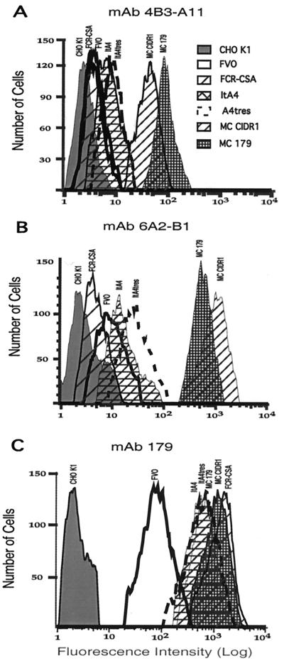
To demonstrate that the mAbs recognize nonhomologous CIDR1s and to obtain confirmatory quantitative data, we measured, by flow cytometry, the reactivity of the mAbs with CHO cells expressing variant CIDR1 domains (Table 2 and Fig. 3). With the exception of FCR3varCSA, all CIDR1 domains on CHO cells bound CD36 (B.G., unpublished data), a coreceptor for endothelial binding. The amount of expression of each CIDR1 was measured by antibody (mAb 179) reactive with an epitope tag expressed by all clones. Expression was found to be similar (within a 2-fold difference), except for FVO CIDR1, which had 1/5th to 1/10th the expression of the others (Table 2). The cross-reactivity was always much lower with the heterologous strains than with MC CIDR1. The reactivity with the CIDR1 from the non-CD36-binding clone, FCR3-CSA, was similar to the control CHO cell reactivity, indicating no cross-reactivity. This result was similar to the finding of no cross-agglutination with this PE. The binding of mAb 6A2-B1 to the different CIDR1 clones was consistently higher than that of mAb 4B3-A11 (Table 2 and Fig. 3), a result that was similar to the higher agglutination of various strains by mAb 6A2-B1 (Table 1). These results clearly demonstrate the cross-reactive nature of both mAbs and independently confirm the agglutination results.
Table 2.
Reactivity of mAb 4B3-A11 and mAb 6A2-B1 with CHO K1 cells expressing various CIDR1 domains
| CHO cells | Fluorescence intensity (median)
|
||
|---|---|---|---|
| mAb 4B3-A11 | mAb 6A2-B1 | Anti-Tag (mAb 179) | |
| Control CHO | 3.3 | 3.0 | 2.5 |
| K1 | |||
| MC 179 | 85.7 | 541 | 599 |
| MC CIDR1 | 41 | 932 | 1,056 |
| ItA4 CIDR1 | 7.7 | 14.5 | 498 |
| A4tres CIDR1 | 9.0 | 26.7 | 569 |
| FVO CIDR1 | 4.3 | 9.8 | 77 |
| FCR3-CSA | 3.9 | 4.7 | 1,259 |
| CIDR1 | |||
Quantitative binding of mAb 4B3-A11 and mAb 6A2-B1 to CHO K1 cells expressing various CIDR1 domains represented by the median fluorescence intensity from Fig. 2. Expression levels of each CHO K1 clone were measured by using mAb 179.
Figure 3.
CIDR1s of various PfEMP1 are recognized by the cross-reactive mAbs. The reactivity of mAb 4B3-A11 and mAb 6A2-B1 with CHO-K1 cells expressing CIDR1 domains of different var genes and the rC1–2 [1–179] region of MCvar 1 CIDR1 (MC 179) was measured by flow cytometry. Expression of the different CIDR1 on the CHO cell surface was measured by mAb 179 that is reactive with an epitope tag shared by all constructs. (A) mAb 4B3-A11 at 100 μg/ml. (B) mAb 6A2-B1 at 100 μg/ml. (C) mAb179 at 6 μg/ml.
mAb 6A2-B1 May Be Reactive with a Conformation-Dependent Epitope.
The cross-reaction of the mAbs to different CIDR1s was unrelated to primary sequence in the rC1–2 (1–179) region. The region recognized by mAb 4B3-A11 (amino acids 81–141) displayed only limited homology among various CIDR1s (Fig. 1; refs. 12 and 24). Although there is more homology in the 1–87 region reactive with mAb 6A2-B1, there are no linear sequences that are completely identical (Fig. 1B). We used three linear peptides from some of the more conserved regions of CIDR1 (amino acids 10–26, 54–63, and 66–77; Fig. 1B), but mAb 6A2-B1 did not react with any of the peptides tested (data not shown). However, it is still possible that these peptides failed to express the complete sequence needed for binding. We also exchanged a small fragment from the MC R+ sequence with the homologous fragment from the FCR3varCSA sequence not recognized by mAb 6A2-B1 (Fig. 1B) without any effect on the binding of mAb 6A2-B1 (data not shown). Thus, it is likely that the mAb recognized a conserved, nonlinear structural epitope that does not depend on disulfide bonds.
Blockade of PE Adhesion.
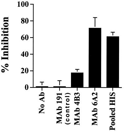
None of the mAbs tested as culture supernatant blocked adhesion. Both mAb 4B3-A11 and 6A2-B1 blocked binding of MC R+ PEs to CD36 when tested at 1 mg/ml (Fig. 4). The percent inhibition when compared with a control mAb 191 was 71.4 ± 12.7 and 17.5 ± 4.2 (mean ± SD, n = 2) for mAbs 6A2-B1 and 4B3-A11, respectively. The blockade of adhesion was not affected by PE agglutination because no agglutination occurred at this Ab concentration. Attempts to block adhesion of PEs of strains ItG-2F6 and ItA4 were not successful, probably because of the low reactivity of mAbs with these strains and because blockade of adhesion usually requires high-Ab titers.
Figure 4.
Blockade of PE adherence. PEs were preincubated with binding buffer alone (no Ab) or binding buffer containing 1 mg/ml unrelated mouse IgG1 (mAb 191), mAb 4B3-A11, mAb 6A2-B1, or pooled human hyperimmune sera (HIS). After 30-min incubation, the PEs were added to immobilized CD36 and tested for binding in duplicate. The results are presented as percent inhibition ± SD from the control mAb 191.
Discussion
The variant antigen of P. falciparum, PfEMP1, contains 50 or more genes in one genome that are expressed singly on the surface of infected red cells and are designed to evade immunity (5, 11, 22, 25–27). As a consequence, antibodies induced by infection are clone-specific. This was shown clearly by acute and convalescent sera from children (1, 4, 6, 9), sera from experimentally infected Aotus monkeys (7), and sera from individuals living in areas of low or seasonal transmission (2, 3, 28). Even antibodies from malaria-immune African adults are variant-specific; the same antibodies only rarely will cross-agglutinate PEs from two variant clones (10). In cases in which the same antibodies recognize more than one PfEMP1, those antibodies do not appear to recognize all variant strains but react with a limited number of clones that may express similar epitopes. The strain specificity is attributed to immunodominant, polymorphic determinants and strong immune selection that leads to primarily discrete, nonoverlapping clonal parasite population structure of PfEMP1 proteins (13, 14). With the vast number of var genes among the general parasite population and the strain-specific immunity to PfEMP1, this protein may seem a hopeless target for vaccine development.
In this study, we present data demonstrating that at least one region of PfEMP1 can induce cross-reactive antibodies. PfEMP1 is divided into domains with specific binding specificity and specific structures (18). We focused on CIDR1 because it binds CD36, a highly conserved function found in most clones of PfEMP1 (9, 12, 29). We have predicted that the majority of the immune response will be to strain-specific determinants. Thus, to identify cross-reactive epitopes we generated singular reagents (mAbs) that can be screened and selected for cross-reactivity. Indeed, of the 16 mice immunized with rC1–2 [1–179] of MC R+, only 1 gave some reactivity with the surface of strains other than MC R+ and only 2 of the mAbs generated from this mouse were cross-reactive. One mAb (6A2-B1) was highly cross-reactive, and another (4B3-A11) recognized fewer P. falciparum PEs tested (Tables 1 and 2 and Fig. 2). Each of the two cross-reactive mAbs recognized a different region of the CIDR1, indicating that there are at least two independent, conserved epitopes in this domain of PfEMP1. These results support the prediction and previous observations that conserved epitopes may be rare and less immunogenic (12, 13, 15).
The reactivity of the mAbs to different CIDR1s expressed on CHO cells excludes the possibility that the cross-reactivity observed by agglutination resulted from recognition of a few PEs that carried epitopes similar to the immunogen, rC1–2 [1–179], from the Malayan Camp parasites. The quantitative differences in the binding of each mAb to various CIDR1s as measured by agglutination and flow cytometry and the observed differences in the CIDR1 sequences suggest that the epitopes are not identical, although they are cross-reactive enough for detection. The 179-aa CD36-binding domain is cysteine-rich, and its function is sensitive to reduction and alkylation and mutagenesis of cysteine residues (12). We predicted that a cross-reactive mAb would recognize the structure involved in binding to CD36. Indeed, both mAbs did not react with the CIDR1 domain of clone FCR3-CSA that does not bind to CD36, and mAb 6A2-B1 failed to recognize several highly conserved, linear peptides from the 1–87 region. Thus, we were surprised that both mAbs bound equally well to the reduced and alkylated domain. We speculate that, although the cysteine-dependent structural integrity of the CIDR1 mediating adhesion to CD36 was not required for epitope recognition, the mAbs may recognize epitopes that are important, but not sufficient, for CD36 binding.
Clonal antigenic variation is an important mechanism of immune evasion maintained by the large and diverse PfEMP1 family (5, 11, 26, 27, 30). For this mechanism to remain effective despite repeated exposure to P. falciparum, the response to PfEMP1 must be kept variant-specific. This is most likely done by diverting the immune response away from potential cross-reactive determinants and directing it to strong, polymorphic, immunodominant epitopes (3, 13, 28). One such example is the CD36-binding fragment of CIDR1 that elicits antibodies by immunization in various animals but does not appear to elicit high-titer antibodies in humans or monkeys exposed to P. falciparum (refs. 11, 12, and 15; D.I.B., unpublished observations).
Multigravid women are protected from placental malaria by antibodies that block the adhesion of parasites from around the world to CSA (20). Thus, functional regions of PfEMP1 involved in particular binding properties may induce cross-reactive immunity (19, 20), although the var genes from these parasites are highly diverse (18). In distinction to the CSA-binding PEs, CD36-adherent parasites generate a strong, variant-specific immune response associated with immune evasion by antigenic variation.
Directing the immune response to conserved epitopes instead of the strain-specific ones, particularly in CD36-adherent parasites, may help to overcome the major obstacle in the development of vaccines against the variant antigen on P. falciparum-infected erythrocytes. The demonstration of conserved epitopes within the CIDR1 domain offers new insight for the potential of the variant antigen as an important target for a malaria vaccine.
Acknowledgments
We thank Dr. David Kaslow for providing us with the y179 yeast recombinant protein.
Abbreviations
- CSA
chondroitin sulfate A
- PfEMP1
Plasmodium falciparum erythrocyte membrane protein 1
- PE
parasitized erythrocyte
- CIDR
cysteine-rich interdomain region
- CHO
Chinese hamster ovary
- GST
glutathione S-transferase
References
- 1.Bull P C, Lowe B S, Kortok M, Molyneux C S, Newbold C I, Marsh K. Nat Med. 1998;4:358–360. doi: 10.1038/nm0398-358. [DOI] [PMC free article] [PubMed] [Google Scholar]
- 2.Barragan A, Kremsner P G, Weiss W, Wahlgren M, Carlson J. Infect Immunol. 1998;66:4783–4787. doi: 10.1128/iai.66.10.4783-4787.1998. [DOI] [PMC free article] [PubMed] [Google Scholar]
- 3.Giha H A, Staalsoe T, Dodoo D, Elhassan I M, Roper C, Satti G M H, Arnot D E, Theander T G, Hviid L. Infect Immunol. 1999;67:4092–4098. doi: 10.1128/iai.67.8.4092-4098.1999. [DOI] [PMC free article] [PubMed] [Google Scholar]
- 4.Bull P C, Lowe B S, Kortok M, Marsh K. Infect Immunol. 1999;67:733–739. doi: 10.1128/iai.67.2.733-739.1999. [DOI] [PMC free article] [PubMed] [Google Scholar]
- 5.Reeder J C, Brown G V. Immunol Cell Biol. 1996;74:546–554. doi: 10.1038/icb.1996.88. [DOI] [PubMed] [Google Scholar]
- 6.Marsh K, Howard R J. Science. 1986;231:150–153. doi: 10.1126/science.2417315. [DOI] [PubMed] [Google Scholar]
- 7.Howard R J, Barnwell J W, Rock E P, Neequaye J, Ofori-Adjei D, Maloy W L, Lyon J A, Saul A. Mol Biochem Parasitol. 1988;27:207–223. doi: 10.1016/0166-6851(88)90040-0. [DOI] [PubMed] [Google Scholar]
- 8.Aguiar J C, Albrecht G R, Cegielski P, Greenwood B M, Jensen J B, Lallinger G, Martinez A, McGregor I A, Minjas J N, Neequaye J, et al. Am J Trop Med Hyg. 1992;47:621–632. doi: 10.4269/ajtmh.1992.47.621. [DOI] [PubMed] [Google Scholar]
- 9.Reeder J C, Rogerson S J, al-Yaman F, Anders R F, Coppel R L, Novakovic S, Alpers M P, Brown G V. Am J Trop Med Hyg. 1994;51:45–55. doi: 10.4269/ajtmh.1994.51.45. [DOI] [PubMed] [Google Scholar]
- 10.Newbold C I, Pinches R, Roberts D J, Marsh K. Exp Parasitol. 1992;75:281–292. doi: 10.1016/0014-4894(92)90213-t. [DOI] [PubMed] [Google Scholar]
- 11.Baruch D I, Pasloske B L, Singh H B, Bi X, Ma X C, Feldman M, Taraschi T F, Howard R J. Cell. 1995;82:77–87. doi: 10.1016/0092-8674(95)90054-3. [DOI] [PubMed] [Google Scholar]
- 12.Baruch D I, Ma X C, Singh H B, Bi X, Pasloske B L, Howard R J. Blood. 1997;90:3766–3775. [PubMed] [Google Scholar]
- 13.Gupta S, Anderson R M. Parasitol Today. 1999;15:497–501. doi: 10.1016/s0169-4758(99)01559-8. [DOI] [PubMed] [Google Scholar]
- 14.Gupta S, Maiden M C, Feavers I M, Nee S, May R M, Anderson R M. Nat Med. 1996;2:437–442. doi: 10.1038/nm0496-437. [DOI] [PubMed] [Google Scholar]
- 15.Baruch D I, Gormley J A, Ma C, Howard R J, Pasloske B L. Proc Natl Acad Sci USA. 1996;93:3497–3502. doi: 10.1073/pnas.93.8.3497. [DOI] [PMC free article] [PubMed] [Google Scholar]
- 16.Newbold C I, Craig A G, Kyes S, Berendt A R, Snow R W, Peshu N, Marsh K. Ann Trop Med Parasitol. 1997;91:551–557. doi: 10.1080/00034989760923. [DOI] [PubMed] [Google Scholar]
- 17.Howard R J, Gilladoga A D. Blood. 1989;74:2603–2618. [PubMed] [Google Scholar]
- 18.Smith J D, Gangadharan S, Gamain B, Baruch D I, Miller L H. Mol Biochem Parasitol. 2000;110:293–310. doi: 10.1016/s0166-6851(00)00279-6. [DOI] [PubMed] [Google Scholar]
- 19.Gupta S, Snow R W, Donnelly C A, Marsh K, Newbold C. Nat Med. 1999;5:340–343. doi: 10.1038/6560. [DOI] [PubMed] [Google Scholar]
- 20.Fried M, Nosten F, Brockman A, Brabin B J, Duffy P E. Nature (London) 1998;395:851–852. doi: 10.1038/27570. [DOI] [PubMed] [Google Scholar]
- 21.Smith J D, Kyes S, Craig A G, Fagan T, Hudson-Taylor D, Miller L H, Baruch D I, Newbold C I. Mol Biochem Parasitol. 1998;97:133–148. doi: 10.1016/s0166-6851(98)00145-5. [DOI] [PubMed] [Google Scholar]
- 22.Scherf A, Hernandez-Rivas R, Buffet P, Bottius E, Benatar C, Pouvelle B, Gysin J, Lanzer M. EMBO J. 1998;17:5418–5426. doi: 10.1093/emboj/17.18.5418. [DOI] [PMC free article] [PubMed] [Google Scholar]
- 23.Kennett R H, Denis K A, Tung A S, Klinman N R. Curr Top Microbiol Immunol. 1978;81:77–91. doi: 10.1007/978-3-642-67448-8_13. [DOI] [PubMed] [Google Scholar]
- 24.Smith J D, Craig A G, Kriek N, Hudson-Taylor D, Kyes S, Fagen T, Pinches R, Baruch D I, Newbold C I, Miller L H. Proc Natl Acad Sci USA. 2000;97:1766–1771. doi: 10.1073/pnas.040545897. . (First Published February 4, 2000; 10.1073/pnas.040545897) [DOI] [PMC free article] [PubMed] [Google Scholar]
- 25.Chen Q, Fernandez V, Sundstrom A, Schlichtherle M, Datta S, Hagblom P, Wahlgren M. Nature (London) 1998;394:392–395. doi: 10.1038/28660. [DOI] [PubMed] [Google Scholar]
- 26.Su X Z, Heatwole V M, Wertheimer S P, Guinet F, Herrfeldt J A, Peterson D S, Ravetch J A, Wellems T E. Cell. 1995;82:89–100. doi: 10.1016/0092-8674(95)90055-1. [DOI] [PubMed] [Google Scholar]
- 27.Newbold C I. Curr Opin Microbiol. 1999;2:420–425. doi: 10.1016/S1369-5274(99)80074-5. [DOI] [PubMed] [Google Scholar]
- 28.Giha H A, Theander T G, Staalso T, Roper C, Elhassan I M, Babiker H, Satti G M H, Arnot D E, Hviid L. Am J Trop Med Hyg. 1998;58:399–405. doi: 10.4269/ajtmh.1998.58.399. [DOI] [PubMed] [Google Scholar]
- 29.Newbold C, Warn P, Black G, Berendt A, Craig A, Snow B, Msobo M, Peshu N, Marsh K. Am J Trop Med Hyg. 1997;57:389–398. doi: 10.4269/ajtmh.1997.57.389. [DOI] [PubMed] [Google Scholar]
- 30.Smith J D, Chitnis C E, Craig A G, Roberts D J, Hudsontaylor D E, Peterson D S, Pinches R, Newbold C I, Miller L H. Cell. 1995;82:101–110. doi: 10.1016/0092-8674(95)90056-x. [DOI] [PMC free article] [PubMed] [Google Scholar]


