This report suggests that single-access laparoscopic splenectomy may be an opportunity to further refine minimally invasive approaches for general surgical disease.
Keywords: Single access surgery, Single incision surgery, Laparoscopic splenectomy, Idiopathic thrombocytopenic purpura
Abstract
Background:
Laparoscopic splenectomy has been performed in a standard fashion with 4 to 5 trocars since the early 1990s. Single access laparoscopy has recently gained interest, but single access laparoscopic splenectomy has not been reported to date. It has the possible benefits of less pain, faster recovery, better cosmesis, with theoretically similar costs to that of traditional trocars.
Methods:
A case is presented and the surgical technique of single access laparoscopic splenectomy is detailed.
Results:
The patient is an otherwise healthy 24-year-old male with medically refractory idiopathic thrombocytopenic purpura and a platelet count of 15 000. A splenectomy was performed using a single incision laparoscopic technique. The patient was placed in a right lateral decubitus position, and a 2.5-cm left upper quadrant incision was made. A multi-instrument flexible single incision port was used that held 3 trocars. A standard splenectomy was performed through this port. A linear stapler was used to transect the splenic hilum. The procedure time was just over 2 hours. The patient did well, was happy with his incision, and was discharged with a platelet count of 108 000.
Conclusions:
Single access laparoscopic splenectomy is feasible in select patients and may provide a less painful, better cosmetic result.
INTRODUCTION
Splenectomy has been used as a treatment for chronic idiopathic thrombocytopenic purpura (ITP) for decades.1 Since its introduction in 1992, laparoscopic splenectomy has been shown to be an equivalently effective and preferred minimally invasive method, decreasing patient discomfort, hospital stays, and recovery times.2–6 In an effort to maximize the benefits of minimally invasive surgery, a new concept in surgery, natural orifice transluminal endoscopic surgery (NOTES), has emerged.7 Its promise is that of totally scarless intraperitoneal surgery with virtually zero patient discomfort. Still under investigation, NOTES has many technical challenges, and its application has been limited to a few centers worldwide.8–10
Another competing minimally invasive approach, single port or single access surgery (SAS) has been described sporadically since the late 1990s.11–13 Since the advent of NOTES, the idea of SAS has been revisited and has gained interest. This approach emphasizes the concept of surgery through one small transabdominal incision rather than the standard multiple trocar sites, with theoretic benefits of less pain and better cosmesis. The allure of SAS is that the surgeon can use relatively standard laparoscopic instruments and skills without the technical difficulties of NOTES surgery. As a result, SAS can be more readily implemented and is likely to have a higher degree of acceptance. The incision can be hidden periumbilically and can be used as the specimen extraction site as well. SAS has successfully been applied to basic laparoscopy (eg, cholecystectomies) as well as advanced procedures, such as colectomy, nephrectomy, and bariatric procedures.14–17 We describe the initial report of SAS laparoscopic splenectomy.
CASE STUDY AND SURGICAL TECHNIQUE
The patient is a thin 24-year-old male with medically recalcitrant idiopathic thrombocytopenic purpura and no other past medical or surgical history. His platelet count remained at 15 000, and a CT scan demonstrated a normalsized 13.5-cm spleen and an adjacent 1.8-cm splenule. Treatment options were thoroughly reviewed with the patient, and after weighing the risks and benefits, the patient elected to undergo a laparoscopic splenectomy.
The patient was placed in a 60-degree right lateral decubitus position on a moldable beanbag mattress with appropriate padding and securing straps. A 25-mm left upper quadrant incision was made just lateral to the rectus muscle. The abdomen was entered under direct vision. The incision site was selected to optimize the linear stapler angle that would be needed to divide the splenic hilum. A special multi-instrument flexible port, SILS Port (Covidien AG, Norwalk, CT), was, then, inserted into the incision. Three low-profile 5-mm trocars were placed through the port (Figure 1). The equipment used throughout the case included a 5-mm, 30-degree laparoscope whose light and camera cords emerged straight from the back of the scope, standard laparoscopic instruments, and one bending grasper.
Figure 1.
Placement of SILS port with three 5-mm low-profile trocars.
Initially, the splenic flexure of the colon was mobilized away from the spleen. Then, the spleen was retracted laterally, and an inferior pole vessel was isolated, clipped with a 5-mm clip applier, and divided with 5-mm ultrasonic shears. At this point, the splenule was visualized, and its attachments were divided. The fundus was mobilized away from the spleen by sequentially dividing the short gastric vessels with ultrasonic shears. The bending grasper was used to retract the spleen laterally and display the splenic hilum.
The avascular plane posterior to the splenic hilum was dissected. Additional blunt dissection was used to open a window superior to the hilum. At this point in the procedure, one of the 5-mm trocars was replaced with a 12-mm trocar to accommodate the stapler. Two fires of a Roticulator laparoscopic linear stapling device carrying a blue load buttressed with a porcine-derived strip were used to transect the splenic hilum. Once the hilum was divided, the patient was transfused 2 units of platelets.
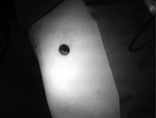
The remaining medial attachments were divided using ultrasonic shears. The bendable grasper was replaced by a snake retractor that was used to retract the spleen medially to allow for division of the lateral attachments. The 12-mm trocar was removed, and a 15-mm Endocatch bag (Covidien, Norwalk, CT) was inserted into the abdomen directly through the SILS port. The spleen and splenule were placed into the bag, and the bag was brought to skin level. The port was removed, and the abdomen desufflated. The spleen was morcellated in a standard fashion and removed. The port was reinserted, and pneumoperitoneum was re-established. The splenic bed was irrigated, and the staple line inspected. Once the port was removed, the resulting incision was remarkably small (Figure 2) and was closed in 2 layers. The patient was extubated and taken to recovery in good condition.
Figure 2.
Final incision prior to closure.
The procedure was completed without complications in 133 minutes with <10cc of blood loss. The patient required minimal pain medications and was discharged on postoperative day number 2 tolerating a regular diet. Two weeks following surgery, the patient was doing well and back to his baseline activity level.
DISCUSSION
Splenectomy is an effective therapy for medically refractory ITP, and a laparoscopic approach has been proven to be of great benefit to patients due to decreased pain, faster recovery, and better cosmesis. NOTES is a new paradigm that has the potential to deliver these benefits to a much greater degree but is still encumbered by many technical limitations. Single access surgery theoretically delivers many of the proposed advantages of NOTES but without its accompanying disadvantages. SAS utilizes standard or slightly modified laparoscopic instrumentation, maintains a sterile environment that is impossible with NOTES, and requires limited additional training for a skilled laparoscopist. In addition, the cost of SAS (such as the specialized port) may be comparable to the cost of standard laparoscopic trocars.
We performed this single access laparoscopic splenectomy safely and effectively and with minimally increased operative time. We took care to maintain the same standards of visualizing anatomic landmarks in proceeding with the steps of the operation. We also selected an ideal patient who was thin without prior abdominal operations and with a normal-sized spleen. Although SAS causes significantly more clashing of instruments, we were able to minimize this problem in a few ways. We used lowprofile trocars and a specialized multi-channel port that allowed greater angled movements for the instruments. The laparoscope was intentionally held further from the abdominal wall, and we utilized a camera that had cords projecting from the back thus away from the surgeons hands. The assistant manipulated the retracting instrument during most of the case, so that the surgeon could hold the camera and the dissecting instrument and move them in conjunction.
Limitations of our current technique include the placement of our incision away from the umbilicus. The selection of our incision site optimized the angle through which we fired our linear stapler when controlling the splenic hilum. Even though the cosmetic result was excellent, we did not hide the scar in the umbilicus. Longer instrumentation with greater roticulation may be needed to perform this operation transumbilically.
The size of spleen that can be resected and extracted may be limited by the single incision technique. Currently, massive splenomegaly increases the risk of conversion from standard laparoscopy to open or requires the addition of a hand-port for splenic manipulation and extraction.18,19 Maneuvering large spleens to safely control the hilum and to place them in the extraction bag will be difficult using a single incision laparoscopic approach. The exact spleen size limitation of single access laparoscopic splenectomy is not yet known.
Another limitation may be the ability to detect an accessory spleen. When performing a splenectomy for ITP, care must be taken not to overlook an accessory spleen that may cause recurrence of thrombocytopenia. Initial reports comparing laparoscopy with open splenectomy suggest that laparoscopic splenectomy may incur more missed accessory spleens.20 Subsequent reports have shown that the 2 approaches are probably equivalent if the surgeons take care to perform a thorough survey of the operative field.6,21 We had preoperative imaging that identified the accessory spleen, and it was also quite close to the splenic hilum and easy to visualize. SAS may make a thorough survey for accessory spleens more challenging.
SAS may be a bridge to NOTES by allowing surgeons to hone their skills in operating within a single incision, but it is also a valid less-invasive alternative to standard laparoscopy. With refinement in surgical technique, operative time will decrease, surgeons' level of comfort with these operations will increase, and SAS may become the routine. This technique yielded clearly visible cosmetic improvements, but we still need to determine with randomized controlled trials whether it improves patient recovery and reduces postoperative pain and complications. Until surgeons become comfortable with this approach, we suggest that a high degree of attention be paid to patient selection. Most importantly, as these new surgical approaches are investigated and adopted, the paramount emphasis must be placed on adherence to safe surgical principles. Clearly, single access laparoscopy has possibly significant limitations on visualization, triangulation of instrumentation, and manipulation of tissue. Just as with traditional laparoscopy, the surgeon must have a very low threshold to add additional trocars or convert to an open procedure. If these principles are followed, the procedure can be done safely, and the theoretical benefits to the patient can be studied.
CONCLUSION
This initial report documents the feasibility of single access laparoscopic splenectomy and introduces another opportunity to further refine minimally invasive approaches in general surgery. With continued evaluation of SAS as well as advancements in instrumentation, SAS may prove to be the favored approach for certain laparoscopic procedures in the future.
Contributor Information
Preeti Malladi, Department of Surgery, Feinberg School of Medicine, Northwestern University, Chicago, Illinois, USA..
Eric Hungness, Department of Surgery, Feinberg School of Medicine, Northwestern University, Chicago, Illinois, USA..
Alex Nagle, Department of Surgery, Feinberg School of Medicine, Northwestern University, Chicago, Illinois, USA..
References:
- 1. Byrne RV. Splenectomy for idiopathic thrombocytopenic purpura. Am J Surg. 1950;79(3): 446–449 [DOI] [PubMed] [Google Scholar]
- 2. Cushieri A, Shimi S, Banting S, Velpen GV. Technical aspects of laparoscopic splenectomy: Hilar segmental devascularization and instrumentation. J R Coll Surg Edinb. 1992;37:414–416 [PubMed] [Google Scholar]
- 3. Carroll BJ, Phillips EH, Semel CJ, Fallas M, Morgenstern L. Laparoscopic splenectomy. Surg Endosc. 1992;6(4): 183–185 [DOI] [PubMed] [Google Scholar]
- 4. LeFor AT, Melvin S, Bailey RW, Flowers JL. Laparoscopic splenectomy in the management of immune thrombocytopenia purpura. Surgery. 1993;114:613–618 [PubMed] [Google Scholar]
- 5. Katkhouda N, Hurwitz MB, Rivera RT, et al. Laparoscopic splenectomy: outcome and efficacy in 103 consecutive patients. Ann Surg. 1998;228(4): 568–578 [DOI] [PMC free article] [PubMed] [Google Scholar]
- 6. Sampath S, Meneghetti AT, Macfarlane JK, Nguyen NH, Benny WB, Panton ON. An 18-year review of open and laparoscopic splenectomy for idiopathic thrombocytopenic purpura. Am J Surg. 2007;193(5): 580–583; discussion 583–584 [DOI] [PubMed] [Google Scholar]
- 7. Rattner D, Kalloo A. ASGE/SAGES working group on natural orifice transluminal endoscopic surgery. Surg Endosc. 2006;20:329. [DOI] [PubMed] [Google Scholar]
- 8. Marescaux J, Dallemagne B, Perretta S, et al. Surgery without scars: report of transluminal cholecystectomy in a human being. Arch Surg. 2007;142:823–826; discussion 826–827 [DOI] [PubMed] [Google Scholar]
- 9. Zornig C, Emmermann A, von Waldenfels HA, Mofid H. Laparoscopic cholecystectomy without visible scar: combined transvaginal and transumbilical approach. Endoscopy. 2007;39:913–915 [DOI] [PubMed] [Google Scholar]
- 10. Zorron R, Filqueiras M, Maggioni LC, et al. Transvaginal cholecystectomy: report of the first case. Surg Innov. 2007;14:279–283 [DOI] [PubMed] [Google Scholar]
- 11. Bresadola F, Pasqualucci A, Donini A, et al. Elective transumbilical compared with standard laparoscopic cholecystectomy. Eur J Surg. 1999;165:29–34 [DOI] [PubMed] [Google Scholar]
- 12. Piskun G, Rajpal S. Transumbilical laparoscopic cholecystectomy utilizes no incisions outside the umbilicus. J Laparoendosc Adv Surg Tech A. 1999;9:361–364 [DOI] [PubMed] [Google Scholar]
- 13. Rispoli G, Armellino MF, Esposito C. One-trocar appendectomy. Surg Endosc. 2002;16:833–835 [DOI] [PubMed] [Google Scholar]
- 14. Reavis KM, Hinojosa MW, Smith BR, Nguyen NT. Single-laparoscopic incision transabdominal surgery sleeve gastrectomy. Obes Surg. 2008;18:1492–1494 [DOI] [PubMed] [Google Scholar]
- 15. Nguyen NT, Hinojosa MW, Smith BR, Reavis KM. Single laparoscopic incision transabdominal (SLIT) surgery – adjustable gastric banding: a novel minimally invasive surgical approach. Obes Surg. 2008;18:1628–1631 [DOI] [PubMed] [Google Scholar]
- 16. Hodgett SE, Hernandez JM, Morton CA, Ross SB, Albrink M, Rosemurgy AS. Laparoendoscopic single site (LESS) cholecystectomy. J Gastrointest Surg. 2009;13(2): 188–192 [DOI] [PubMed] [Google Scholar]
- 17. Remzi FH, Kirat HT, Kaouk JH, Geisler DP. Single-port laparoscopy in colorectal surgery. Colorectal Dis. 2008;10(8): 823–826 [DOI] [PubMed] [Google Scholar]
- 18. Kaban GK, Czerniach DR, Cohen R, et al. Hand-assisted laparoscopic splenectomy in the setting of splenomegaly. Surg Endosc. 2004;18(9): 1340–1343 Epub 2004 Jun 23 [DOI] [PubMed] [Google Scholar]
- 19. Mahon D, Rhodes M. Laparoscopic splenectomy: size matters. Ann R Coll Surg Engl. 2003;85(4): 248–251 [DOI] [PMC free article] [PubMed] [Google Scholar]
- 20. Morris KT, Horvath KD, Jobe BA, Swanstrom LL. Laparoscopic management of accessory spleens in immune thrombocytopenic purpura. Surg Endosc. 1999;13(5): 520–522 [DOI] [PubMed] [Google Scholar]
- 21. Casaccia M, Torelli P, Squarcia S, et al. Laparoscopic splenectomy for hematologic diseases: a preliminary analysis performed on the Italian Registry of Laparoscopic Surgery of the Spleen (IRLSS). Surg Endosc. 2006; 20(8): 1214–1220 Epub 2006 Jul 3 [DOI] [PubMed] [Google Scholar]


