Abstract
Although mosquito genome projects uncovered orthologues of many known developmental regulatory genes, extremely little is known about the development of vector mosquitoes. Here, we investigate the role of the Netrin receptor frazzled (fra) during embryonic nerve cord development of two vector mosquito species. Fra expression is detected in neurons just prior to and during axonogenesis in the embryonic ventral nerve cord of Aedes aegypti (dengue vector) and Anopheles gambiae (malaria vector). Analysis of fra function was investigated through siRNA-mediated knockdown in Ae. aegypti embryos. Confirmation of fra knockdown, which was maintained throughout embryogenesis, indicated that microinjection of siRNA is an effective method for studying gene function in Ae. aegypti embryos. Loss of fra during Ae. aegypti development results in thin and missing commissural axons. These defects are qualitatively similar to those observed in Dr. melanogaster fra null mutants. However, the Aa. aegypti knockdown phenotype is stronger and bears resemblance to the Drosophila commissureless mutant phenotype. The results of this investigation, the first targeted knockdown of a gene during vector mosquito embryogenesis, suggest that although Fra plays a critical role during development of the Ae. aegypti ventral nerve cord, mechanisms regulating embryonic commissural axon guidance have evolved in distantly related insects.
Introduction
Completion of the Aedes aegypti and Anopheles gambiae genome projects uncovered orthologues of many known developmental regulatory genes in these two important mosquito vectors of dengue and malaria, respectively [1], [2]. Although characterization of the function of these genes could provide insight into the evolution of insect development or potentially reveal novel strategies for vector control, extremely little is known about the genetic regulation of mosquito development [3], [4]. Excellent descriptive analyses of Ae. aegypti embryogenesis were completed in the 1970's [5], [6], and additional developmental analyses in this species were recently published [7], [8]. Still, expression of only a handful of mosquito embryonic genes has been described in Ae. aegypti or other vector mosquitoes [9], [10], [11], [12], [13], [14], [15], [16]. This is likely a result of the technical challenges historically encountered by those performing developmental analyses in mosquitoes. In fact, Christophers [17], author of the most comprehensive text on the biology of Ae. aegypti, indicated that the eggs of this species are not the most suitable form on which to study mosquito embryology.
Given the many known advantages of studying the biology of Ae. aegypti [3], [18], we recently published a series of protocols for the study of its development [19], [20], [21], [22], [23]. These methodologies, in addition to those published previously [9], [11], will promote analysis of mosquito developmental genetics. We are presently employing these techniques to examine mosquito nervous system development. Analysis of mosquito neural development will lead to a better understanding of the developmental basis of motor function, sensory processing, and behavior, key aspects of mosquito host location.
During Drosophila melanogaster nervous system development, midline cells secrete guidance molecules such as Netrin (Net) proteins that regulate the growth of commissural axons [24], [25], [26]. The Dr. melanogaster Net proteins are expressed at the midline and are required for proper commissural axon guidance in the embryonic ventral nerve cord. Frazzled (Fra), the Drosophila homolog of the vertebrate Deleted in Colorectal Cancer (DCC) Net receptor, guides axons in response to Net signaling [27] and also controls Net distribution in flies [28]. Previous studies indicated that deletion of netA and B or fra results in defective guidance of commissural axons in Drosophila [27], [29], [30]. More recent data suggest that Drosophila Nets function as short-range guidance cues that promote midline crossing [31].
Although data support the homology of axon-guiding midline cells [16], [32], [33], [34], [35], [36], homology of midline cells, which form differently in various arthropod species (discussed in [32]) has been debated. To address whether common molecular mechanisms regulate nerve cord formation during arthropod nervous system development, we recently analyzed patterns of axon tract formation and the putative homology of midline cells in distantly related arthropods. These comparative analyses were aided by a cross-reactive antibody generated against the Netrin (Net) protein, a midline cell marker and regulator of axonogenesis [16]. Despite divergent mechanisms of midline cell formation and nerve cord development in arthropods, detection of conserved Net accumulation patterns suggests that Net-Fra signaling plays a conserved role in the regulation of ventral nerve cord development of Tetraconata [16]. Here, we continue to examine this hypothesis through examination of the expression of the Net receptor frazzled in both Ae. aegypti and An. gambiae. Moreover, for the first time, we use siRNA-mediated knockdown to functionally test this hypothesis in Ae. aegypti.
Results and Discussion
Development of the mosquito embryonic ventral nerve cord
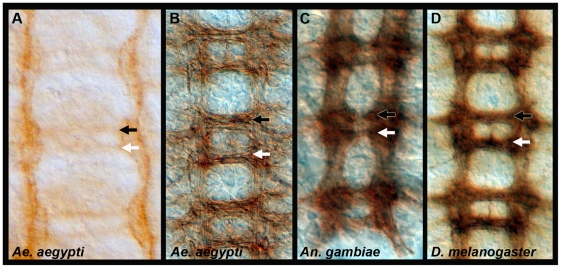
A scaffold of axon pathways develop in Dr. melanogaster and give rise to the embryonic ventral nerve cord, which has a ladder-like appearance (Fig. 1D). Within each segment of the developing fruit fly embryo, a pair of bilaterally symmetrical longitudinal axon tracts are pioneered separately on either side of the midline in each segment. A number of early growth cones project only on their own side, but most CNS interneurons will project their axons across the midline in either the anterior or posterior commissural axon tracts before extending rostrally or caudally in the developing longitudinals ([24], [25]; Fig 1D). Nerve cord development was assessed during mosquito embryogenesis with an anti-acetylated tubulin antibody (Fig. 1A–C). Acetylated tubulin is first detected in Ae. aegypti at 52 hrs. after egg laying (AEL) when the longitudinal axon tracts have begun to form and the commissural axon tracts are initiating (Fig. 1A). During the next several hours, the axon tracts thicken as additional neurons project their axons (Fig. 1B). Anterior and posterior commissures are initially fused (not shown), as observed in Dr. melanogaster [37]. At 56 hrs. AEL, the commissures have separated, and the mature ventral embryonic nerve cord of Ae. aegypti (Fig. 1B) resembles that of An. gambiae (33 hrs. AEL shown in Fig. 1C) and a St. 16 Drosophila embryo (Fig. 1D).
Figure 1. Development of the Ae. aegypti embryonic ventral nerve cord.
Anti-acetylated tubulin staining (A–C) marks the developing axon tracts in 52 hr. (A) and 56 hr. (B) Ae. aegypti embryos. By 56 hrs. (C), the Ae. aegypti nerve cord resembles that of a 33 hr. An. gambiae embryo and a St. 16 Dr. melanogaster nerve cord (BP102 staining is shown in D). These time points in the three respective species correspond to germ-band retracted embryos in which segmentation is obvious and organogenesis has initiated. Filleted nerve cords are oriented anterior up in all panels. The anterior commissure is marked by a black arrowhead, and a white arrowhead marks the posterior commissure.
Expression of fra in the developing mosquito CNS
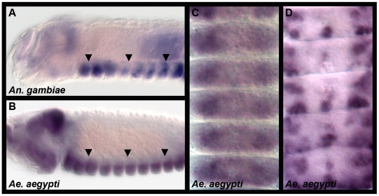
Net accumulation data have indicated that Net-Fra signaling may play conserved roles during insect ventral nerve cord development [16], [36]. However, in insects, fra expression has not been examined outside of Drosophila, where it is expressed on developing axons of the commissural and longitudinal axon pathways, including the earliest commissural axons [27]. Expression of Ae. aegypti fra (Aae fra) and An. gambiae fra (Aga fra) were therefore analyzed through whole-mount in situ hybridization at the onset of nerve cord development in both species. Aae fra expression initiates in developing neurons, including the earliest commissural axons, just prior to establishment of the axonal scaffold and is maintained during ventral nerve cord formation (Fig. 2B–D). Comparable fra expression patterns are detected in the developing nervous system of An. gambiae (Fig. 2A). These data are consistent with the hypothesis that Fra functions to regulate growth of commissural axons in mosquitoes.
Figure 2. Expression of fra in the developing mosquito CNS.
Comparable fra expression patterns are detected in lateral views of the developing nervous systems (arrows) of An. gambiae (33 hrs., A) and Ae. aegypti (52 hrs., B). Ventral views of Aae fra expression in 52 hr. (C, segments T3–A5) and 54 hr. (D; segments A2–A6) Ae. aegypti embryos are shown. Anterior is oriented left in A and B and up in C and D.
si-RNA mediated knockdown of fra during Ae. aegypti development
Analysis of fra expression (Fig. 2) suggested that this gene may regulate ventral nerve cord development in mosquitoes. Functional testing of this hypothesis required the development of a strategy to selectively inhibit gene function during mosquito development. RNA interference (RNAi) technology, which has emerged as an effective method for inhibiting gene function in many organisms, was therefore combined with previously described Ae. aegypti microinjection techniques [38], [39] to knockdown fra during Ae. aegypti development. Two separate siRNAs corresponding to different regions of Aae fra, fra siRNA-A and fra siRNA-B, as well as a scrambled control version of siRNA-A, were used in these experiments.
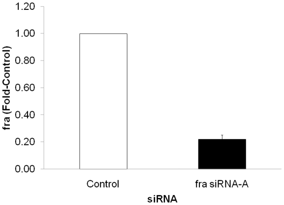
siRNAs were injected pre-cellular blastoderm, and knockdown was assessed through both quantitative real-time PCR (qRT-PCR) and whole-mount in situ hybridization. Multiple qRT-PCR replicates at three different time points, including 24, 48 (not shown), and 72 hrs. (Fig. 3), confirmed knockdown of fra that was maintained through the end of embryogenesis. At 72 hrs., the time point that was typically assayed once injection protocols and knockdown strategies had been optimized, fra transcript levels were reduced by 80% on average (Fig. 3, p<0.0001), and a maximum of 90% knockdown was achieved in one replicate. Knockdown in the developing CNS was verified through in situ hybridization, which confirmed reduced levels of fra transcripts in the embryonic CNS at levels comparable to those detected by qRT-PCR, and which revealed nearly complete knockdown in the developing nervous systems of embryos bearing strong phenotypes (Fig. 4C). These studies suggest that siRNA methodology can be used for targeted disruption of embryonic gene function in Ae aegypti.
Figure 3. Confirmation of fra knockdown in Ae. aegypti.
qRT-PCR was used to assess fra levels following microinjection of fra siRNA-A. A scrambled version of fra siRNA-A was injected as a control. At 72 hrs. post injection, levels of fra were 80% less than that of the control-injected group (N = 3, p<0.0001).
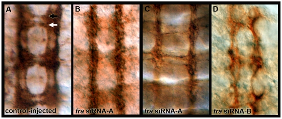
Figure 4. Ae. aegypti fra knockdown CNS phenotypes.
Anti-acetylated tubulin staining (reddish brown) marks the axons of the ventral nerve cords of scrambled control (A) and fra siRNA injected embryos (B–D). Knockdown phenotypes characterized by thinning or loss of commissural axons were observed at 54 hrs. (B–D). Comparable results were obtained with two different siRNAs (fra siRNA-A in B,C; fra siRNA-B in D). Knockdown of fra was confirmed by double-labeling to detect fra mRNA expression (dark blue in C). Nerve cords are oriented anterior up in each panel. The anterior commissure is marked by a black arrowhead, and a white arrowhead marks the posterior commissure.
Ae. aegypti fra knockdown CNS phenotypes
The impact of fra knockdown on Ae. aegypti embryonic nerve cord development was assessed through anti-acetylated tubulin staining at 54 hrs. AEL. In embryos injected with fra siRNA-A, 71% of anterior commissures and 80% of posterior commissures are thin or absent (Fig. 4B, C, Table 1). As observed in Drosophila [27], the posterior commissure is more severely disrupted than the anterior, with 51% of the embryos displaying a severe phenotype in the posterior commissure and 36% of embryos displaying a severe anterior commissure phenotype (Table 1). Occasional breaks in the longitudinal tracts were also noted in fra knockdown embryos. Injection of either fra siRNA-A (Fig. 4B,C) or siRNA-B (Fig. 4D), which correspond to two separate Aae fra sequences, produced similar phenotypes. This result indicates that the knockdown phenotypes described are due to loss of fra and are not the result of off-site targeting. Injection of the scrambled control siRNA did not disrupt nerve cord development (Fig. 4A, Table 1).
Table 1. Quantification of Aae fra knockdown phenotype penetrance and severity.
| AnteriorWT | Mild | Strong | PosteriorWT | Mild | Strong | |
| Control | 91 (100%) | 0 (0%) | 0 (0%) | 91(100%) | 0 (0%) | 0 (0%) |
| fra siRNA-A | 22 (29%) | 26 (35%) | 27 (36%) | 15 (20%) | 22 (29%) | 38 (51%) |
Embryos stained with anti-acetylated tubulin were scored at 54 hrs. post-injection of fra siRNA-A or scrambled control siRNA. The number and percentage of total segments bearing wild-type (WT), mild, or strong phenotypes in the anterior and posterior commissures are reported. Mild phenotypes correspond to thinning commissures, and severe phenotypes correspond to near or complete absence of commissural axons.
It should be noted that the penetrance and severity of the Aae fra knockdown phenotype are higher than that reported for the Drosophila fra null, in which only 12% of the anterior commissures and 43% of the posterior commissures are reportedly thin or absent [27]. In fact, in embryos in which CNS transcripts are nearly depleted, the Aae fra knockdown phenotype (Fig. 4B,C) bears strong resemblance to the Drosophila commissureless phenotype, in which commissure formation is entirely blocked [40]. These results suggest that Net-Fra signaling may play a more critical role in formation of the Ae. aegypti ventral nerve cord, and that the guidance cues postulated to compensate for loss of Net-Fra signaling in Dr. melanogaster [29] may not be present in mosquitoes. These observations suggest that further analysis of embryonic nerve cord development in mosquitoes may uncover underlying differences between Dr. melanogaster and mosquito nervous system development. In support of this concept, our ongoing analysis of semaphorin knockdown in Ae. aegypti suggests that the function of this gene in nerve cord development has evolved in insects (data not shown).
Developmental Genetics in Vector Mosquitoes
Although we have made great advances in understanding developmental genetics in Drosophila, comparatively little is known about the genetic basis for development in mosquitoes and other arthropods. In this investigation, we examined the role of Fra during development of two vector mosquitoes. Expression of fra in the developing ventral nerve cord was found to be conserved between the two mosquitoes and Dr. melanogaster. However, the results of this investigation, the first targeted knockdown of a gene during vector mosquito embryogenesis, illustrate that although Fra plays a critical role during development of the Ae. aegypti ventral nerve cord, mechanisms regulating embryonic commissural axon guidance may have evolved in distantly related insects. This is a somewhat unexpected finding given the many similarities in insect CNS development that have been observed (for example, see [34], [41]). Given these findings in Ae. aegypti, it would also be interesting to apply the siRNA-mediated knockdown strategies utilized here to An. gambiae and to formally assess the function of Aga fra.
Characterizing the function of additional developmental genes in mosquitoes is critical. To date, expression patterns of only a handful of mosquito developmental genes [9], [10], [11], [12], [13], [14], [15], [16] have been reported. Adelman et al. [13] showed that control sequences for one of these genes, nanos [11], demonstrated promise as part of a transposable element-based gene drive system that may be used to spread and fix antipathogen effector genes in natural populations. Their investigations illustrate the exciting potential for the application of evo-devo approaches in efforts to develop strategies for vector control. The methodologies used in this investigation, in particular the siRNA-mediated knockdown strategy for functional analysis of developmental genes in Ae. aegypti embryos, will broaden and enhance these efforts.
Materials and Methods
Ethics statement
This study was performed in accordance with the recommendations in the Guide for the Care and Use of Laboratory Animals of the National Institutes of Health. The animal use protocol was approved by the University of Notre Dame Institutional Animal Care and Use Committee (Study # 11-036).
Mosquito Rearing, Egg Collection, and Fixation
The Ae. aegypti Liverpool-IB12 (LVP-1B12) strain and An. gambiae (M Form) were used in these investigations. Procedures for mosquito rearing and egg collection [22], [42], which was performed at 26°C, have been described. Ae. aegypti embryos were fixed as described [20]. An. gambiae embryos were fixed using a comparable procedure, except that eggs were fixed at room temperature.
Immunohistochemistry
Immunohistochemistry was performed as described [19]. Anti-acetylated tubulin (Zymed, San Francisco, CA) was used at a concentration of 1∶100, and HRP-conjugated secondary antibodies (Jackson Immunoresearch, West Grove, PA) were used at a final concentration of 1∶200.
In situ hybridization
Riboprobes corresponding to Aae fra (AAEL014592) and Aga fra (AGAP006083) were synthesized according to the Patel [43] protocol. In situ hybridization was performed as previously described [23].
RNA interference
Knockdown was performed through embryonic microinjection of siRNAs targeting Aae fra. siRNA design and microinjection were performed as described [21]. The following siRNAs were synthesized by Dharmacon RNAi Technologies (Lafayette, CO): siRNA-A sense: CCA GAT GGG TAT GGG AGA T and antisense: GGT CTA CCC ATA CCC TCT A (corresponding to base pairs 2011–2032 of Aae fra) and siRNA-B sense: TCC ATA CAC CTA CGA AGG A and antisense: AGG TAT GTG GAT GCT TCCT (corresponds to base pairs 3862–3883 of Aae fra). A scrambled version of siRNA-A was used as a control: sense GAT TAG ACG AAT ACC ACT A and antisense: CTA ATC TGC TTA TGG TGA T. siRNAs were injected at a concentration of 6 ug/uL.
Measurement of knockdown effectiveness was determined through in situ hybridization (see above) and through qRT-PCR. qRT-PCR was performed as previously described [44]. In short, total RNA was extracted from ∼30 pooled siRNA-microinjected mosquito embryos using Trizol (Invitrogen, Carlsbad, CA). cDNA was prepared with the High Capacity RNA to cDNA Kit (Applied Biosystems, Foster City, CA), which includes a blend of random and oligo(dT) primers, according to the manufacturer's instructions. Real-time quantification was performed using the SYBR Green I PCR kit (Applied Biosystems, Foster City, CA) in conjunction with an Applied BioSystems Step One Plus Real-Time PCR System. Primer sets for Aae fra were: For 5′ GCG ACC CAA CAC TCA ATA TG 3′ and Rev 5′ GTC GTA GGA TAC CGT GAG AT 3′. Primer sets for the housekeeping gene rpS17, which was included as the internal standard for data normalization as previously described [44], were as follows: For 5′ AGA CAA CTA CGT GCC GGA AG 3′ and Rev 5′ TTG GTG ACC TGG ACA ACG ATG 3′. Three independent biological replicates were conducted, and all PCR reactions were performed in triplicate. Quantification of results was accomplished by standardizing reactions to rpS17 levels, and then using the ΔΔCt method as described [45]. Results were expressed as fold-difference compared with the scrambled control-injected embryos. qRT-PCR data from replicate experiments were statistically analyzed with the Student T Test.
Acknowledgments
We thank Frank Collins for advice, encouragement, and use of equipment. Many thanks to Nora Besansky and Marcy Kern for An. gambiae eggs and to Nipam Patel for suggestions about fixing An. gambiae eggs. We are extremely grateful to Sun Longhua of the Malcolm Frasier lab who taught us to microinject mosquito embryos. William Browne suggested that we use siRNAs and gave expert advice on their design. Thanks to Becky DeBruyn and Diane Lovin for their technical assistance. We are grateful to the members of the Scheel and Severson labs and the EIGH for their advice during the course of this investigation.
Footnotes
Competing Interests: The authors have declared that no competing interests exist.
Funding: Christy Le and Michael Tomchaney were supported by the University of Notre Dame College of Science Summer Undergraduate Research Fellowship program. This work was supported by the following awards to MDS: NIAID Award R01AI081795-01, NINDS Award R15 NS 048904-0, and an IUSM Research Support Funds Grant. The funders had no role in study design, data collection and analysis, decision to publish, or preparation of this manuscript.
References
- 1.Holt RA, Subramanian GM, Halpern A, Sutton GG, Charlab R, et al. The genome sequence of the malaria mosquito Anopheles gambiae. Science. 2002;298:129–149. doi: 10.1126/science.1076181. [DOI] [PubMed] [Google Scholar]
- 2.Nene V, Wortman JR, Lawson D, Haas B, Kodira C, et al. Genome sequence of Aedes aegypti, a major arbovirus vector. Science. 2007;316:1718–1723. doi: 10.1126/science.1138878. [DOI] [PMC free article] [PubMed] [Google Scholar]
- 3.Clemons A, Haugen M, Flannery E, Tomchaney M, Kast K, et al. Aedes aegypti: an emerging model for vector mosquito development. Cold Spring Harb Protoc. 2010;2010:pdb emo141. doi: 10.1101/pdb.emo141. [DOI] [PMC free article] [PubMed] [Google Scholar]
- 4.Chen XG, Mathur G, James AA. Gene expression studies in mosquitoes. Adv Genet. 2008;64:19–50. doi: 10.1016/S0065-2660(08)00802-X. [DOI] [PMC free article] [PubMed] [Google Scholar]
- 5.Raminani LN, Cupp, W E. Early embryology of Aedes aegypti (L.) (Diptera: Culicdae). Int J Insect Morphol Embryol. 1975;4:517–528. [Google Scholar]
- 6.Raminani LN CE. Embryology of Aedes aegypti (L.) (Diptera: Culicidae): organogenesis. Int J Insect Morphol & Embryol. 1978;7:273–296. [Google Scholar]
- 7.Farnesi LC, Martins AJ, Valle D, Rezende GL. Embryonic development of Aedes aegypti (Diptera: Culicidae): influence of different constant temperatures. Mem Inst Oswaldo Cruz. 2009;104:124–126. doi: 10.1590/s0074-02762009000100020. [DOI] [PubMed] [Google Scholar]
- 8.Vital W, Rezende GL, Abreu L, Moraes J, Lemos FJ, et al. Germ band retraction as a landmark in glucose metabolism during Aedes aegypti embryogenesis. BMC Dev Biol. 2010;10:25. doi: 10.1186/1471-213X-10-25. [DOI] [PMC free article] [PubMed] [Google Scholar]
- 9.Goltsev Y, Hsiong W, Lanzaro G, Levine M. Different combinations of gap repressors for common stripes in Anopheles and Drosophila embryos. Dev Biol. 2004;275:435–446. doi: 10.1016/j.ydbio.2004.08.021. [DOI] [PubMed] [Google Scholar]
- 10.Calvo E, Walter M, Adelman ZN, Jimenez A, Onal S, et al. Nanos (nos) genes of the vector mosquitoes, Anopheles gambiae, Anopheles stephensi and Aedes aegypti. Insect Biochem Mol Biol. 2005;35:789–798. doi: 10.1016/j.ibmb.2005.02.007. [DOI] [PubMed] [Google Scholar]
- 11.Juhn J, James AA. oskar gene expression in the vector mosquitoes, Anopheles gambiae and Aedes aegypti. . Insect Mol Biol. 2006;15:363–372. doi: 10.1111/j.1365-2583.2006.00655.x. [DOI] [PubMed] [Google Scholar]
- 12.Juhn J, Marinotti O, Calvo E, James AA. Gene structure and expression of nanos (nos) and oskar (osk) orthologues of the vector mosquito, Culex quinquefasciatus. . Insect Mol Biol. 2008;17:545–552. doi: 10.1111/j.1365-2583.2008.00823.x. [DOI] [PMC free article] [PubMed] [Google Scholar]
- 13.Adelman ZN, Jasinskiene N, Onal S, Juhn J, Ashikyan A, et al. nanos gene control DNA mediates developmentally regulated transposition in the yellow fever mosquito Aedes aegypti. . Proc Natl Acad Sci U S A. 2007;104:9970–9975. doi: 10.1073/pnas.0701515104. [DOI] [PMC free article] [PubMed] [Google Scholar]
- 14.Goltsev Y, Fuse N, Frasch M, Zinzen RP, Lanzaro G, et al. Evolution of the dorsal-ventral patterning network in the mosquito, Anopheles gambiae. . Development. 2007;134:2415–2424. doi: 10.1242/dev.02863. [DOI] [PubMed] [Google Scholar]
- 15.Goltsev Y, Rezende GL, Vranizan K, Lanzaro G, Valle D, et al. Developmental and evolutionary basis for drought tolerance of the Anopheles gambiae embryo. Dev Biol. 2009;330:462–470. doi: 10.1016/j.ydbio.2009.02.038. [DOI] [PMC free article] [PubMed] [Google Scholar]
- 16.Simanton W, Clark S, Clemons A, Jacowski C, Farrell-VanZomeren A, et al. Conservation of arthropod midline netrin accumulation revealed with a cross-reactive antibody provides evidence for midline cell homology. Evol Dev. 2009;11:260–268. doi: 10.1111/j.1525-142X.2009.00328.x. [DOI] [PMC free article] [PubMed] [Google Scholar]
- 17.Christophers SR. Cambridge, UK: Cambridge University Press; 1960. Aedes aegypti, The yellow fever mosquito: its life history, bionomics, and structure. [Google Scholar]
- 18.Severson DW, DeBruyn B, Lovin DD, Brown SE, Knudson DL, et al. Comparative genome analysis of the yellow fever mosquito Aedes aegypti with Drosophila melanogaster and the malaria vector mosquito Anopheles gambiae. . J Hered. 2004;95:103–113. doi: 10.1093/jhered/esh023. [DOI] [PubMed] [Google Scholar]
- 19.Clemons A, Flannery E, Kast K, Severson D, Duman-Scheel M. Immunohistochemical analysis of protein expression during Aedes aegypti development. Cold Spring Harb Protoc. 2010;2010:pdb prot5510. doi: 10.1101/pdb.prot5510. [DOI] [PMC free article] [PubMed] [Google Scholar]
- 20.Clemons A, Haugen M, Flannery E, Kast K, Jacowski C, et al. Fixation and preparation of developing tissues from Aedes aegypti. . Cold Spring Harb Protoc. 2010;2010:pdb prot5508. doi: 10.1101/pdb.prot5508. [DOI] [PMC free article] [PubMed] [Google Scholar]
- 21.Clemons A, Haugen M, Severson D, Duman-Scheel M. Functional analysis of genes in Aedes aegypti embryos. Cold Spring Harb Protoc. 2010;2010:pdb prot5511. doi: 10.1101/pdb.prot5511. [DOI] [PMC free article] [PubMed] [Google Scholar]
- 22.Clemons A, Mori A, Haugen M, Severson DW, Duman-Scheel M. Culturing and egg collection of Aedes aegypti. Cold Spring Harb Protoc. 2010;2010:pdb prot5507. doi: 10.1101/pdb.prot5507. [DOI] [PMC free article] [PubMed] [Google Scholar]
- 23.Haugen M, Tomchaney M, Kast K, Flannery E, Clemons A, et al. Cold Spring Harbor Protocols; 2010. Whole-mount in situ hybridization for analysis of gene expression during Aedes aegypti development. [DOI] [PMC free article] [PubMed] [Google Scholar]
- 24.Tessier-Lavigne M, Goodman CS. The molecular biology of axon guidance. Science. 1996;274:1123–1133. doi: 10.1126/science.274.5290.1123. [DOI] [PubMed] [Google Scholar]
- 25.Kaprielian Z, Runko E, Imondi R. Axon guidance at the midline choice point. Dev Dyn. 2001;221:154–181. doi: 10.1002/dvdy.1143. [DOI] [PubMed] [Google Scholar]
- 26.Duman-Scheel M. Netrin and DCC: axon guidance regulators at the intersection of nervous system development and cancer. Curr Drug Targets. 2009;10:602–610. doi: 10.2174/138945009788680428. [DOI] [PMC free article] [PubMed] [Google Scholar]
- 27.Kolodziej PA, Timpe LC, Mitchell KJ, Fried SR, Goodman CS, et al. frazzled encodes a Drosophila member of the DCC immunoglobulin subfamily and is required for CNS and motor axon guidance. Cell. 1996;87:197–204. doi: 10.1016/s0092-8674(00)81338-0. [DOI] [PubMed] [Google Scholar]
- 28.Hiramoto M, Hiromi Y, Giniger E, Hotta Y. The Drosophila netrin receptor frazzled guides axons by controlling netrin distribution. Nature. 2000;406:886–889. doi: 10.1038/35022571. [DOI] [PubMed] [Google Scholar]
- 29.Harris R, Sabatelli LM, Seeger MA. Guidance cues at the Drosophila CNS midline: identification and characterization of two Drosophila Netrin/UNC-6 homologs. Neuron. 1996;17:217–228. doi: 10.1016/s0896-6273(00)80154-3. [DOI] [PubMed] [Google Scholar]
- 30.Mitchell KJ, Doyle JL, Serafini T, Kennedy TE, Tessier-Lavigne M, et al. Genetic analysis of netrin genes in Drosophila: netrins guide CNS commissural axons and peripheral motor axons. Neuron. 1996;17:203–215. doi: 10.1016/s0896-6273(00)80153-1. [DOI] [PubMed] [Google Scholar]
- 31.Brankatschk M, Dickson BJ. Netrins guide Drosophila commissural axons at short range. Nat Neurosci. 2006;9:188–194. doi: 10.1038/nn1625. [DOI] [PubMed] [Google Scholar]
- 32.Gerberding M, Scholtz G. Cell lineage of the midline cells in the amphipod crustacean Orchestia cavimana (Crustacea, Malacostraca) during formation and separation of the germ band. Dev Genes Evol. 1999;209:91–102. doi: 10.1007/s004270050231. [DOI] [PubMed] [Google Scholar]
- 33.Gerberding M, Scholtz G. Neurons and glia in the midline of the higher crustacean Orchestia cavimana are generated via an invariant cell lineage that comprises a median neuroblast and glial progenitors. Dev Biol. 2001;235:397–409. doi: 10.1006/dbio.2001.0302. [DOI] [PubMed] [Google Scholar]
- 34.Duman-Scheel M, Patel NH. Analysis of molecular marker expression reveals neuronal homology in distantly related arthropods. Development. 1999;126:2327–2334. doi: 10.1242/dev.126.11.2327. [DOI] [PubMed] [Google Scholar]
- 35.Browne WE, Schmid BG, Wimmer EA, Martindale MQ. Expression of otd orthologs in the amphipod crustacean, Parhyale hawaiensis. Dev Genes Evol. 2006;216:581–595. doi: 10.1007/s00427-006-0074-7. [DOI] [PubMed] [Google Scholar]
- 36.Duman-Scheel M, Clark SM, Grunow ET, Hasley AO, Hill BL, et al. Delayed onset of midline netrin expression in Artemia franciscana coincides with commissural axon growth and provides evidence for homology of midline cells in distantly related arthropods. Evol Dev. 2007;9:131–140. doi: 10.1111/j.1525-142X.2007.00144.x. [DOI] [PMC free article] [PubMed] [Google Scholar]
- 37.Doe CQ, Goodman C. Embryonic development of the Drosophila central nervous system. In: Martinez Arias A, editor. The Development of Drosophila melanogaster. Plainview, NY: Cold Spring Harbor Laboratory Press; 1993. pp. 1091–1130. [Google Scholar]
- 38.Lobo NF, Clayton JR, Fraser MJ, Kafatos FC, Collins FH. High efficiency germ-line transformation of mosquitoes. Nat Protoc. 2006;1:1312–1317. doi: 10.1038/nprot.2006.221. [DOI] [PubMed] [Google Scholar]
- 39.Jasinskiene N, Juhn J, James A. Microinjection of A. aegypti embryos to obtain transgenic mosquitoes. Journal of Visualized Experiments. 2007;5 doi: 10.3791/219. [DOI] [PMC free article] [PubMed] [Google Scholar]
- 40.Seeger M, Tear G, Ferres-Marco D, Goodman CS. Mutations affecting growth cone guidance in Drosophila: genes necessary for guidance toward or away from the midline. Neuron. 1993;10:409–426. doi: 10.1016/0896-6273(93)90330-t. [DOI] [PubMed] [Google Scholar]
- 41.Thomas JB, Bastiani MJ, Bate M, Goodman CS. From grasshopper to Drosophila: a common plan for neuronal development. Nature. 1984;310:203–207. doi: 10.1038/310203a0. [DOI] [PubMed] [Google Scholar]
- 42.Benedict MQ. Crampton JM, Beard CB, Louis C, editors. The Molecular Biology of Insect Vectors of Disease. London: Chapman and Hall. 1997. pp. 3–12.
- 43.Patel N. In situ hybridization to whole mount Drosophila embryos; In: Krieg PA, editor. New York: Wiley-Liss; 1996. pp. 357–370. [Google Scholar]
- 44.Morlais I, Mori A, Schneider JR, Severson DW. A targeted approach to the identification of candidate genes determining susceptibility to Plasmodium gallinaceum in Aedes aegypti. . Mol Genet Genomics. 2003;269:753–764. doi: 10.1007/s00438-003-0882-7. [DOI] [PubMed] [Google Scholar]
- 45.Livak KJ, Schmittgen TD. Analysis of relative gene expression data using real-time quantitative PCR and the 2(-Delta Delta C(T)) Method. Methods. 2001;25:402–408. doi: 10.1006/meth.2001.1262. [DOI] [PubMed] [Google Scholar]


