Abstract
Alternative splicing plays critical roles in differentiation, development and disease and is a major source for protein diversity in higher eukaryotes. Traditionally, analysis of alternative splicing regulation has focused on RNA sequence elements and their associated factors, but recent provocative studies point to a key function of chromatin structure and histone modifications in alternative splicing regulation. These insights suggest that epigenetic regulation not only determines what parts of the genome are expressed, but also how they are spliced.
Introduction
The tenth anniversary of the publication of the first draft of the human genome sequence has sparked a renewed and expanded interest in alternative pre-mRNA splicing. Alternative splicing explains how the vast mammalian proteomic complexity can be achieved with the limited number of genes found in higher eukaryotes. Current estimates based on deep sequencing methodologies indicate that more than 90% of human genes undergo alternative splicing (Croft et al., 2000; Pan et al., 2008; Wang et al., 2008). Alternative splicing is an integral part of differentiation and developmental programs and contributes to cell lineage and tissue identity as indicated by the mapping of more than 22,000 tissue-specific alternative transcript events in a recent genome-wide sequencing study of tissue-specific alternative splicing (Wang, 2008). The importance of alternative splicing is dramatically highlighted by the numerous diseases that are caused by mutations in either cis-acting RNA elements or trans-acting protein splicing factors (Caceres and Kornblihtt, 2002; Cooper et al., 2009). Prominent splicing diseases include cystic fibrosis, frontotemporal dementia, Parkinsonism, retinitis pigmentosa, spinal muscular atrophy, myotonic dystrophy, premature aging, and cancer.
Traditionally, alternative splicing has been thought to be predominantly regulated by splicing enhancers and silencers (Chasin, 2007). These short, conserved RNA sequences are typically 10 nt in length, are located either in exons or introns, acting either isolated or in clusters, and stimulate (enhancers) or inhibit (silencers) the use of splice sites through the specific binding of regulatory proteins such as SR proteins (serine/arginine rich proteins) or heterogeneous nuclear ribonucleoproteins (hnRNPs) (Long and Caceres, 2009; Han et al., 2010). In addition, some silencers, instead of recruiting regulatory proteins, act by determining a particular pre-mRNA secondary structure that hinders the recognition of a neighboring splicing enhancer by SR proteins (Buratti and Baralle, 2004). Disease mutations often affect the use of constitutive or alternative splice sites by cis-acting mutations that disrupt regulatory RNA sequence elements and by trans-acting mutations that affect the quality or quantity of alternative or constitutive splicing factors.
It has long been clear that a full understanding of alternative splicing regulation will require the molecular characterization and structural modeling of the spliceosome and the analysis of RNA regulatory elements. However, the emerging complexity of alternative splicing regulation makes it apparent that information from those approaches will not be sufficient to decipher how alternative splicing is regulated. Here we discuss mechanisms and implications of the recently uncovered role of epigenetic components such as chromatin structure and histone modifications to alternative splicing regulation.
Coupling of transcription and splicing
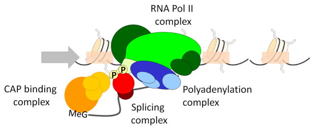
More than twenty years ago, visualization of Drosophila embryo nascent transcripts by electron microscopy showed that splicing can occur co-transcriptionally (Beyer and Osheim, 1988) (Figure 1). Co-transcriptional splicing was later directly demonstrated for the human dystrophin gene (Tennyson et al., 1995), where it appears a very intuitive concept given that transcription of this 2,400 kb-gene would take ~16 hours to complete. A quantitative study of the c-Src and fibronectin mRNAs, comparing chromatin-bound and nucleoplasmic RNA fractions, shows that most introns are excised efficiently in the chromatin-bound fractions, with a gradient of co-transcriptional splicing efficiency from promoter-proximal to promoter-distal introns, suggesting co-transcriptional splicing (Pandya-Jones and Black, 2009). However, co-transcriptionality of splicing is not strict, in the sense that introns are not necessarily removed in the exact order they are transcribed (Attanasio et al., 2003; Bauren and Wieslander, 1994; Kessler et al., 1993; LeMaire and Thummel, 1990). If that were the case, the competition between splicing sites that leads to alternative splicing would be impossible.
Figure 1. Coupling of transcription and RNA processing.
RNA polymerase II (green) recruits RNA processing factors such as the 5′ cap-binding complex (CAP) (yellow), splicing and pre-spliceosome factors (red) and the polyadenylation complex (blue) in the context of nucleosome-containing chromatin. Recruitment of RNA processing factors occurs via the RNA Pol II C-terminal domain (CTD; light green) and much of RNA processing occurs co-transcriptionally.
Splicing complexes are recruited to all introns and exons in a time window that begins when the target sequence is transcribed and extends to the moment of splicing catalysis. For the entire splicing reaction to be co-transcriptional, both recruitment and catalysis must occur before transcription termination and transcript release. Alternatively, recruitment of some or all splicing factors may occur co-transcriptionally but the catalysis itself may occur post-transcriptionally. Co-transcriptional pre-mRNA splicing appears to be a general rule for long mammalian genes. It is unclear how prevalent it is in organisms with shorter introns, such as yeast, although several studies support the notion that recruitment of spliceosomal components is also mostly co-transcriptional in this organism (Gornemann et al., 2005; Kotovic et al., 2003; Lacadie and Rosbash, 2005; Tardiff et al., 2006) (Figure 1). Completion of intron removal appears to be post-transcriptional in most cases, and only in genes with relatively long downstream exons does it occur prior to transcript release (Tardiff et al., 2006). The message from these studies is that co-transcriptional recruitment of splicing factors is largely preferred, but that co-transcriptional completion of intron removal is not mandatory and depends on the specific kinetics of transcription and splicing. In other words, the selective pressure in favor of co-transcriptional splicing acts on the association of splicing factors, which can be viewed as the “commitment to splice” rather than on the catalysis itself. This might not apply to other RNA processing events like capping and cleavage/polyadenylation (McCracken et al., 1997a; McCracken et al, 1997b; Hirose et al., 1998; Maniatis and Reed, 2002; Moore and Proudfoot, 2009), where both the recruitment of the factors and enzymes involved as well as the catalysis appear to be co-transcriptional.
Although co-transcriptionality of splicing is a pre-requisite for coupling, it does not necessarily mean the two events are coupled. Co-transcriptionality simply means that splicing takes place, or is committed to occur, before the nascent RNA is released from RNA Pol II. Coupling implies that the transcription and splicing machineries interact with each other or that the kinetics of one process determines the outcome of the other. Efficient coordination between transcription and processing may be a specific feature of RNA Pol II and particularly of the carboxy-terminal domain (CTD) of its catalytic subunit given that a phosphorylated CTD is required for co-transcriptional splicing (Bird et al., 2004) (Figure 1). When protein-coding genes are placed under the control of either RNA Pol I, RNA Pol III or T7 RNA polymerase promoters, transcription takes place, but pre-mRNA processing is impaired and the resulting transcripts are poorly spliced (Dower and Rosbash, 2002; McCracken et al., 1998; Sisodia et al., 1987; Smale and Tjian, 1985). In fact, association of splicing factors to sites of transcription is dependent on RNA Pol II CTD (Misteli and Spector, 1999) and deletion of the CTD causes defects in capping, cleavage/polyadenylation, and splicing of the β-globin transcript (McCracken et al., 1997b) (Figure 1). Many splicing factors are able to interact with RNA Pol II in vivo, including almost all known SR proteins and U1snRNP and in nuclear extracts that support both transcription and splicing in vitro, SR proteins appear to be much more effective in promoting splicing when the latter is co-transcriptional than when it is post-transcriptional (Das et al., 2007). However, SR proteins are not delivered to splicing sites by RNA Pol II alone, but rather require ongoing pre-mRNA synthesis (Sapra et al., 2009), demonstrating that recruitment is not dependent on pre-assembled SR-RNA Pol II complexes. Coupled in vitro transcription/splicing assays, although not necessarily reflecting functional coupling as it would occur in vivo (Lazarev and Manley, 2007), show that nascent pre-mRNA synthesized by RNA Pol II is stabilized and efficiently spliced (Hicks et al., 2006). This is likely because it is immediately and quantitatively directed into the spliceosome assembly pathway, instead of being assembled into non-specific hnRNP complexes, which are inhibitory for spliceosome assembly (Das et al., 2006).
Strong evidence for functional coupling between transcription and pre-mRNA processing comes from analyzing how modulation of transcription affects alternative splicing events. It has been demonstrated that the outcome of alternative splicing is influenced by the promoter used to drive transcription (Cramer et al., 1999; Cramer et al., 1997; Pagani et al., 2003), by hormone-responsive elements (Auboeuf et al., 2002) or by recruitment of different transcription factors or co-activators to the promoter (Auboeuf et al., 2004a; Auboeuf et al., 2004b; Nogues et al., 2002). The effects are not the trivial consequence of different mRNA levels produced by each promoter, but depend on qualitative properties conferred by promoters to the transcription/RNA processing machinery.
Control of alternative splicing by elongation rate
The standard experimental approach to study splicing mechanisms is by in vitro splicing assays. This methodology employs in vitro synthesized pre-mRNA substrates in splicing reactions carried out in cell-free nuclear extracts. Although these conditions are appropriate to identify splicing factors and RNA intermediates, they are not ideally suited to obtain an accurate picture of the timing of splicing in relation to the generation of nascent RNA during transcription. These limitations can be overcome by in vivo experiments using either transfected reporter minigenes or endogenous genes as templates for splicing reactions. It was in fact differences in the behavior of a splicing event in vivo compared to in vitro that first hinted at a kinetic role for transcription on splicing. Eperon et al. (1988) found that the use of an alternative 5′ splice site sequestered within a short stem of RNA secondary structure was determined by the length of the loop in vivo. Above a threshold loop length, the alternative site was used despite the potential structure. In contrast, the alternative site was not used during splicing in vitro with all lengths of loop tested (Eperon et al., 1988). The simplest interpretation of these experiments is that the rate of RNA synthesis affects its secondary structure, which in turn affects splicing. Further evidence for a kinetic link between transcription and splicing came from experiments in which a MAZ sequence, which leads to RNA Pol II pausing, inserted into the tropomyosin gene promoted higher inclusion of tropomyosin exon 3 (Roberts et al., 1998). Conclusive evidence for a role of elongation on alternative splicing regulation was finally revealed by the finding that the nature of the promoter affects alternative splicing outcome (Cramer et al., 1997, 1999; Kornblihtt, 2005). The original observation of the promoter effect involved transient transfection of mammalian cells with reporter minigenes for the alternatively spliced cassette exon 33 (E33, also referred to as EDI or EDA) of human fibronectin (FN) under the control of different RNA Pol II promoters. When transcription of the minigene was driven by the β-globin promoter, for example, E33 inclusion levels in the mature mRNA were about 10 times lower than when transcription was driven from the FN or cytomegalovirus (CMV) promoter. These effects were not the consequence of the promoter strength but depended on some qualitative properties conferred by promoters to the transcription/RNA processing machinery. Two non-exclusive mechanisms could explain the promoter effect: differential promoter occupation could affect the recruitment of splicing factors by the transcription machinery (recruitment coupling) or determine different rates of RNA Pol II elongation (kinetic coupling).
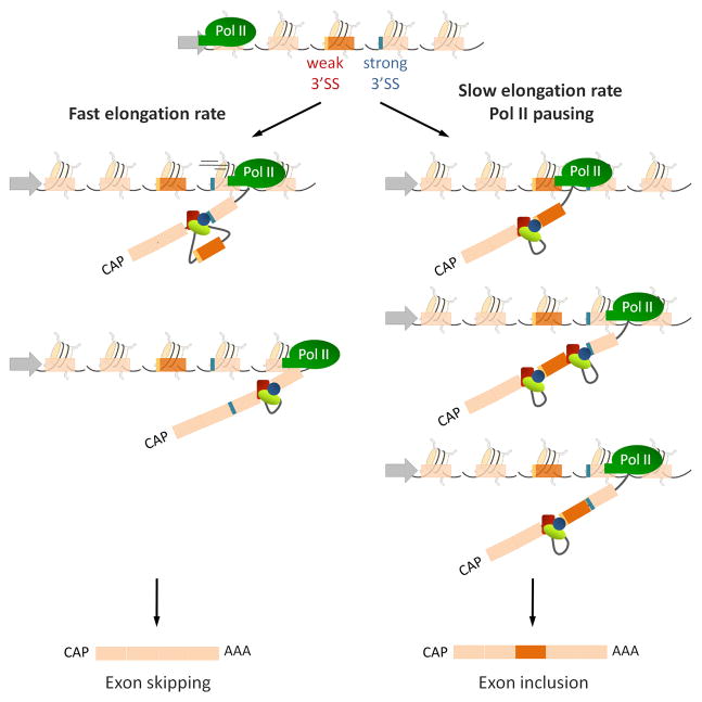
Several lines of evidence support the idea that RNA Pol II elongation can affect alternative splicing through kinetic coupling (Figure 2). Replication of reporter plasmids for alternative splicing in transiently transfected cells greatly stimulated E33 inclusion. This effect was counteracted by treating the cells with trichostatin A (TSA), a potent inhibitor of histone deacetylation and therefore a chromatin “opener”, allowing for the possibility that replication conveys a more compact chromatin structure to the template, thus slowing elongation and leading to higher E33 inclusion (Kadener et al., 2001). Furthermore, drugs that inhibit elongation, like DRB (Kadener et al. 2001, Nogués et al. 2002), flavopiridol, or camptothecin (de la Mata et al., 2010), favor E33 inclusion. On the other hand, activation of transcription by Sp1, a transcription factor that promotes initiation, has no effect on E33 inclusion, whereas activation by VP16, a factor that promotes both initiation and elongation, decreases E33 inclusion (Nogués et al. 2002). The strongest evidence for a kinetic role of RNA pol II elongation comes from a slow mutant of RNA Pol II, which increases E33 inclusion in human cells (de la Mata et al., 2003). Interestingly, the homologous mutation in Drosophila (C4 pol II) is viable but shows changes in the alternative splicing pattern of ultrabithorax (Ubx) mRNA, that are consistent with the only conspicuous phenotype of the C4 flies, which is an enlargement of the halteres that resembles the Ubx mutants. Why slowing elongation would only affect the Ubx gene is not known but a clue might be that this gene has the longest introns in Drosophila (17 and 50 kb) flanking the alternative exons affected in the C4 genotype, suggesting that elongation becomes more critical when introns are long. Similar effects of elongation on splicing have been reported in yeast on an artificially created alternative exon when transcription is carried out by a slow RNA Pol II mutant or when the elongation factor TFIIS is mutated (Howe et al., 2003). Finally, DNA damage signaling following irradiation of cells with UV light affects alternative splicing of fibronectin, caspase 9, Bcl-x and other human genes as a consequence of the inhibition of RNA Pol II elongation caused by UV-dependent hyperphosphorylation of the CTD (Muñoz et al., 2009).
Figure 2. The RNA Polymerase II kinetic model for alternative splicing.
Rapid elongation of RNA Polymerase II (Pol II) leads to simultaneous availability to the splicing machinery of a weak (red) and a strong (blue) splice site which compete for the recruitment of splicing factors (red, blue and green ovals) resulting in skipping of the weaker exon (orange rectangle). Pausing or slowing down of the RNA Pol II favors the recruitment of the splicing machinery to the first transcribed, weaker exon leading to its subsequent inclusion in a “first served, first committed” model.
These data support a “first come, first served” model for regulation of alternative splicing (Aebi, 1987) (Figure 2). In one version of this model, slow elongation favors removal of the intron upstream of an alternative cassette exon before removal of the downstream intron. In an alternative version, slow elongation favors recruitment of splicing factors to the upstream intron before the downstream intron is synthesized, which in turn would promote exon inclusion. Once commitment is achieved, the order of intron removal becomes irrelevant (Figure 2). The latter model is supported by recent evidence showing that there is a preferential removal of the intron downstream of the fibronectin cassette exon 33 before the upstream intron has been removed (de la Mata et al., 2010). Most importantly, whereas cis-acting mutations and trans-acting factors that upregulate E33 inclusion act by changing the relative order of intron removal, reduction of elongation, which also causes higher E33 inclusion, does not affect the order of intron removal, suggesting that slow elongation favors commitment to exon inclusion during spliceosome assembly (de la Mata et al., 2010). According to this, “first served” would not be equivalent to “first excised” but to “first committed”, in agreement with the observed preferential co-transcriptionality of spliceosome recruitment rather than catalysis.
Chromatin and histone modifications as regulators of alternative splicing
As we delve deeper into the regulation of alternative splicing, it is becoming clear that control of splice site choice is far more complex than anticipated. Neither RNA-binding elements nor control by RNA Pol II elongation rate appear sufficient to fully explain the faithful regulation of alternative splicing. RNA binding motifs are not always conserved between genes, and even when motifs are transcribed containing errors, they often still accurately recruit the appropriate set of splicing factors to the exon (Fox-Walsh and Hertel, 2009). Similarly, although RNA Pol II elongation rate affects splicing outcome in different scenarios (de la Mata et al., 2003; Muñoz, 2009), it remains unclear to what extent RNA pol II processivity can be modulated in vivo, how RNA Pol II elongation rate is controlled and whether regulation of alternative splicing patterns through RNA Pol II kinetics is a commonly used mechanism in vivo. These considerations indicate that other mechanisms contribute to the control of alternative splicing. A major recent discovery is that chromatin structure and epigenetic histone modifications act as key regulators of alternative splicing.
Chromatin structure
The first indirect evidence that chromatin structure participates in the regulation of alternative splicing was the finding that fibronectin exon E33 inclusion was sensitive to replication-mediated chromatinization status of the plasmid and to the histone deacetylase inhibitor TSA (Kadener et al., 2001; Nogués et al, 2002). Further support came from the study of hormone-sensitive promoters that were tested for their effects on alternative splicing of a CD44 reporter gene (Auboeuf et al., 2002). Treatment with different steroid hormones induced changes in CD44 alternative splicing only if the minigene was under the control of the appropriate steroid-dependent promoter and in the presence of the specific hormone receptor, even though strong constitutive promoters were used (Auboeuf et al., 2002). Importantly, the effect on splicing was not due to changes in transcription rate, the density of the RNA Pol II, the strength of the promoter or saturation of the splicing machinery, but appeared mediated by the recruitment of specific hormone receptor co-regulators that remodeled chromatin (Auboeuf et al., 2002). In addition, the histone acetyltransferase Gcn5 in yeast (Gunderson and Johnson, 2009) and STAGA in humans (Martinez et al., 2001) physically interact with U2 snRNPs, and the histone arginine methyltransferase CARM1 interacts with U1 snRNP proteins (Cheng et al., 2007; Ohkura et al., 2005), suggesting a role of chromatin complexes in facilitating the correct assembly of the pre-spliceosome on pre-mRNA. These effects are independent of elongation rate, arguing for a more direct role for chromatin structure on splicing factor recruitment (Gunderson and Johnson, 2009). Furthermore, chromatin remodelers of the SWI/SNF family in humans and Drosophila also have an effect on alternative splicing that is independent of their ATPase remodeling activity and dependent on physical interaction and recruitment of snRNPs U1 and U5 (Batsche et al., 2006; Tyagi et al., 2009).
The recent advent of methods to map chromatin structure at a genome-wide scale further supports a role for chromatin structure in alternative splicing. Genome-wide mapping of nucleo-some positioning by micrococcal nuclease (MNase) digestion from various species has shown that nucleosomes are positioned non-randomly along genes and are particularly enriched at intron-exon junctions thus marking exons (Andersson et al., 2009; Chodavarapu et al., 2010; Dhami et al., 2010; Kolasinska-Zwierz et al., 2009; Nahkuri et al., 2009; Ponts et al., 2008; Schwartz et al., 2009; Spies et al., 2009; Tilgner et al., 2009). Nucleosomes, defined as a stretch of ~ 147 bp of DNA wrapped around an octamer of histone proteins, are structural units of chromatin that determine chromatin conformation and compaction. Intriguingly, the average size of a mammalian exon is similar to the length of DNA wrapped around a nucleosome, possibly pointing to a protective role of the nucleosome and a function in exon definition (Schwartz et al., 2009; Tilgner et al., 2009). Indeed, nucleosome enrichment around exons is conserved in evolution from plants to mammals and found both in somatic cells and gametes (Nahkuri et al., 2009), suggesting an essential role of nucleosome positioning in exon definition.
The marking of exons by nucleosomes may play a role in splicing regulation given that they are positioned irrespective of gene expression levels (Andersson et al., 2009; Tilgner et al., 2009). Moreover, isolated exons in the middle of long introns display higher nucleosome positioning than clustered exons separated by small introns (Spies et al., 2009), whereas pseudo-exons, which are non-included intronic sequences flanked by strong splice sites, are depleted of nucleosomes (Tilgner et al., 2009). More tellingly, included alternatively spliced exons are more highly enriched in nucleosomes than excluded ones (Schwartz et al., 2009) and nucleosome density varies according to splice site strength with stronger positioning at exons defined by weaker splice elements (Spies et al., 2009; Tilgner et al., 2009), arguing for a role of nucleosome positioning not only in exon definition but also in the regulation of splicing.
Along with nucleosomes, RNA Pol II is also differentially distributed along genes in plants and humans with preferential accumulation at exons relative to introns (Brodsky et al., 2005; Chodavarapu et al., 2010; Schwartz et al., 2009). Nucleosomes have been shown to behave as barriers that can locally modulate RNA Pol II density by inducing its pausing (Hodges et al., 2009a). Together with the ability of RNA Pol II to interact with histone modifiers, such as the histone 3 lysine 36 (H3K36) methyltransferase Set2 (Xiao et al., 2003), and to recruit splicing regulators, such as SR proteins or U2 snRNP subunits (de la Mata and Kornblihtt, 2006; Listerman et al., 2006), nucleosome positioning may be modulating RNA Pol II density at exons and therefore splicing efficiency. In agreement, RNA Pol II is more highly enriched at alternatively spliced exons than at constitutive ones (Brodsky et al., 2005). Furthermore, overexpression of the ATPase-dependent chromatin remodeling complex SWI/SNF subunit Brm in human cells induces accumulation of phospho-RNA Pol II in a central block of alternative exons of the CD44 gene, and causes increased inclusion of these exons into mature mRNA (Batsche et al., 2006).
Although these observations point to a role of nucleosome positioning and chromatin structure in alternative splicing regulation, a caveat of these studies is their correlative nature. Directed experiments to test the effect on alternative splice site selection upon modulation of chromatin and nucleosome positioning in a targeted fashion will be required to distinguish direct from indirect effects on alternative splicing.
Histone modifications in alternative splicing regulation
Histone modifications are also emerging as potential regulators of alternative splicing. Genome-wide analysis of the distribution of 42 histone modifications reveals that histone marks are non-randomly distributed in the genome and that several modifications are enriched specifically in exons relative to their flanking intronic regions (Kolasinska-Zwierz et al., 2009; Spies et al., 2009; Andersson et al., 2009; Schwartz et al., 2009). Even though the enrichment of many histone modifications is a reflection of the higher density of nucleosomes at exons, some histone marks such as trimethylated H3K36 (H3K36me3), H3K4me3 and H3K27me2, are elevated even after normalization for nucleosome enrichment, whereas others, such as H3K9me3, are depleted (Dhami et al., 2010; Spies et al., 2009). In support of a splicing regulatory role of histone marks, histone modification levels do not correlate with transcriptional activity (Spies et al., 2009) and in active genes the transcription-associated H3K36me3 modification is less prominently enriched in alternatively spliced exons than in constitutive exons (Andersson et al., 2009; Kolasinska-Zwierz et al., 2009).
An additional indication for a role of histone modifications in alternative splicing is the observation that treatment of cells with the histone deacetylase inhibitor TSA induces skipping of the alternatively spliced fibronectin E33 and the neural cell adhesion molecule (NCAM) exon 18 (Nogués et al., 2002; Alló et al., 2009; Schor et al., 2009). In a more physiological context, depolarization of human neuronal cells increases H3K9 acetylation and H3K36 methylation locally around the alternatively spliced exon 18 of NCAM and induces exon skipping (Schor et al., 2009). Noticeably, no changes in histone acetylation are observed at the NCAM promoter. This reversible effect is likely due to an intragenic and local modulation of the RNA Pol II elongation rate (Schor et al., 2009). Furthermore, targeting of an intronic sequence upstream of the alternatively spliced E33 of fibronectin with small interfering RNAs induces local heterochromatinization and increased E33 inclusion without affecting general transcription levels (Allo et al., 2009). Consistently, inhibition of histone deacetylation, DNA methylation, H3K9 methylation and downregulation of heterochromatin protein 1 α (HP1α) abolishes the siRNA-mediated effect on exon E33 splicing (Allo et al., 2009), suggesting a role of these modifications in alternative splicing regulation.
Further evidence for histone-mediated alternative splicing control comes from observations on the human fibroblast growth factor receptor 2 (FGFR2) gene. FGFR2 is alternatively spliced into two mutually exclusive and highly tissue-specific isoforms, FGFR2-IIIb and -IIIc. According to the pattern of splicing, the gene is enriched in a particular subset of histone modifications with H3K36me3 and H3K4me1 accumulating along the alternatively spliced region in mesenchymal cells, where exon IIIc is included, and H3K27me3 and H3K4me3 enriched in epithelial cells, where exon IIIb is used (Luco et al., 2010). Importantly, modulation of H3K36me3 or H3K4me3 levels by overexpression or downregulation of their respective histone methyltransferases changes the tissue-specific alternative splicing pattern in a predictable fashion (Luco et al., 2010). Taken together these observations suggest that localized changes in chromatin conformation and histone modification signatures along an alternatively spliced region can change splicing outcome.
Although there is no experimental evidence at present, it is possible that DNA methylation may also, directly or indirectly via histone modifications, affect splice site choice. DNA methylation patterns correlate better with histone methylation patterns than with genome sequence context (Meissner et al., 2008). Mapping in plants and human cells of DNA methylation levels by single-molecule whole-genome bisulfite-sequencing reveal that DNA methylation is also non-randomly distributed along the genome, specifically marking exons (Chodavarapu et al., 2010; Hodges et al., 2009b) and correlating well with H3K36me3, but inversely correlating with H3K4me2 levels (Hodges et al., 2009b).
These observations point to a role for histone modifications in the regulation of alternative splicing and this regulation may involve the modulation of RNA Pol II elongation rate. However, an additional mechanism has recently emerged involving direct physical crosstalk between chromatin and the splicing machinery via an adaptor complex (Sims et al., 2007; Luco et al., 2010) (Figure 3).
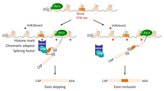
Figure 3. The chromatin-adaptor recruiting model of alternative splicing.
Histone modifications along the gene determine the binding of an adaptor protein which reads specific histone marks and in turn recruits splicing factors. In the case of exons whose alternative splicing is dependent on polypyrimidine tract binding protein (PTB) splicing factor, high levels of trimethylated histone 3 lysine 36 (H3K36me3, blue) attract the chromatin-binding factor MRG15 that acts as an adaptor protein and by protein-protein interaction helps to recruit PTB to its weaker binding site inducing exon skipping. If the PTB-dependent gene is hypermethylated in H3K4me3 (red), MRG15 does not accumulate along the gene, and PTB is not recruited to its target pre-mRNA, thus favoring exon inclusion.
Chromatin-splicing adaptor systems
A hint towards a direct role for histone modifications in alternative splicing regulation came from comparative mapping of a set of histone modifications along several genes whose alternative splicing is dependent on the polypyrimidine tract binding protein (PTB) splicing factor. These studies revealed a strong correlation between several histone modifications across the alternatively spliced regions and splicing outcome (Luco et al., 2010). PTB-dependent genes were found to be enriched in H3K36me3 and depleted in H3K4me3 in the alternatively spliced regions. Modulation of these histone marks was sufficient to switch the pattern of PTB-dependent exon inclusion (Luco et al., 2010). The molecular mechanism by which H3K36me3 acts in this case does not appear to involve modulation of RNA Pol II elongation rate, but rather the creation of a platform on chromatin for the recruitment of PTB to the nascent RNA (Figure 3) (Luco et al., 2010). This occurs via an adaptor protein, MRG15, that specifically binds to H3K36me3. The high levels of H3K36me3 along the alternatively spliced region of the gene attract MRG15, which in turn interacts with PTB recruiting it to the nascent RNA (Luco et al., 2010). In contrast, in cell types where H3K36me3 levels are low, the splicing repressor PTB is only poorly recruited to the newly forming RNA favoring inclusion of the PTB-dependent exon (Luco et al., 2010) (Figure 3). H3K36me3, MRG15 and PTB thus establish a chromatin-splicing adaptor system. In line with this interpretation, increasing H3K36me3 levels in the absence of the MRG15 adaptor protein has no effect on FGFR2 alternative splicing (Luco et al., 2010).
Although histone modifications clearly play a direct role in splicing regulation in this system, interestingly, the histone modifications do not appear to be the sole determinant of splicing outcome, but rather appear to act as a modifier. Genome-wide analysis of PTB-dependent alternative splicing patterns reveal that the splicing events most sensitive to changes in histone modifications relied on weak PTB-binding sites whereas alternative splicing events involving strong PTB-binding sites are not dependent on H3K36me3, suggesting that epigenetic modifications act in concert with RNA-binding elements to strengthen their effect (Luco et al., 2010).
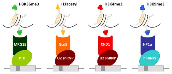
There is reason to believe that the combination of H3K36me3/MRG15/PTB is not the only chromatin-splicing adaptor system in mammalian cells (Figure 4). It is known that H3K4me3 levels play a role in the recruitment of the early spliceosome to human cyclin D1 pre-mRNA via binding of the chromatin-adaptor protein CHD1 (Sims et al., 2007). CHD1 contains a chromo-domain that specifically recognizes H3K4me3 and interacts with components of the U2 snRNP complex, but not U1 snRNP (Sims et al., 2007). Consistent with a role in splicing regulation, downregulation of H3K4me3 or CHD1 alters the efficiency of pre-mRNA splicing and reduces association of splicing factor 3a (SF3a) subcomplexes and U2 snRNP with pre-mRNA in vitro and in vivo (Sims et al., 2007). Interestingly, CHD1 is also a component of the histone acetyltransferase SAGA complex (Pray-Grant et al., 2005) in which Gcn5, which binds to acetylated H3 (Li and Shogren-Knaak, 2009), also interacts and recruits U2 snRNP components to the exon (Gunderson and Johnson, 2009; Figure 4). Another example of a possible chromatin-splicing adaptor system is H3K9 tri-methylation and HP1 proteins, which appear to recruit hnRNPs in Drosophila (Piacentini et al., 2009; Figure 4). Mass spectrometry analysis of proteins that bind to H3K9me identified the chromatin-binding protein HP1α/β and the splicing factors SRp20 and ASF/SF2 as interaction partners (Loomis et al., 2009). Co-immunoprecipitation experiments confirmed that HP1β interacts with ASF/SF2 in humans (Loomis et al., 2009) and HP1α with hnRNP proteins in Drosophila (Piacentini et al., 2009). These results point to a possible role for H3K9me3 in the regulation of recruitment of splicing factors mediated by the chromatin-adaptor protein HP1, although their functional relevance to splice site selection remains to be determined (Figure 4). Finally, other combinations of interacting histone modifications, chromatin-binding proteins and splicing factors may exists, possibly constituting a complex network of communication between chromatin and RNA. Genome-wide mapping of histone modifications and comparison to alternative splicing patterns should reveal such additional chromatin-splicing adaptor systems.
Figure 4. Chromatin-adaptor complexes.
Several histone modification-binding chromatin proteins interact with splicing factors (Luco et al., 2010; Sims et al., 2007; Gunderson and Johnson, 2009; Piacentini et al., 2009; Loomis et al., 2009).
An integrated model for alternative splice site selection
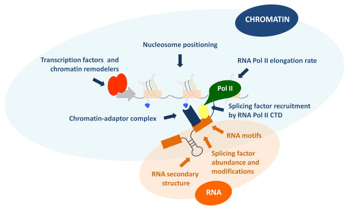
Regulation of alternative splicing has long been thought to involve mostly cis-acting RNA elements. However the picture becomes far more complex when one considers that RNA processing is coupled to transcription (Figure 5). The ultimate driving factor in determining splicing outcome will always be the recruitment of splicing regulators to the target RNA (Barash et al., 2010). However, when and which factors are recruited is not only dependent on the combination of RNA motifs, the tissue- or developmental-specific pattern of expression of the splicing factors or their post-translational modifications, but is also greatly influenced by chromatin architecture and histone modifications. The contribution of transcription regulators and histone modifiers to splicing regulation is likely two-fold: On the one hand, they remodel and open chromatin for the recruitment of elongation factors that activate RNA Pol II elongation kinetics. On the other hand, the positioning of nucleosomes along exons, plus the enrichment in particular subsets of histone modifications, may modulate the recruitment of splicing regulators (Kolasinska-Zwierz et al., 2009; Schwartz et al., 2009; Spies et al., 2009). This could occur either through pausing of RNA Pol II, which favors the formation of the spliceosome by protein-protein interactions (de la Mata and Kornblihtt, 2006; Listerman et al., 2006), or through specific recruitment of splicing factors via adaptor complexes to weak RNA binding sites (Luco et al., 2010; Sims et al., 2007; Gunderson and Johnson, 2009; Piacentini et al., 2009). These observations suggests an integrated model for the regulation of transcription and splicing in which the factors involved in the transcription control and chromatin maintenance are also involved in the recruitment and assembly of the spliceosome (Figure 5).
Figure 5. An integrated model for the regulation of alternative splicing.
Alternative splicing patterns are determined by a combination of parameters including cis-acting RNA regulatory elements and RNA secondary structures (highlighted in orange) together with transcriptional and chromatin properties (highlighted in blue) that modulate the recruitment of splicing factors to the pre-mRNA.
The role of chromatin in other RNA processing events
The role of chromatin and histone modifications likely goes beyond alternative splicing. There are some indications that other RNA processing events are similarly modulated by chromatin and epigenetic modifications.
In S. cerevisiae, the 3′ region near the polyadenylation site is depleted of nucleosomes (Mavrich et al., 2008) and in human T-cells nucleosome density dips noticeably within ~200nt of the canonical polyadenylation signal (Spies et al., 2009). Functional relevance for nucleosome density in polyadenylation is suggested by the fact that in genes containing multiple polyadenylation sites, the most highly used site preferentially falls within a nucleosome-depleted region (Spies et al., 2009). Bioinformatic analysis also suggests that nucleosome affinity is reduced near highly used polyadenylation sites but markedly increases just downstream of them (Spies et al., 2009). Altered nucleosome density may affect RNA Pol II elongation kinetics, which is known to affect polyadenylation, or the recruitment of the polyadenylation machinery to the nascent transcript (McCracken et al., 1997b).
Further evidence for a role of chromatin in RNA processing comes from the surprising finding in Drosophila that several histone variants including the core histones H3.3A, H3.3B, H2a.V and the H3-histone chaperone Asf1 are required for processing of the metazoan histone RNAs (Marzluff et al., 2008). Histone RNAs are unique in that they lack introns, are not polyadeny-lated and their 3′ ends are processed in a single step by formation of a 3′ stem-loop structure (Wagner et al., 2007). Loss of H2a.V, the functional ortholog of human H2A.X and H2A.Z, results in read-through and failure to process histone pre-mRNAs. The reason appears to be failure to recruit histone RNA processing factors to nascent histone RNAs (Marzluff et al., 2008). Although it is currently unknown how H2a.V affects processing factor recruitment, one intriguing possibility is that the altered chromatin structure at the histone loci interferes with the proper assembly of histone RNA processing machinery, thus decreasing RNA processing efficiency.
Another RNA metabolic event that appears to be influenced by chromatin is RNA degradation. In an attempt to uncover novel functions for the S.pombe histone variant H2A.Z, Grewal and colleagues noted that absence of H2A.Z leads to an increase in antisense transcripts in 5–8% of loci, whereas the level of sense-transcripts is only modestly affected (Zofall et al., 2009). The accumulating antisense messages were mostly read-through transcripts, and run-on experiments indicated that the elevated transcript levels are not due to increased transcription but are the consequence of failed degradation by the exosome (Zofall et al., 2009). In support of these conclusions, deletion of the exosome subunit rrp6 resulted in an antisense RNA profile similar to that observed upon loss of H2A.Z. These observations point to a role of chromatin structure in antisense RNA degradation by the exosome. The absence of H2A.Z may directly, or indirectly through loading of factors involved in maintaining chromatin structure, change chromatin conformation, which interferes with RNA Pol II progression, leading to RNA Pol II stalling and consequent degradation of transcripts. However this view is not fully consistent with the observation of read-through transcripts generated in the absence of H2A.Z. Alternatively, H2A.Z may play a role in communicating to an RNA Pol II-associated exosome that read-through transcripts have been generated. Intriguing questions are whether similar mechanisms are also at play in higher eukaryotes and whether H2A.Z, other histone variants and chromatin structure in general, play a more universal role in stability and controlled degradation of regular sense-transcripts.
Chromatin as a memory of alternative splicing patterns
Many alternative splicing events occur in a tissue- and/or cell-type specific fashion. How tissue- and cell-type specific alternative splicing patterns are established, propagated and maintained is only poorly understood. One mechanism involves the tissue-specific expression of alternative splicing regulators, the classical example being the neuron-specific splicing factor NOVA-1/2, which regulates an extensive network of alternatively spliced target genes (Ule et al., 2005). However, given the scarcity of dedicated alternative splicing master controllers such regulation is likely the exception rather than the rule.
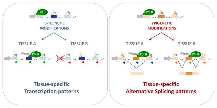
Cell- and tissue-specific alternative splicing patterns are obviously not determined solely by RNA-binding motifs, as they are present identically in all the cell types. Although cell- and tissue-specific differences in expression of constitutive splicing factors have been reported (Hanamura et al., 1998), it is difficult to envision how global changes in abundance of general splicing factors in a cell-type or tissue can account for the intricate regulation of individual exons, some of which need to be included, some skipped and others requiring activation of cryptic splice sites, even within the same gene. Given the realization that chromatin structure and histone modifications can affect alternative splicing, it is attractive to speculate that, in analogy to the histone indexing mechanisms used to specify which genes are expressed and which ones are silenced, the histone-based system may also encode information that specifies alternative splicing patterns in cell types and tissues (Figure 6). Such a system may involve marking of chromatin stretches encoding alternatively spliced regions by histone modifications to either alter RNA Pol II elongation rate locally as the polymerase passes through, or to recruit splicing factors via adaptor complexes. An advantage of such a histone-based alternative splicing regulatory system is that it would provide an epigenetic memory for splicing decisions which could be passed on during proliferation of a cell population and could be modified during differentiation without the requirement to establish a new set of alternative splicing rules at each step of differentiation. Obviously, an epigenetic alternative splicing memory would still require the proper expression of splicing factors, a process which itself may be controlled by epigenetic mechanisms. Regardless of mechanisms, it appears that epigenetic regulation is not limited to controlling what regions of the genome are expressed, but also how they are spliced.
Figure 6. The epigenetics of alternative splicing.
The combination of histone modifications along a gene establishes and maintains tissue-specific transcription patterns (left panel), as well as heritable tissue-specific alternative splicing patterns (right panel).
Conclusions and outlook
The realization that chromatin and histone modifications contribute to RNA processing, particularly alternative pre-mRNA splicing, has recently inspired many avenues of investigation, but at the same time raises many key questions. At this point, we do not even have a comprehensive view of how histone modifications relate to alternative splicing outcome. For that, histone modifications must be comprehensively mapped across the genome in as many cell types and tissues as possible and compared to genome-wide alternative splicing patterns. This information should be readily forthcoming from genome-wide histone modification mapping projects and the parallel characterization of the transcriptome in those systems by deep sequencing. These approaches will also answer the fundamental question of whether histone modifications that have been implicated in alternative splicing regulation act alone or in a combinatorial fashion with other epigenetic marks. Systematic genome-wide studies should also resolve the issue of how extensive the regulation of alternative splicing by histone modifications is. Are all alternative splicing events sensitive to histone modifications, or is only a subset of exons affected? If so, what are their characteristics? An intriguing extension of these considerations is the possibility that non-coding RNAs might play a role in alternative splicing regulation (Kishore and Stamm, 2006; Khanna and Stamm, 2010). ncRNAs are now known to be involved in heterochromatin structure and it is possible that some ncRNAs are specifically transcribed and associate with alternatively spliced regions of genes.
Having established a role for histone modifications in alternative splicing, and given the intimate linkage between transcription and RNA processing, the question of whether splicing in turn also affects histone modifications must be asked. It is possible that in the same way histone modifications modulate recruitment of splicing factors, splicing regulators also modulate the recruitment of histone modifiers and chromatin remodelers to the nucleosomes regulating chromatin conformation in a feedback mechanism. In support of such cross-talk, inhibition of splicing abolishes transcription and splicing factors stimulate elongation (O’Keefe et al., 1994; Fong and Zhou, 2001; Lin et al., 2008), suggesting that transcription, chromatin and splicing are intimately dependent on each other. Considering the still preliminary but tantalizing evidence that chromatin may also affect other RNA processing events, it will be important to probe in more detail the effect of chromatin on 3′ processing, RNA stability and other RNA processing steps.
Finally, we must consider the possible physiological and pathological consequences and opportunities of histone mediated RNA processing effects. First and foremost, it will be important to determine whether histone modification effects on RNA processing are heritable and therefore truly epigenetic or whether they are merely transient modulators. To address this question, we will have to systematically analyze changes in RNA processing patterns during differentiation and development and compare them to histone modification fingerprints of cells and tissues. We should also comprehensively probe for aberrant RNA processing in diseases caused by epigenetic defects. In addition, we may have to reconsider the expected effects of drugs targeting epigenetic mechanisms such as the clinically used histone deacetylase and DNA methyltransferase inhibitors and we might want to think about designing new drugs targeting chromatin for the treatment of splicing-diseases. Clearly the emerging role of epigenetics in RNA processing provides many new challenges, and even more opportunities.
Acknowledgments
This work was supported by grants to A.R.K. from the Agencia Nacional de Promoción de Ciencia y Tecnología of Argentina, the Universidad de Buenos Aires and the European Alternative Splicing Network (EURASNET). I.E.S. is a recipient of a post-doctoral fellowships and A.R.K. is a career investigator from the Consejo Nacional de Investigaciones Científicas y Técnicas of Argentina (CONICET). A.R.K. is an international research scholar of the Howard Hughes Medical Institute. Work in the Misteli laboratory is supported by the Intramural Research Program of the National Institutes of Health (NIH), NCI, Center for Cancer Research.
Footnotes
Publisher's Disclaimer: This is a PDF file of an unedited manuscript that has been accepted for publication. As a service to our customers we are providing this early version of the manuscript. The manuscript will undergo copyediting, typesetting, and review of the resulting proof before it is published in its final citable form. Please note that during the production process errors may be discovered which could affect the content, and all legal disclaimers that apply to the journal pertain.
References
- Aebi M, Weissmann C. Precision and orderliness in splicing. Trends Genet. 1987;3:102–107. [Google Scholar]
- Allo M, Buggiano V, Fededa JP, Petrillo E, Schor I, de la Mata M, Agirre E, Plass M, Eyras E, Abou Elela S, Klinck R, Chabot B, Kornblihtt AR. Control of alternative splicing through siRNA-mediated transcriptional gene silencing. Nat Struct Mol Biol. 2009;16:717–724. doi: 10.1038/nsmb.1620. [DOI] [PubMed] [Google Scholar]
- Andersson R, Enroth S, Rada-Iglesias A, Wadelius C, Komorowski J. Nucleosomes are well positioned in exons and carry characteristic histone modifications. Genome Res. 2009;19:1732–1741. doi: 10.1101/gr.092353.109. [DOI] [PMC free article] [PubMed] [Google Scholar]
- Attanasio C, David A, Neerman-Arbez M. Outcome of donor splice site mutations accounting for congenital afibrinogenemia reflects order of intron removal in the fibrinogen alpha gene (FGA) Blood. 2003;101:1851–1856. doi: 10.1182/blood-2002-03-0853. [DOI] [PubMed] [Google Scholar]
- Auboeuf D, Honig A, Berget SM, O’Malley BW. Coordinate regulation of transcription and splicing by steroid receptor coregulators. Science. 2002;298:416–419. doi: 10.1126/science.1073734. [DOI] [PubMed] [Google Scholar]
- Auboeuf D, Dowhan DH, Kang YK, Larkin K, Lee JW, Berget SM, O’Malley BW. Differential recruitment of nuclear receptor coactivators may determine alternative RNA splice site choice in target genes. Proc Natl Acad Sci U S A. 2004a;101:2270–2274. doi: 10.1073/pnas.0308133100. [DOI] [PMC free article] [PubMed] [Google Scholar]
- Auboeuf D, Dowhan DH, Li X, Larkin K, Ko L, Berget SM, O’Malley BW. CoAA, a nuclear receptor coactivator protein at the interface of transcriptional coactivation and RNA splicing. Mol Cell Biol. 2004b;24:442–453. doi: 10.1128/MCB.24.1.442-453.2004. [DOI] [PMC free article] [PubMed] [Google Scholar]
- Barash Y, Calarco JA, Gao WJ, Pan Q, Wang XC, Shai O, Blencowe BJ, Frey BJ. Deciphering the splicing code. Nature. 2010;465:53–59. doi: 10.1038/nature09000. [DOI] [PubMed] [Google Scholar]
- Batsche E, Yaniv M, Muchardt C. The human SWI/SNF subunit Brm is a regulator of alternative splicing. Nat Struct Mol Biol. 2006;13:22–29. doi: 10.1038/nsmb1030. [DOI] [PubMed] [Google Scholar]
- Bauren G, Wieslander L. Splicing of Balbiani ring 1 gene pre-mRNA occurs simultaneously with transcription. Cell. 1994;76:183–192. doi: 10.1016/0092-8674(94)90182-1. [DOI] [PubMed] [Google Scholar]
- Beyer AL, Osheim YN. Splice site selection, rate of splicing, and alternative splicing on nascent transcripts. Genes Dev. 1988;2:754–765. doi: 10.1101/gad.2.6.754. [DOI] [PubMed] [Google Scholar]
- Bird G, Zorio DA, Bentley DL. RNA polymerase II carboxy-terminal domain phosphorylation is required for cotranscriptional pre-mRNA splicing and 3′-end formation. Mol Cell Biol. 2004;24:8963–8969. doi: 10.1128/MCB.24.20.8963-8969.2004. [DOI] [PMC free article] [PubMed] [Google Scholar]
- Brodsky AS, Meyer CA, Swinburne IA, Giles H, Keenan BJ, Liu XLS, Fox EA, Silver PA. Genomic mapping of RNA polymerase II reveals sites of co-transcriptional regulation in human cells. Genome Biol. 2005;6 doi: 10.1186/gb-2005-6-8-r64. [DOI] [PMC free article] [PubMed] [Google Scholar]
- Buratti E, Baralle FE. Influence of RNA secondary structure on the pre-mRNA splicing process. Mol Cell Biol. 2004;24:10505–10514. doi: 10.1128/MCB.24.24.10505-10514.2004. [DOI] [PMC free article] [PubMed] [Google Scholar]
- Caceres JF, Kornblihtt AR. Alternative splicing: multiple control mechanisms and involvement in human disease. Trends Genet. 2002;18:186–193. doi: 10.1016/s0168-9525(01)02626-9. [DOI] [PubMed] [Google Scholar]
- Chasin LA. Searching for splicing motifs. Adv Exp Med Biol. 2007;623:85–106. doi: 10.1007/978-0-387-77374-2_6. [DOI] [PubMed] [Google Scholar]
- Cheng DH, Cote J, Shaaban S, Bedford MT. The arginine methyltransferase CARM1 regulates the coupling of transcription and mRNA processing. Mol Cell. 2007;25:71–83. doi: 10.1016/j.molcel.2006.11.019. [DOI] [PubMed] [Google Scholar]
- Chodavarapu RK, Feng S, Bernatavichute YV, Chen PY, Stroud H, Yu Y, Hetzel JA, Kuo F, Kim J, Cokus SJ, Casero D, Bernal M, Huijser P, Clark AT, Kramer U, Merchant SS, Zhang X, Jacobsen SE, Pellegrini M. Relationship between nucleosome positioning and DNA methylation. Nature. 2010;15:388–392. doi: 10.1038/nature09147. [DOI] [PMC free article] [PubMed] [Google Scholar]
- Cooper TA, Wan L, Dreyfuss G. RNA and disease. Cell. 2009;136:777–93. doi: 10.1016/j.cell.2009.02.011. [DOI] [PMC free article] [PubMed] [Google Scholar]
- Cramer P, Caceres JF, Cazalla D, Kadener S, Muro AF, Baralle FE, Kornblihtt AR. Coupling of transcription with alternative splicing: RNA pol II promoters modulate SF2/ASF and 9G8 effects on an exonic splicing enhancer. Mol Cell. 1999;4:251–258. doi: 10.1016/s1097-2765(00)80372-x. [DOI] [PubMed] [Google Scholar]
- Cramer P, Pesce CG, Baralle FE, Kornblihtt AR. Functional association between promoter structure and transcript alternative splicing. Proc Natl Acad Sci U S A. 1997;94:11456–11460. doi: 10.1073/pnas.94.21.11456. [DOI] [PMC free article] [PubMed] [Google Scholar]
- Croft L, Schandorff S, Clark F, Burrage K, Arctander P, Mattick JS. ISIS, the intron information system, reveals the high frequency of alternative splicing in the human genome. Nat Genet. 2000;24:340–341. doi: 10.1038/74153. [DOI] [PubMed] [Google Scholar]
- Das R, Dufu K, Romney B, Feldt M, Elenko M, Reed R. Functional coupling of RNAP II transcription to spliceosome assembly. Genes Dev. 2006;20:1100–1109. doi: 10.1101/gad.1397406. [DOI] [PMC free article] [PubMed] [Google Scholar]
- Das R, Yu J, Zhang Z, Gygi MP, Krainer AR, Gygi SP, Reed R. SR proteins function in coupling RNAP II transcription to pre-mRNA splicing. Mol Cell. 2007;26:867–881. doi: 10.1016/j.molcel.2007.05.036. [DOI] [PubMed] [Google Scholar]
- de la Mata M, Alonso CR, Kadener S, Fededa JP, Blaustein M, Pelisch F, Cramer P, Bentley D, Kornblihtt AR. A slow RNA polymerase II affects alternative splicing in vivo. Mol Cell. 2003;12:525–532. doi: 10.1016/j.molcel.2003.08.001. [DOI] [PubMed] [Google Scholar]
- de la Mata M, Kornblihtt AR. RNA polymerase II C-terminal domain mediates regulation of alternative splicing by SRp20. Nat Struct Mol Biol. 2006;13:973–980. doi: 10.1038/nsmb1155. [DOI] [PubMed] [Google Scholar]
- de la Mata M, Lafaille C, Kornblihtt AR. First come, first served revisited: factors affecting the same alternative splicing event have different effects on the relative rates of intron removal. RNA. 2010;16:904–912. doi: 10.1261/rna.1993510. [DOI] [PMC free article] [PubMed] [Google Scholar]
- Dhami P, Saffrey P, Bruce AW, Dillson SC, Chiang K, Bonhoure N, Koch CM, Bye J, James K, Foad NS, Ellis P, Watkins NA, Ouwehand WH, Langford C, Andrews RM, Dunham I, Vetire D. Complex exon-intron marking by histone modifications is not determined solely by nucleosome distribution. PloS One. 2010;5:e12339. doi: 10.1371/journal.pone.0012339. [DOI] [PMC free article] [PubMed] [Google Scholar]
- Dower K, Rosbash M. T7 RNA polymerase-directed transcripts are processed in yeast and link 3′ end formation to mRNA nuclear export. RNA. 2002;8:686–697. doi: 10.1017/s1355838202024068. [DOI] [PMC free article] [PubMed] [Google Scholar]
- Eperon LP, Graham IR, Griffiths AD, Eperon IC. Effects of RNA secondary structure on alternative splicing of pre-mRNA: is folding limited to a region behind the transcribing RNA polymerase? Cell. 1988;54:393–401. doi: 10.1016/0092-8674(88)90202-4. [DOI] [PubMed] [Google Scholar]
- Fong YW, Zhou Q. Stimulatory effect of splicing factors on transcriptional elongation. Nature. 2001;414:929–933. doi: 10.1038/414929a. [DOI] [PubMed] [Google Scholar]
- Fox-Walsh KL, Hertel KJ. Splice-site pairing is an intrinsically high fidelity process. Proc Natl Acad Sci U S A. 2009;106:1766–1771. doi: 10.1073/pnas.0813128106. [DOI] [PMC free article] [PubMed] [Google Scholar]
- Gornemann J, Kotovic KM, Hujer K, Neugebauer KM. Cotranscriptional spliceosome assembly occurs in a stepwise fashion and requires the cap binding complex. Mol Cell. 2005;19:53–63. doi: 10.1016/j.molcel.2005.05.007. [DOI] [PubMed] [Google Scholar]
- Gunderson FQ, Johnson TL. Acetylation by the Transcriptional Coactivator Gcn5 Plays a Novel Role in Co-Transcriptional Spliceosome Assembly. Plos Genet. 2009;5 doi: 10.1371/journal.pgen.1000682. [DOI] [PMC free article] [PubMed] [Google Scholar]
- Han SP, Tang YH, Smith R. Functional diversity of the hnRNPs: past, present and perspectives. Biochem J. 2010;430:379–92. doi: 10.1042/BJ20100396. [DOI] [PubMed] [Google Scholar]
- Hanamura A, Caceres JF, Mayeda A, Franza BR, Krainer AR. Regulated tissue-specific expression of antagonistic pre-mRNA splicing factors. RNA. 1998;4:430–444. [PMC free article] [PubMed] [Google Scholar]
- Hicks MJ, Yang CR, Kotlajich MV, Hertel KJ. Linking splicing to Pol II transcription stabilizes pre-mRNAs and influences splicing patterns. PLoS Biol. 2006;4:e147. doi: 10.1371/journal.pbio.0040147. [DOI] [PMC free article] [PubMed] [Google Scholar]
- Hirose Y, Manley JL. RNA polymerase II is an essential mRNA polyadenylation factor. Nature. 1998;395:93–6. doi: 10.1038/25786. [DOI] [PubMed] [Google Scholar]
- Hodges C, Bintu L, Lubkowska L, Kashlev M, Bustamante C. Nucleosomal Fluctuations Govern the Transcription Dynamics of RNA Polymerase II. Science. 2009a;325:626–628. doi: 10.1126/science.1172926. [DOI] [PMC free article] [PubMed] [Google Scholar]
- Hodges E, Smith AD, Kendall J, Xuan ZY, Ravi K, Rooks M, Zhang MQ, Ye K, Bhattacharjee A, Brizuela L, McCombie WR, Wigler M, Hannon GJ, Hicks JB. High definition profiling of mammalian DNA methylation by array capture and single molecule bisulfite sequencing. Genome Res. 2009b;19:1593–1605. doi: 10.1101/gr.095190.109. [DOI] [PMC free article] [PubMed] [Google Scholar]
- Howe KJ, Kane CM, Ares M., Jr Perturbation of transcription elongation influences the fidelity of internal exon inclusion in Saccharomyces cerevisiae. RNA. 2003;9:993–1006. doi: 10.1261/rna.5390803. [DOI] [PMC free article] [PubMed] [Google Scholar]
- Kadener S, Cramer P, Nogues G, Cazalla D, de la Mata M, Fededa JP, Werbajh SE, Srebrow A, Kornblihtt AR. Antagonistic effects of T-Ag and VP16 reveal a role for RNA pol II elongation on alternative splicing. EMBO J. 2001;20:5759–5768. doi: 10.1093/emboj/20.20.5759. [DOI] [PMC free article] [PubMed] [Google Scholar]
- Kessler O, Jiang Y, Chasin LA. Order of intron removal during splicing of endogenous adenine phosphoribosyltransferase and dihydrofolate reductase pre-mRNA. Mol Cell Biol. 1993;13:6211–6222. doi: 10.1128/mcb.13.10.6211. [DOI] [PMC free article] [PubMed] [Google Scholar]
- Khanna A, Stamm S. Regulation of alternative splicing by short non-coding nuclear RNAs. RNA Biol. 2010;7 doi: 10.4161/rna.7.4.12746. [DOI] [PMC free article] [PubMed] [Google Scholar]
- Kishore S, Stamm S. The snoRNA HBII-52 regulates alternative splicing of the serotonin receptor 2C. Science. 2006;311:230–2. doi: 10.1126/science.1118265. [DOI] [PubMed] [Google Scholar]
- Kolasinska-Zwierz P, Down T, Latorre I, Liu T, Liu XS, Ahringer J. Differential chromatin marking of introns and expressed exons by H3K36me3. Nat Genet. 2009;41:376–381. doi: 10.1038/ng.322. [DOI] [PMC free article] [PubMed] [Google Scholar]
- Kornblihtt AR. Promoter usage and alternative splicing. Curr Opin Cell Biol. 2005;17:262–268. doi: 10.1016/j.ceb.2005.04.014. [DOI] [PubMed] [Google Scholar]
- Kotovic KM, Lockshon D, Boric L, Neugebauer KM. Cotranscriptional recruitment of the U1 snRNP to intron-containing genes in yeast. Mol Cell Biol. 2003;23:5768–5779. doi: 10.1128/MCB.23.16.5768-5779.2003. [DOI] [PMC free article] [PubMed] [Google Scholar]
- Lacadie SA, Rosbash M. Cotranscriptional spliceosome assembly dynamics and the role of U1 snRNA:5′ss base pairing in yeast. Mol Cell. 2005;19:65–75. doi: 10.1016/j.molcel.2005.05.006. [DOI] [PubMed] [Google Scholar]
- Lazarev D, Manley JL. Concurrent splicing and transcription are not sufficient to enhance splicing efficiency. RNA. 2007;13:1546–57. doi: 10.1261/rna.595907. [DOI] [PMC free article] [PubMed] [Google Scholar]
- LeMaire MF, Thummel CS. Splicing precedes polyadenylation during Drosophila E74A transcription. Mol Cell Biol. 1990;10:6059–6063. doi: 10.1128/mcb.10.11.6059. [DOI] [PMC free article] [PubMed] [Google Scholar]
- Li SS, Shogren-Knaak MA. The Gcn5 Bromodomain of the SAGA Complex Facilitates Cooperative and Cross-tail Acetylation of Nucleosomes. J Biol Chem. 2009;284:9411–9417. doi: 10.1074/jbc.M809617200. [DOI] [PMC free article] [PubMed] [Google Scholar]
- Lin S, Coutinho-Mansfield G, Wang D, Pandit S, Fu XD. The splicing factor SC35 has an active role in transcriptional elongation. Nat Struct Mol Biol. 2008;15:819–826. doi: 10.1038/nsmb.1461. [DOI] [PMC free article] [PubMed] [Google Scholar]
- Listerman I, Sapra AK, Neugebauer KM. Cotranscriptional coupling of splicing factor recruitment and precursor messenger RNA splicing in mammalian cells. Nat Struct Mol Biol. 2006;13:815–822. doi: 10.1038/nsmb1135. [DOI] [PubMed] [Google Scholar]
- Loomis RJ, Naoe Y, Parker JB, Savic V, Bozovsky MR, Macfarlan T, Manley JL, Chakravarti D. Chromatin binding of SRp20 and ASF/SF2 and dissociation from mitotic chromosomes is modulated by histone H3 serine 10 phosphorylation. Mol Cell. 2009;33:450–461. doi: 10.1016/j.molcel.2009.02.003. [DOI] [PMC free article] [PubMed] [Google Scholar]
- Luco RF, Pan Q, Tominaga K, Blencowe BJ, Pereira-Smith OM, Misteli T. Regulation of Alternative Splicing by Histone Modifications. Science. 2010;327:996–1000. doi: 10.1126/science.1184208. [DOI] [PMC free article] [PubMed] [Google Scholar]
- Maniatis T, Reed R. An extensive network of coupling among gene expression machines. Nature. 2002;416:499–506. doi: 10.1038/416499a. [DOI] [PubMed] [Google Scholar]
- Martinez E, Palhan VB, Tjernberg A, Lymar ES, Gamper AM, Kundu TK, Chait BT, Roeder RG. Human STAGA complex is a chromatin-acetylating transcription coactivator that interacts with pre-mRNA splicing and DNA damage-binding factors in vivo. Mol Cel Biol. 2001;21:6782–6795. doi: 10.1128/MCB.21.20.6782-6795.2001. [DOI] [PMC free article] [PubMed] [Google Scholar]
- Marzluff WF, Wagner EJ, Duronio RJ. Metabolism and regulation of canonical histone mRNAs: life without a poly(A) tail. Nat Rev Genet. 2008;9:843–854. doi: 10.1038/nrg2438. [DOI] [PMC free article] [PubMed] [Google Scholar]
- Mavrich TN, Ioshikhes IP, Venters BJ, Jiang C, Tomsho LP, Qi J, Schuster SC, Albert I, Pugh BF. A barrier nucleosome model for statistical positioning of nucleosomes throughout the yeast genome. Genome Res. 2008;18:1073–1083. doi: 10.1101/gr.078261.108. [DOI] [PMC free article] [PubMed] [Google Scholar]
- McCracken S, Fong N, Rosonina E, Yankulov K, Brothers G, Siderovski D, Hessel A, Foster S, Shuman S, Bentley DL. 5′-Capping enzymes are targeted to pre-mRNA by binding to the phosphorylated carboxy-terminal domain of RNA polymerase II. Genes Dev. 1997a;11:3306–18. doi: 10.1101/gad.11.24.3306. [DOI] [PMC free article] [PubMed] [Google Scholar]
- McCracken S, Fong N, Yankulov K, Ballantyne S, Pan G, Greenblatt J, Patterson SD, Wickens M, Bentley DL. The C-terminal domain of RNA polymerase II couples mRNA processing to transcription. Nature. 1997b;385:357–361. doi: 10.1038/385357a0. [DOI] [PubMed] [Google Scholar]
- McCracken S, Rosonina E, Fong N, Sikes M, Beyer A, O’Hare K, Shuman S, Bentley D. Role of RNA polymerase II carboxy-terminal domain in coordinating transcription with RNA processing. Cold Spring Harb Symp Quant Biol. 1998;63:301–309. doi: 10.1101/sqb.1998.63.301. [DOI] [PubMed] [Google Scholar]
- Meissner A, Mikkelsen TS, Gu HC, Wernig M, Hanna J, Sivachenko A, Zhang XL, Bernstein BE, Nusbaum C, Jaffe DB, Gnirke A, Jaenisch R, Lander ES. Genome-scale DNA methylation maps of pluripotent and differentiated cells. Nature. 2008;454:766–770. doi: 10.1038/nature07107. [DOI] [PMC free article] [PubMed] [Google Scholar]
- Misteli T, Spector DL. RNA polymerase II targets pre-mRNA splicing factors to transcription sites in vivo. Mol Cell. 1999;3:697–705. doi: 10.1016/s1097-2765(01)80002-2. [DOI] [PubMed] [Google Scholar]
- Moore MJ, Proudfoot NJ. Pre-mRNA processing reaches back to transcription and ahead to translation. Cell. 2009;136:688–700. doi: 10.1016/j.cell.2009.02.001. [DOI] [PubMed] [Google Scholar]
- Munoz MJ, Perez Santangelo MS, Paronetto MP, de la Mata M, Pelisch F, Boireau S, Glover-Cutter K, Ben-Dov C, Blaustein M, Lozano JJ, et al. DNA damage regulates alternative splicing through inhibition of RNA polymerase II elongation. Cell. 2009;137:708–720. doi: 10.1016/j.cell.2009.03.010. [DOI] [PubMed] [Google Scholar]
- Nahkuri S, Taft RJ, Mattick JS. Nucleosomes are preferentially positioned at exons in somatic and sperm cells. Cell Cycle. 2009;8:3420–3424. doi: 10.4161/cc.8.20.9916. [DOI] [PubMed] [Google Scholar]
- Nogues G, Kadener S, Cramer P, Bentley D, Kornblihtt AR. Transcriptional activators differ in their abilities to control alternative splicing. J Biol Chem. 2002;277:43110–43114. doi: 10.1074/jbc.M208418200. [DOI] [PubMed] [Google Scholar]
- Ohkura N, Takahashi M, Yaguchi H, Nagamura Y, Tsukada T. Coactivator-associated arginine methyltransferase 1, CARM1, affects pre-mRNA splicing in an isoform-specific manner. J Biol Chem. 2005;280:28927–28935. doi: 10.1074/jbc.M502173200. [DOI] [PubMed] [Google Scholar]
- O’Keefe RT, Mayeda A, Sadowski CL, Krainer AR, Specotr DL. Disruption of pre-mRNA splicing in vivo results in reorganization of splicing factors. J Cell Biol. 1994;124:249–60. doi: 10.1083/jcb.124.3.249. [DOI] [PMC free article] [PubMed] [Google Scholar]
- Pagani F, Stuani C, Zuccato E, Kornblihtt AR, Baralle FE. Promoter architecture modulates CFTR exon 9 skipping. J Biol Chem. 2003;278:1511–1517. doi: 10.1074/jbc.M209676200. [DOI] [PubMed] [Google Scholar]
- Pan Q, Shai O, Lee LJ, Frey BJ, Blencowe BJ. Deep surveying of alternative splicing complexity in the human transcriptome by high-throughput sequencing. Nat Genet. 2008;40:1413–5. doi: 10.1038/ng.259. [DOI] [PubMed] [Google Scholar]
- Pandya-Jones A, Black DL. Co-transcriptional splicing of constitutive and alternative exons. RNA. 2009;15:1896–1908. doi: 10.1261/rna.1714509. [DOI] [PMC free article] [PubMed] [Google Scholar]
- Piacentini L, Fanti L, Negri R, Del Vescovo V, Fatica A, Altieri F, Pimpinelli S. Heterochromatin Protein 1 (HP1a) Positively Regulates Euchromatic Gene Expression through RNA Transcript Association and Interaction with hnRNPs in Drosophila. Plos Genet. 2009;5 doi: 10.1371/journal.pgen.1000670. [DOI] [PMC free article] [PubMed] [Google Scholar]
- Ponts N, Yang JF, Chung DWD, Prudhomme J, Girke T, Horrocks P, Le Roch KG. Deciphering the Ubiquitin-Mediated Pathway in Apicomplexan Parasites: A Potential Strategy to Interfere with Parasite Virulence. Plos One. 2008;3 doi: 10.1371/journal.pone.0002386. [DOI] [PMC free article] [PubMed] [Google Scholar]
- Pray-Grant MG, Daniel JA, Schieltz D, Yates JR, 3rd, Grant PA. Chd1 chromodomain links histone H3 methylation with SAGA- and SLIK-dependent acetylation. Nature. 2005;433:434–438. doi: 10.1038/nature03242. [DOI] [PubMed] [Google Scholar]
- Roberts GC, Gooding C, Mak HY, Proudfoot NJ, Smith CW. Co-transcriptional commitment to alternative splice site selection. Nucleic Acids Res. 1998;26:5568–5572. doi: 10.1093/nar/26.24.5568. [DOI] [PMC free article] [PubMed] [Google Scholar]
- Sapra AK, Anko ML, Grishina I, Lorenz M, Pabis M, Poser I, Rollins J, Weiland EM, Neugebauer KM. SR protein family members display diverse activities in the formation of nascent and mature mRNPs in vivo. Mol Cell. 2009;34:179–190. doi: 10.1016/j.molcel.2009.02.031. [DOI] [PubMed] [Google Scholar]
- Schor IE, Rascovan N, Pelisch F, Allo M, Kornblihtt AR. Neuronal cell depolarization induces intragenic chromatin modifications affecting NCAM alternative splicing. Proc Natl Acad Sci U S A. 2009;106:4325–4330. doi: 10.1073/pnas.0810666106. [DOI] [PMC free article] [PubMed] [Google Scholar]
- Schwartz S, Meshorer E, Ast G. Chromatin organization marks exon-intron structure. Nat Struct Mol Biol. 2009;16:990–995. doi: 10.1038/nsmb.1659. [DOI] [PubMed] [Google Scholar]
- Long JC, Caceres JF. The SR protein family of splicing factors: master regulators of gene expression. Biochem J. 2009;417:15–27. doi: 10.1042/BJ20081501. [DOI] [PubMed] [Google Scholar]
- Sims RJ, Millhouse S, Chen CF, Lewis BA, Erdjument-Bromage H, Tempst P, Manley JL, Reinberg D. Recognition of trimethylated histone h3 lysine 4 facilitates the recruitment of transcription postinitiation factors and pre-mRNA splicing. Mol Cell. 2007;28:665–676. doi: 10.1016/j.molcel.2007.11.010. [DOI] [PMC free article] [PubMed] [Google Scholar]
- Sisodia SS, Sollner-Webb B, Cleveland DW. Specificity of RNA maturation pathways: RNAs transcribed by RNA polymerase III are not substrates for splicing or polyadenylation. Mol Cell Biol. 1987;7:3602–3612. doi: 10.1128/mcb.7.10.3602. [DOI] [PMC free article] [PubMed] [Google Scholar]
- Smale ST, Tjian R. Transcription of herpes simplex virus tk sequences under the control of wild-type and mutant human RNA polymerase I promoters. Mol Cell Biol. 1985;5:352–362. doi: 10.1128/mcb.5.2.352. [DOI] [PMC free article] [PubMed] [Google Scholar]
- Spies N, Nielsen CB, Padgett RA, Burge CB. Biased Chromatin Signatures around Polyadenylation Sites and Exons. Mol Cell. 2009;36:245–254. doi: 10.1016/j.molcel.2009.10.008. [DOI] [PMC free article] [PubMed] [Google Scholar]
- Tardiff DF, Lacadie SA, Rosbash M. A genome-wide analysis indicates that yeast pre-mRNA splicing is predominantly posttranscriptional. Mol Cell. 2006;24:917–929. doi: 10.1016/j.molcel.2006.12.002. [DOI] [PMC free article] [PubMed] [Google Scholar]
- Tennyson CN, Klamut HJ, Worton RG. The human dystrophin gene requires 16 hours to be transcribed and is cotranscriptionally spliced. Nat Genet. 1995;9:184–190. doi: 10.1038/ng0295-184. [DOI] [PubMed] [Google Scholar]
- Tilgner H, Nikolaou C, Althammer S, Sammeth M, Beato M, Valcarcel J, Guigo R. Nucleosome positioning as a determinant of exon recognition. Nat Struct Mol Biol. 2009;16:996–1001. doi: 10.1038/nsmb.1658. [DOI] [PubMed] [Google Scholar]
- Tyagi A, Ryme J, Brodin D, Farrants AKO, Visa N. SWI/SNF Associates with Nascent Pre-mRNPs and Regulates Alternative Pre-mRNA Processing. Plos Genet. 2009;5 doi: 10.1371/journal.pgen.1000470. [DOI] [PMC free article] [PubMed] [Google Scholar]
- Ule J, Ule A, Spencer J, Williams A, Hu JS, Cline M, Wang H, Clark T, Fraser C, Ruggiu M, Zeeberg BR, Kane D, Weinstein JN, Blume J, Darnell RB. Nova regulates brain-specific splicing to shape the synapse. Nat Genet. 2005;37:844–852. doi: 10.1038/ng1610. [DOI] [PubMed] [Google Scholar]
- Wagner EJ, Burch BD, Godfrey AC, Salzler HR, Duronio RJ, Marzluff WF. A genome-wide RNA interference screen reveals that variant histones, are necessary for replication-dependent histone pre-mRNA processing. Mol Cell. 2007;28:692–699. doi: 10.1016/j.molcel.2007.10.009. [DOI] [PubMed] [Google Scholar]
- Xiao TJ, Hall H, Kizer KO, Shibata Y, Hall MC, Borchers CH, Strahl BD. Phosphorylation of RNA polymerase II CTD regulates H3 methylation in yeast. Genes Dev. 2003;17:654–663. doi: 10.1101/gad.1055503. [DOI] [PMC free article] [PubMed] [Google Scholar]
- Zofall M, Fischer T, Zhang K, Zhou M, Cui BW, Veenstra TD, Grewal SIS. Histone H2A.Z cooperates with RNAi and heterochromatin factors to suppress antisense RNAs. Nature. 2009;461:419–422. doi: 10.1038/nature08321. [DOI] [PMC free article] [PubMed] [Google Scholar]


