Abstract
Slope-related differences in hindlimb movements and activation of the soleus and tibialis anterior muscles were studied during treadmill locomotion in intact rats and in rats 4 and 10 weeks following transection and surgical repair of the sciatic nerve. In intact rats, the tibialis anterior and soleus muscles were activated reciprocally at all slopes, and the overall intensity of activity in tibialis anterior and the mid-step activity in soleus increased with increasing slope. Based on the results of principal components analysis, the pattern of activation of soleus, but not of tibialis anterior, changed significantly with slope. Slope-related differences in hindlimb kinematics were found in intact rats, and these correlated well with the demands of walking up or down slopes. Following recovery from sciatic nerve injury, the soleus and tibialis anterior were co-activated throughout much of the step cycle and there was no difference in intensity or pattern of activation with slope for either muscle. Unlike intact rats, these animals walked with their feet flat on the treadmill belt through most of the stance phase. Even so, during downslope walking limb length and limb orientation throughout the step cycle were not significantly changed from values found in intact rats. This conservation of hindlimb kinematics was not observed during level or upslope walking. These findings are interpreted as evidence that the recovering animals adopt a novel locomotor strategy that involves stiffening of the ankle joint by antagonist co-activation and compensation at more proximal joints. Their movements are most suitable to the requirements of downslope walking but the recovering rats lack the ability to adapt to the demands of level or upslope walking.
Keywords: EMG activity, global limb vector, locomotion, peripheral nerve injury, principal components analysis (PCA)
INTRODUCTION
Terrestrial animals utilize a variety of locomotor styles (e.g. walking, trotting and galloping) and speeds to respond to environmental challenges and negotiate different terrains. Slope is a unique environmental feature that is accommodated through an interaction between neuronal circuits, primarily in the spinal cord and the musculoskeletal system. In rhythmic movements such as locomotion, the timing and intensity of activity of different muscle groups is based on the timing of the outputs of spinal circuits and their spatial orientation to groups of motoneurons (Yakovenko et al., 2002), as well as by integration of afferent feedback from skin and the muscles themselves (Donelan et al., 2009; Pearson, 2000). Several aspects of the neural control of slope walking have been described in the cat (Carlson-Kuhta et al., 1998; Fowler et al., 1993; Gregor et al., 2006; Smith et al., 1998). It has emerged from these studies that up and downslope locomotion pose opposite challenges to the sensorimotor system that are met by mechanisms specific to each direction of slope.
Slope locomotion thus may have promise as a means to evaluate functional recovery following injury to the peripheral nervous system. When peripheral nerves are injured, especially injuries involving axotomy, the relationship between spinal circuitry and the elements of the musculoskeletal system is changed. Even though axons in the proximal segments of cut nerves can regenerate, functional recovery following such injuries is poor (Frostick et al., 1998; Scholz et al., 2009). Several strategies that enhance axon regeneration after peripheral nerve transection have been identified, e.g. proteoglycan degradation (Groves et al., 2005), electrical stimulation (Al-Majed et al., 2000; English et al., 2007) and exercise (Sabatier et al., 2008), but far less is understood about whether function is restored when axon regeneration is enhanced (Varejao et al., 2004). A better understanding of the slope-related specificity of muscle activation and hindlimb control in intact and injured animals may help in efforts to identify strategies that ultimately enhance function.
In several recent studies of limb function during locomotion, a shift from analyses of movements about individual joints (Thota et al., 2005) to a more global, limb-level function has been advocated in rats (Hamilton et al., 2011), cats (Chang et al., 2009), humans (Auyang et al., 2009) and helmeted guinea fowl (Daley et al., 2007). Global representation of the hindlimb as a single vector varied less from step to step and between animals than movements about individual joints (Hamilton et al., 2011; Chang et al., 2009) and this was conserved during recovery from ankle extensor nerve injury in cats (Chang et al., 2009). Because this finding correlates well with evidence that whole limb function is linked to specific populations of neurons in the spinal circuits (Auyang et al., 2009; Bosco and Poppele, 2000; Poppele et al., 2002), such a streamlined approach, in combination with electromyographic (EMG) analysis, might provide the sensitivity required to test experimental treatments for peripheral nerve injuries.
The aim of this study was to characterize the effects of up and downslope walking on the timing and intensity of EMG activity from the soleus and tibialis anterior muscles (i.e. a primary ankle extensor and a primary ankle flexor) and limb-level function of the hindlimb in healthy rats, and to determine how these measures change after sciatic nerve transection and subsequent muscle reinnervation.
MATERIALS AND METHODS
Procedures
Female Sprague Dawley rats [Rattus norvegicus (Berkenhout, 1769)] with an average mass of 270–315 g were tested in this study. Three groups of rats were used, with eight rats in the intact group and three rats in each of the two post-injury groups. They were tested at 4 weeks and 10 weeks after injury. Rats were housed one per cage in a temperature- and humidity-controlled room with 12:12 h light:dark cycles. They were allowed normal cage activities under standard laboratory conditions and fed with standard chow and water ad libitum and evaluated daily by clinical veterinarians for signs of discomfort and pain. All procedures were approved by the Institutional Animal Care and Use Committee of Emory University.
Rats were anesthetized using sodium pentobarbital (60 mg kg–1) or ketamine (75 mg kg–1) and xylazine (10 mg kg–1) administered intraperitoneally and supplemented as needed. Each rat was then implanted with EMG wires in muscles of the right hindlimb only. All surgical procedures were performed under aseptic conditions. Pairs of multi-stranded (10×50 gauge, Cooner, AS631, Chatsworth, CA, USA) stainless steel fine-wire electrodes stripped of their final 1 mm of Teflon insulation were implanted in the right soleus (Sol) and tibialis anterior (TA) muscles. The wires were lead to a pedestal connector mounted on the head (Plastics One, Inc., Roanoke, VA, USA; part no. MS363).
Prior to implantation, each rat was trained to walk on a single lane Plexiglas-enclosed treadmill (53×10×14 cm; Columbus Instruments, Columbus, OH, USA) intermittently for 3–4 days, for 5–10 min day–1, at 11 m min–1, and given a Fruit Loop reward at each of three slopes [downslope (Ds), level (Lv) and upslope (Us)]. Stress was minimized as much as possible with low noise levels and gentle handling of the rats in order to help ensure acclimatization and to obtain locomotion under normal conditions. However, in some rats mild intensities of foot shock were used as negative reinforcement to promote walking.
In three rats the right sciatic nerve was transected just proximal to the branch point of the sural nerve using sharp scissors. For all nerve repairs, the proximal and distal segments of the cut nerves were aligned as carefully as possible on a small piece of Gor-Tex and secured in place under very little tension using fibrin glue (English, 2005; MacGillivray, 2003; Menovsky and Beek, 2001). Nerve transection and repair was performed at the time of implantation of recording electrodes.
Data collection and analysis
Hair was closely clipped from around the right hindlimb while the rat was anesthetized with isoflurane. Marks for digitizing were applied with a permanent marker over the right greater trochanter, lateral malleolus and fifth metatarsophalangeal (MTP) joints. Each rat walked on the treadmill while it was level (0 deg) and at two different slope conditions, i.e. + and –20 deg (±36.4% grade), and slope-order during each experiment was randomized. Data from many 15-s locomotion trials were collected for each slope at a treadmill speed of 11 m min–1 to ensure that at least six step cycles were recorded where the rat was walking at a constant speed. Steady-state step cycles were selected for analysis from video records obtained during locomotor trials (see below). A cycle was selected if the rat was walking in the same place on the belt and did not ride the belt backward or accelerate forward in the preceding or following step cycles. The beginning of a step cycle was defined as the first video frame where the foot was touching the belt and ended at the last video frame of the following swing phase. The stance phase begins when the toe touches the treadmill belt and ends when the swing phase starts (i.e. the first video frame where the foot is off the belt). Further subdivisions of the step cycle are described below. The Plexiglas treadmill enclosure helped control parallax error by minimizing lateral position variation on the treadmill belt.
Kinematics
Sagittal plane gait kinematics were obtained from two-dimensional videos of treadmill locomotion. A single Dragonfly Express high-speed digital camera (Point-Grey Research, Vancouver, BC, Canada) was used to record motion pictures (120 frames s–1) of the right side of the rat while walking on the treadmill. The camera was placed orthogonal to the treadmill and magnification was calibrated to cover the entire length of the belt. Video was streamed through an IEEE-1394 port and recorded to the computer's hard drive at 620×480 pixels and codified in 256 gray levels. MaxTRAQ (Innovision Systems, Inc., Columbiaville, MI, USA) or ImageJ (http://rsbweb.nih.gov/ij/) software was used to digitize the markers over the hip, ankle and MTP joints for each frame of a step cycle. To study hindlimb movements during sloped walking, a single global kinematic variable, the vector between the MTP and hip marker positions (Fig. 2), was computed from the digitized points using custom LabView software (National Instruments, Austin, TX, USA). The magnitude of this vector is referred to as limb length and the direction of this vector, measured as the rostral orientation of the extensible hindlimb to the treadmill belt, is referred to as limb angle. The digitized points were also used to determine the included angle of the foot to the treadmill belt (measured caudally).
Fig. 2.
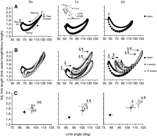
Kinematics of the hindlimb. (A) The adjusted limb length (i.e. quotient of limb length and femur length) plotted against limb angle for Ds, Lv and Us walking of intact rats (black bubbles). The locations of the hip, knee, ankle and MTP joints are shown in the inset in the Lv panel. Limb length (dot-dash line) and limb angle (dotted line) are the magnitude and direction, respectively, of the position vector between the hip and MTP joints. Limb length was calculated as the distance between the hip and MTP joint in every video frame. Limb angle was calculated as the angle between the treadmill belt and the calculated limb on the rostral side. The foot angle was calculated as the angle between the line intersecting the ankle and MTP joints and the treadmill belt on the caudal side. (B) Data from injured animals (gray bubbles) overlaid on the data from intact animals. Each trace forms a complete loop and is the average of seven rats walking at a treadmill speed of 11 m min–1. Each value is the inter-rat average. The size of the bubbles is proportional to the s.e.m. Bubble size is a general measure of variability about each data point and is not intended to be a precise depiction of the s.e.m. in either direction. The step cycle starts at paw contact and advances counter-clockwise, as indicated by dotted arrows above and beneath the object in A Ds. The upper portion of the object defined by these points is stance and the lower portion is swing. Circular black and white targets identify maximal adjusted femur lengths from intact rats where post-operative data obscure intact data. (C) The average (± s.e.m.) of these measures of hindlimb position from the data in B. Significant changes in average position after nerve injury (4 weeks and 10 weeks compared with Intact) were found for Lv and Us walking. 4 and 10 weeks were not significantly different from each other. There were no significant differences for Ds walking. *P<0.05 vs Ds (angle); †P<0.05 vs Lv and Us (length); ‡P<0.05 vs Ds and Lv (length); §P<0.05 vs intact (angle); ¶P<0.05 vs intact (length); NS, not significant.
Electromyography
Locomotor EMG activity was recorded from the Sol and TA muscles and synchronized to concurrently collected digitized video with a common pulse. Potentials were amplified (×1000) using a bandpass of 30–1000 Hz and then digitized for each 15-s locomotion trial using custom written LabView software. Frame numbers corresponding to the onset (paw contact) and termination (next contact) of selected step cycles were recorded from videos and then used to extract the corresponding EMG potentials. The extracted EMG potentials from individual step cycles were rectified and low-pass filtered with a time constant of 20 ms, and then time-normalized to 100 time bins. Therefore, all figures showing EMG activity have a time scale of 1–100.
For each rat, the amplitude of the EMG value for each time bin has been normalized to the maximum locomotor EMG value across all time bins from all step cycles recorded during a testing session (Gregor et al., 2006; Maas et al., 2010). This value always occurred during upslope walking and was always considerably larger than the vast majority of EMG values from other time bins. Thus, the magnitudes of the EMG profiles illustrated in Fig. 3 are considerably smaller than 100% on the ordinate. This scaling was consistent across all rats as noted by the small between-rat variance, which is also illustrated in Fig. 3. Average EMG activity during muscle activation was calculated as the average of such normalized EMG activity during the step cycle that was ≥5% of the maximum EMG activity recorded from each rat.
Fig. 3.
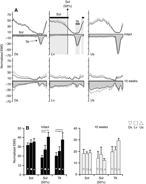
Muscle activity during walking at different slopes before and after injury. (A) Normalized electromyographic (EMG) activity recorded from the soleus (Sol) and tibialis anterior (TA) during downslope (Ds), level (Lv) and upslope (Us) treadmill locomotion in intact (N=8) and injured rats at 10 weeks following transection and surgical repair of the sciatic nerve (N=3). The Sol profiles are on the top (no fill) and the TA profiles are inverted (bottom) and shaded gray for clarity. The variance is illustrated as a corresponding gray tracing for both the Sol and TA (average + s.e.m.). (B) Selected components of normalized (as a percentage of the maximum activity recorded) Sol and TA EMG amplitudes were averaged and compared across slopes (Ds, inverted triangle; Lv, square; Us, triangle). Average overall EMG activity for the Sol was obtained during the portion of the step cycle indicated by the solid horizontal line and light gray shading in the top middle panel in A. These data are labeled Sol in B. The average Sol activity measured at the midpoint in the step cycle is labeled Sol (50%) and is indicated by the dotted line in A. Average overall EMG activity for the TA was obtained during the portion of the step cycle labeled TA with a double underline and shaded light gray in A.
Principal components analysis
Principal components analysis (PCA) was used to help evaluate differences in patterns of locomotor EMG activity between muscles (Sol and TA), slopes (downslope, level and upslope) and groups (intact, 4 weeks post-injury, 10 weeks post-injury). A total of 84 EMG profiles were incorporated into the data set (supplementary material Fig. S1). PCA is a data reduction method that uses structure detection to express two or more correlated variables with one or more uncorrelated variables that are termed principal components (Bishop, 1995). The proportion of overall variance in a data set that is explained by each principal component is termed its eigenvalue. To obtain the maximum variance explained by each principal component, eigenvalues were obtained using varimax normalization (Widmer et al., 2003). The first principal component necessarily counts for the largest proportion of the variance in the data (i.e. it has the largest eigenvalue). Each succeeding principal component explains the maximum amount of the remaining variance and is uncorrelated with previous principal components.
Principal component (factor) loading values resulting from PCA were used in subsequent statistical analysis. Factor loading values represent the correlation coefficient between the EMG activity profile and the regression line representing each principal component. They were used as a quantitative representation of the shape of the EMG profiles (i.e. one loading value for each principal component for each EMG profile).
Statistical analysis
Basic statistics were calculated for variables of interest, including those representing aspects of the step cycle, hindlimb kinematics, EMG magnitude and timing, and inter-slope variability of the Sol and TA. The data are reported as means ± s.e.m. unless otherwise noted. Analysis of variance and post hoc tests (Fisher's LSD) were used to statistically analyze the data. The significance level was set at 0.05 for all statistical tests.
RESULTS
The step cycle
The step cycle in this study was delineated by paw contact with the treadmill belt. Several temporal aspects of the step cycle were significantly affected by slope (Table 1). Step cycle duration was significantly longer during Us walking than Ds walking, but not Lv walking. The proportion of the step cycle consisting of stance increased and that consisting of swing decreased significantly with Us walking compared with Ds and Lv walking. The duration of stance was significantly longer for Us than Ds and Lv walking, and there was no change in swing duration with slope. It is apparent from the changes in stance period and swing duration that the increase in step cycle duration during Us walking came about solely from an increase in stance duration.
Table 1.
Average timing of the step cycle during slope walking
After transection and repair of the sciatic nerve, step cycle durations were still affected by slope. At 4 and 10 weeks post-injury, the duration of steps during Ds and Lv walking were significantly shorter than those during Us walking. At the 10-week time point, step duration during Ds and Lv walking were also significantly different from each other. The Us step cycle duration at 10 weeks was significantly longer than during Us walking in intact rats. Despite generally longer step cycles, stance phase durations were not significantly changed after injury when compared with stance durations of intact rats. The stance duration found during Us walking was significantly longer than that found for both Lv and Ds walking at both the 4- and 10-week sampling times. At 4 weeks, the stance phase durations associated with Ds and Lv walking were significantly different from one another. After injury, swing phase durations were significantly longer than in intact rats at both the 4- and 10-week sampling times. As in intact animals, there was no slope effect on swing phase duration in injured animals. Although some slope-related modulation of the timing of the step cycle attributable to the stance phase remains after sciatic nerve injury, more of the step cycle is composed of the swing phase after injury.
Hindlimb kinematics
Foot posture
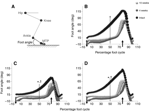
Several aspects of hindlimb movements differ significantly with slope. Changes in position of the foot are illustrated in Fig. 1B–D for intact rats (black symbols) and at 4 and 10 weeks following transection and repair of the sciatic nerve (gray symbols). At the 4-week sample time, muscles crossing the ankle joint are just beginning to receive innervation from regenerating axons. By 10 weeks after nerve transection and repair, muscle reinnervation is nearly complete (Hamilton et al., 2011). Following paw contact in intact rats, foot angle increases gradually through the stance phase and then decreases rapidly as the foot is removed from the treadmill belt. Although the contours of the traces in this figure are similar for all slopes, the Ds tracing is distinctly diminutive compared with the Lv and Us traces for intact rats. The foot angle at the mid-point of the step cycle (vertical dashed lines) was also computed. This measure is convenient to tabulate and is significantly smaller for Ds walking than Us or Lv walking for intact rats. A larger mid-step foot angle means that the muscles of the hindlimb have been used indirectly (as in the knee extensors) and/or directly (as in the ankle extensors) to produce sufficient ankle joint moments to advance the ankle forward with respect to the MTP joint.
Fig. 1.
Kinematics of the foot. (A) Position of the entire hindlimb at the beginning of stance (as a frame of reference). The foot angle is shown by the dotted line. The hip, knee, ankle and metatarsophalangeal (MTP) joints are also shown. (B–D) Values of the foot angle throughout the step cycle starting with paw contact are shown for intact (N=8) and injured rats at 4 and 10 weeks (N=3 for both injured groups) after sciatic nerve transection and repair for upslope (Us; B), downslope (Ds; C) and level (Lv; D) walking. Paw contact is indicated in B–D with a downward pointing arrow that crosses the x-axis. An upward pointing arrow indicates where the swing phase generally begins. A vertical dashed line indicates midway through the step cycle. Each value (bubble) is the inter-rat average. The size of the bubbles is proportional to the s.e.m. (i.e. larger bubbles indicate larger variance about the mean). The average foot angle at the midpoint of the step is significantly greater in Lv and Us walking compared with Ds walking for the intact group. For each slope the average intact foot angle at the line inset is significantly larger than the same position on the abscissa for both injury groups. *Slope effect, P<0.01 vs Us for intact group; †injury effect, P<0.05 vs intact for both injury groups.
Following injury to the sciatic nerve and subsequent reinnervation of the muscles crossing the ankle joint, the movements of the foot are quite different. The foot is placed flat on the treadmill belt at paw contact and held in that position until late in the stance phase, when it is lifted (Fig. 1B–D). Mid-step foot angle was significantly smaller for both groups of injured rats at both post-injury times and for each slope. Injured rats at 4 and 10 weeks were not significantly different from one another. Thus the greater muscle reinnervation found 10 weeks post-injury had no effect on mid-step foot position during locomotion.
Global limb vector
We used a global measure of the entire jointed hindlimb to study the kinematics of limb movements. This measure is a position vector linking the hip and MTP joints in a sagittal plane. The magnitude of this vector is termed limb length and its direction or orientation is termed limb angle. We expressed limb length as a proportion of the length of the femur to minimize inter-animal and inter-trial variations attributable to size differences. Femur length was measured from video images. This new variable is referred to as ‘adjusted limb length’ in figures and legends (see inset in Fig. 2A, Lv). When the adjusted limb length is plotted against limb angle across the step cycle, an enclosed parabolic object is described as found in Fig. 2A,B. The data constituting this object advance in time in a counter-clockwise manner. The right- and left-most inflection points roughly correspond with the onset of stance (paw contact) and swing (paw off), respectively. The limb shortens, or yields, initially during stance and then lengthens towards the conclusion of stance as it propels the animal forward. The limb shortens again at the onset of the swing phase to clear the treadmill belt and reposition the foot in preparation for the next bout of ground support. The latter part of swing consists of lengthening as the limb reaches to the rostral position of belt contact.
Although the overall shape of this enclosed parabola is similar at different slopes, there are some differences: (1) its overall position in this global hindlimb space changes, (2) it rotates and (3) it becomes markedly skewed with slope. The average limb angle (abscissa) and adjusted limb length (ordinate) were computed to evaluate the overall position of the object in global hindlimb space at different slopes (Fig. 2C). During Us walking, average limb angle was shifted significantly leftward, towards smaller limb angles. Average adjusted limb length during Ds walking was significantly longer compared with that found during Lv walking. No significant changes in average adjusted limb length were found between Us and Ds walking.
The object defined by the limb angle–adjusted limb length relationship is rotated clockwise during Us walking and counter-clockwise with Ds walking. The rotation is due to opposite changes in limb length at the right- and left-most inflection points on the object. Adjusted limb length is significantly longer at paw contact during Ds walking, compared with Lv and Us, and significantly increased at paw off during Us walking compared with Lv and Ds. Limb angle at paw contact was similar at all slopes studied, whereas limb angle significantly decreased at paw off during upslope compared with downslope walking.
With only two exceptions, there were no significant differences between intact and injured rats for Ds walking for any of these measures. The exceptions were limb angle at paw contact and adjusted limb length at paw off. In contrast, significant changes in all three measures of adjusted limb length and limb angle were noted for Lv and Us walking at both 4 and 10 weeks following sciatic nerve transection and repair (Fig. 2). No significant slope differences were found between the two times studied, despite the more extensive muscle reinnervation found at 10 weeks. Both limb angle and adjusted limb length are conserved remarkably after sciatic nerve injury during Ds walking but not Lv and Us walking. This conservation is achieved despite the marked changes in foot posture during locomotion described above. Presumed adaptive changes in hindlimb use with slope walking found in intact rats were not present in lesioned animals.
Patterns of locomotor muscle activity
The average normalized Sol and TA EMG activity for all intact rats and rats 10 weeks after sciatic nerve transection and repair is shown in Fig. 3A. The timing of activity in these two muscles is reciprocal in intact rats. Activity in TA begins near the time of paw off and is found during the swing phase. Activity in Sol begins in late swing and continues for most of the stance phase. In reference to the step cycle subdivisions originally defined by Philippson and later popularized by Goslow and colleagues, EMG activity in the TA begins during the F epoch of the step cycle and tapers off during the E1 epoch (Philippson, 1905), and is quiescent during the E2 and E3 epochs. Soleus EMG activity begins during the E1 epoch and persists throughout the E2 and E3 epochs (Philippson, 1905; Goslow et al., 1973). Following sciatic nerve transection and repair and muscle reinnervation, locomotor EMG activity in TA and Sol is no longer reciprocal, but appears as co-activation (Fig. 3A, 10 weeks).
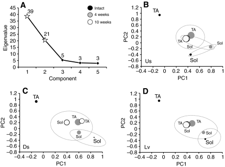
We evaluated the significance of any slope-related differences in muscle activation pattern using PCA. The average locomotor EMG profile for each rat was incorporated in the data set and subjected to PCA (see supplementary material Fig. S1). The results of this analysis for intact rats are shown in Fig. 4. The first two principal components explained 75.7% of the variance in the data set. The first principal component (PC1) alone explained 49.4% of the variance. Both PCs had eigenvalues that were much greater than 1.0 (38.5 and 20.5; Fig. 5A), which means that each of them alone, especially PC1, explained a much larger proportion of overall variance than any single raw variable. The eigenvalue for PC3 fell sharply at 5.3, and explained only 6.8% of the variance in the data set. Therefore, we only considered PC1 and PC2 in further analysis. Factor loadings, the correlation of eigenvalues to the original data set, were calculated for each PC for each muscle, for each slope condition, in intact rats and rats at 4- and 10-week survival times. Significance of the differences in factor loadings between the different groups was determined using ANOVA and post hoc paired testing.
Fig. 4.
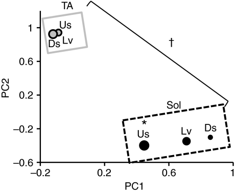
Profile analysis of distal hindlimb muscle activity from intact rats. Average loading values for principal components one and two (PC1 and PC2) are illustrated as bubble plots. Symbol size is proportional to the s.e.m. (i.e. larger bubbles indicate larger variance about the mean). All intra-slope comparisons of Sol vs TA were significantly different on PC1 or PC2, or both (data enclosed by gray line vs data enclosed by dotted line). Upslope PC1 loadings were significantly different from Lv and Ds PC1 loadings for the Sol. PC loadings did change with slope for the TA. *P<0.05 vs Lv and Ds; †P<0.05, TA vs Sol, PC1 and PC2.
Fig. 5.
Profile analysis of distal hindlimb muscle activity from intact and injured rats. (A) A scree plot showing the eigenvalues associated with the first five principal components. Eigenvalues for the first two principal components are represented by stars. (B–D) Average loading factor values for PC1 and PC2 illustrated as bubble plots for Us, Ds and Lv walking. Symbol size is proportional to the s.e.m. (i.e. larger bubbles indicate larger variance about the mean). Black, gray and white bubbles represent data from intact rats, rats at 4 weeks post-injury and rats at 10 weeks post-injury, respectively. Dotted lines enclose sets of bubbles that are not significantly different from one another.
In intact rats, factor loadings for PC1 were greatest for Sol and factor loadings for PC2 were greatest for TA. This suggests that the Sol pattern of activity is most influential in the data set because PC1 explains the majority of the variance in the data set. For PC1, mean factor loadings for Sol from Ds and Lv walking were significantly different from Us walking, but not from each other. We interpret this outcome as evidence that the pattern of EMG activity in Sol in intact rats is significantly different during Us walking, but not Ds and Lv walking.
The average TA loading values for PC1 and PC2 were highly conserved across slopes, and values from different slopes did not differ significantly (Fig. 4). We conclude from these data that, in intact animals, the patterns of EMG activity from the TA do not change with slope. All average TA loading values for PC1 and PC2 were significantly different from all average Sol loading values for PC1 and PC2 (P≤0.05). We believe that this finding is consistent with the highly reciprocal pattern of activation of these two muscles in intact rats.
Average factor loadings resulting from PCA analysis that included data from rats at 4 and 10 weeks following sciatic nerve transection and repair are shown in Fig. 5. A scree plot that shows eigenvalues for the first five principal components is shown in Fig. 5A. Because the third PC is so much smaller than the first two, factor loadings were studied only in association with the first two factors (Fig. 5A, stars). Mean factor loadings for PC1 and PC2 for Lv, Us and Ds walking are shown in Fig. 5B–D. No significant effect of slope on any post-injury EMG activity profiles was found. For each of the slopes and at both survival times studied, the mean factor loading from the intact TA is significantly different from all other means, for both PC1 and PC2. For all slopes, the mean factor loading from the intact Sol is significantly different from the means for TA at all times and from that for Sol at 10 weeks, but not from Sol at 4 weeks. Thus the pattern of activation of TA during walking in post-injury rats is significantly different from that of the intact rats at the earliest time of muscle reinnervation, and this novel pattern persists for at least the next 6 weeks. A novel activity pattern in Sol evolves more gradually and is not evident until 10 weeks after injury. For all slopes and at all times, factor loadings for the reinnervated TA and Sol muscles are not significantly different from each other. We interpret this finding to mean that the novel and persistent pattern of activity found after sciatic nerve transection and repair and subsequent muscle reinnervation is a co-activation of these functional antagonists.
Intensity of locomotor EMG activity
When expressed as a proportion of the entire burst of activity, the amplitude of the rectified and integrated EMG activity increases significantly with slope in TA but not Sol in intact rats (Fig. 3B). However, the period of the Sol EMG activity constitutes a much larger portion of the step cycle than does the TA EMG activity. Most of the EMG activity in Sol occurs early in the stance phase during Ds walking but more intense EMG activity occurs later in the step cycle with increasingly positive slope (Fig. 3A). This observation prompted us to measure the intensity of Sol EMG activity at the mid-point in the step cycle, as described above for foot angle. Average values of this mid-step Sol activity were significantly increased during Lv and Us walking compared with Ds in intact rats [Fig. 3B, Intact: Sol (50%)]. Thus the intensity of activation of TA and Sol each vary significantly with slope. In the 4-week (data not shown) and 10-week post-injury groups (Fig. 3B) there were no significant differences with slope in any of these measures of EMG activity. We interpret this finding as evidence that the modulation of TA and Sol EMG intensity with slope is lost in the reinnervated muscles.
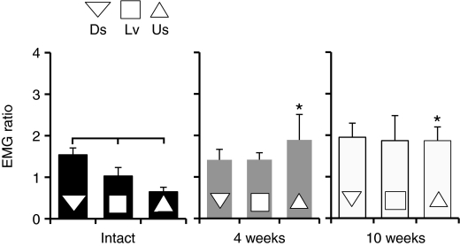
The amount of EMG activity in the Sol that occurs just before paw contact (i.e. during the E1 epoch), during the latter swing phase while the limb is still suspended and its path is unobstructed, has been used as an index of central control of Sol muscle activity (Gorassini et al., 1994; Maas et al., 2010) because autogenic afferent modulation is not active while the paw is not in contact with the walking surface (Pearson and Collins, 1993; Prochazka et al., 1989). We thought that a decline or increase in pre-contact EMG activity that occurs as part of a general trend of increased or decreased EMG activity, respectively, would have a tenuous association with central control of muscle activity. Therefore, to overcome this drawback we expressed the average pre-contact EMG intensity as a function of the mean Sol EMG intensity at the mid-step. The pre-contact EMG activity ratio thus expresses pre-contact EMG activity as a proportion of the peak EMG amplitude during the stance phase. A decline in this metric (as reported below for increasing slope) could result, hypothetically, from a decline in pre-contact EMG intensity, an increase in EMG activity at the mid-step, or both. However, we would argue that any of these three possibilities would result in a decline in preparatory motor output for the impending stance phase.
The EMG ratios are shown for locomotion at different slopes in all three groups of rats in Fig. 6. All slope comparisons were significantly different for intact rats (P<0.05). There was a progressive decrease in the pre-contact EMG ratio in Sol with increasing slope. After injury, EMG ratios did not change with slope. For Us walking the EMG ratios at 4 and 10 weeks were significantly larger than for intact rats (P≤0.01). We interpret these results to mean that central control of Sol muscle activation declines with increasing slope in intact rats and is elevated at all slopes after sciatic nerve transection. This may mean that modulation of Sol EMG activity is more dependent on central control after sciatic nerve transection.
Fig. 6.
Pre-contact Sol muscle activation ratio. The EMG ratio was calculated as the EMG activity before paw on divided by EMG activity at the midstep. The average EMG ratio (+s.e.m.) is shown for all slopes for intact and post-injury rats at 4 weeks and 10 weeks. The bracket indicates P<0.05, for all slope comparisons; *P<0.05 vs intact at same slope.
DISCUSSION
Several comprehensive studies of cats inform much of our current understanding of the neural control of walking on slopes (Carlson-Kuhta et al., 1998; Donelan et al., 2009; Gregor et al., 2006; Smith et al., 1998). Hindlimb kinematics and muscle EMG activity have also been studied in rats during level (Thota et al., 2005) and upslope walking (Hodson-Tole and Wakeling, 2008; Roy et al., 1991). What has emerged from these studies is that up and downslope locomotion pose opposite challenges to the sensorimotor system that are met by mechanisms specific to each direction of slope. We believe that slope walking could be a useful model with which to study functional recovery following injuries to peripheral nerves.
Adaptive changes during locomotion on slopes
One of the main findings of this study is that changes in both the movements of the hindlimb and Sol and TA activation patterns occur when rats walk up or down slopes. The pattern of activation of TA does not change with slope, but significant increases in TA EMG amplitudes were found as slope increased, similar to results reported for cats during slope walking (Carlson-Kuhta et al., 1998; Smith et al., 1998). These changes are highly correlated with the mechanical demands of upslope walking. Ankle flexion excursions during the swing phase are greater with increasingly positive slope (Carlson-Kuhta et al., 1998) and our finding that swing duration decreases during Us walking means that these movements need to be generated faster.
Both the pattern and the amplitude of EMG activity in Sol change with slope, and similar profiles of ankle-extensor force production (Gabaldon et al., 2004) and ground reaction forces (Gregor et al., 2006) have been reported for slope-walking in cats. As has been shown in the cat (Carlson-Kuhta et al., 1998; Fowler et al., 1993; Gregor et al., 2006; Kaya et al., 2003; Smith et al., 1998), these changes in the Sol activity pattern are related to the requirements for propulsion at different slopes. Thus, slope-related changes in EMG activity in both TA and Sol could be considered adaptations to the demands of slope walking in rats.
Slope-related changes in hindlimb kinematics were largely opposite during upslope compared with downslope walking. The objects formed by the adjusted limb length-angle plots are skewed towards increased limb length at paw off during Us walking and toward increased limb length at paw contact with Ds walking. Longer step cycles were found during Us walking and this increase is almost entirely attributable to an increased stance duration. Decreased limb angles at paw off during upslope walking reflect a significant amount of rotation of the hip about the foot. The effects of the positive slope used in this study were internally consistent and similar to the patterns of hindlimb kinematics reported in cats (Smith and Carlson-Kuhta, 1995).
During Ds walking, an increase in the extension of the limb in the latter portion of the swing phase is necessary to reach for the low contact position on the treadmill. This swing phase extension is aided by gravity and largely passive. The similarity of limb angles at paw contact and paw off during downslope walking suggest that there was less rotation of the foot about the hip during the stance phase than during Us walking. In contrast, there was a great deal of limb shortening during the stance phase of Ds walking. These changes in hindlimb movements are consistent with diminished propulsive requirements for Ds walking (Smith et al., 1998) and with diminished EMG activity in the Sol during the latter portion of the EMG burst found in this study. Thus, changes in the movements of the hindlimb during Us and Ds walking relative to Lv walking could be considered adaptive to the demands imposed by those tasks.
Slope walking in response to sciatic nerve injury
The changes in muscle activation patterns and hindlimb kinematics with slope were used as a standard against which to study functional recovery following sciatic nerve transection and repair. Our main finding was that fundamental differences in both the timing of activation of the TA and Sol muscles and foot and hindlimb movements occur following muscle reinnervation, but changes in these novel patterns that might adapt the animal for slope walking are limited.
Functionally antagonistic flexor and extensor muscles about a joint are generally activated reciprocally during rhythmic behaviors such as locomotion, and this was true for the intact rats tested in this study, even across slopes. After transection and repair of the sciatic nerve, the timing of locomotor EMG activity in the reinnervated Sol and TA muscles shifted from reciprocal to co-active. The change was nearly immediate for the TA muscle, but took longer for Sol. We think that this shift reflects a deliberate co-activation to increase ankle joint stiffness during the stance phase (Enoka, 2008), and is not the result of a loss of myospecificity resulting from misdirection of regenerating axons to reinnervate functionally inappropriate muscle targets because a similar co-activation results when the extent of axon misdirection during regeneration is either decreased (Sabatier et al., in press) or increased (Hamilton et al., 2011). We view the co-activation of these functional antagonists as part of a novel locomotor strategy that is adopted by the rats in response to the sciatic nerve lesion. The absence of any change in either the pattern or the amplitude of activity with slope is consistent with such an interpretation.
Following recovery from sciatic nerve injury there is a nearly complete conservation of hindlimb kinematics during Ds walking. Both hindlimb length and orientation are not significantly different from those of intact rats during Ds locomotion. Since the posture of the foot, especially during stance, is markedly different in these animals, this conservation must mean that changes in the movements at the knee and hip joints, and perhaps in movements of the axial skeleton, compensate remarkably. A similar observation was made for Lv walking following smaller peripheral nerve injuries in cats (Chang et al., 2009), and it was this conservation of global limb movements that prompted us to use this approach in rats.
However, adaptive changes in these movements of the hindlimb during Lv and Us walking, such as those found in intact rats, are not found after injury. The animals use a strategy for limb movements that is the same for all slopes and is similar to that used during downslope walking. The net effect of this inability to change hindlimb movements with slope is that movements are less like those found in intact rats as slope increases. The basis for this limited ability to adapt to the demands of slope walking is not yet clear. Following recovery from a peripheral nerve injury, a new relationship between neural circuitry and the musculoskeletal system exists (Alvarez et al., 2010). We think that the similarity of both hindlimb movements and EMG activity at different slopes following recovery from sciatic nerve injury is a reflection of such a new relationship. If the fundamental changes observed are evidence of the adoption of a novel locomotor strategy, then they reflect changes in the outputs of spinal circuitry that have occurred in response to the injury. The simplest explanation of the inability to adapt these changes to the demands of slope walking would be a limited ability for integration of afferent feedback following peripheral nerve injury (Alvarez et al., 2010; Cope et al., 1994; Haftel et al., 2005). Until the nature of the proprioceptive feedback from reinnervated muscles during slope walking can be established and until the effects of such feedback on neural circuits in the CNS can be studied, such explanations must remain speculative.
Supplementary Material
A.W.E., B.N.T., J.N. and M.J.S. were funded by HD32571 and HD053669. M.J.S. was also funded by a USPHS NIH Institutional Research and Academic Career Development grant (K12 GM00680-05). Deposited in PMC for release after 12 months.
Supplementary material available online at http://jeb.biologists.org/cgi/content/full/214/6/1007/DC1
REFERENCES
- Al-Majed A. A., Neumann C. M., Brushart T. M., Gordon T. (2000). Brief electrical stimulation promotes the speed and accuracy of motor axonal regeneration. J. Neurosci. 20, 2602-2608 [DOI] [PMC free article] [PubMed] [Google Scholar]
- Alvarez F. J., Bullinger K. L., Titus H. E., Nardelli P., Cope T. C. (2010). Permanent reorganization of Ia afferent synapses on motoneurons after peripheral nerve injuries. Ann. NY Acad. Sci. 1198, 231-241 [DOI] [PMC free article] [PubMed] [Google Scholar]
- Auyang A. G., Yen J. T., Chang Y. H. (2009). Neuromechanical stabilization of leg length and orientation through interjoint compensation during human hopping. Exp. Brain Res. 192, 253-264 [DOI] [PubMed] [Google Scholar]
- Bishop C. (1995). Neural Networks for Pattern Recognition. Oxford: Oxford University Press; [Google Scholar]
- Bosco G., Poppele R. E. (2000). Reference frames for spinal proprioception: kinematics based or kinetics based? J. Neurophysiol. 83, 2946-2955 [DOI] [PubMed] [Google Scholar]
- Carlson-Kuhta P., Trank T. V., Smith J. L. (1998). Forms of forward quadrupedal locomotion. II. A comparison of posture, hindlimb kinematics, and motor patterns for upslope and level walking. J. Neurophysiol. 79, 1687-1701 [DOI] [PubMed] [Google Scholar]
- Chang Y. H., Auyang A. G., Scholz J. P., Nichols T. R. (2009). Whole limb kinematics are preferentially conserved over individual joint kinematics after peripheral nerve injury. J. Exp. Biol. 212, 3511-3521 [DOI] [PMC free article] [PubMed] [Google Scholar]
- Cope T. C., Bonasera S. J., Nichols T. R. (1994). Reinnervated muscles fail to produce stretch reflexes. J. Neurophysiol. 71, 817-820 [DOI] [PubMed] [Google Scholar]
- Daley M. A., Felix G., Biewener A. A. (2007). Running stability is enhanced by a proximo-distal gradient in joint neuromechanical control. J. Exp. Biol. 210, 383-394 [DOI] [PMC free article] [PubMed] [Google Scholar]
- Donelan J. M., McVea D. A., Pearson K. G. (2009). Force regulation of ankle extensor muscle activity in freely walking cats. J. Neurophysiol. 101, 360-371 [DOI] [PubMed] [Google Scholar]
- English A. W. (2005). Enhancing axon regeneration in peripheral nerves also increases functionally inappropriate reinnervation of targets. J. Comp. Neurol. 490, 427-441 [DOI] [PubMed] [Google Scholar]
- English A. W., Schwartz G., Meador W., Sabatier M. J., Mulligan A. (2007). Electrical stimulation promotes peripheral axon regeneration by enhanced neuronal neurotrophin signaling. Dev. Neurobiol. 67, 158-172 [DOI] [PMC free article] [PubMed] [Google Scholar]
- Enoka R. M. (2008). Neuromechanics of Human Movement. Champaign, IL: Human Kinetics; [Google Scholar]
- Fowler E. G., Gregor R. J., Hodgson J. A., Roy R. R. (1993). Relationship between ankle muscle and joint kinetics during the stance phase of locomotion in the cat. J. Biomech. 26, 465-483 [DOI] [PubMed] [Google Scholar]
- Frostick S. P., Yin Q., Kemp G. J. (1998). Schwann cells, neurotrophic factors, and peripheral nerve regeneration. Microsurgery 18, 397-405 [DOI] [PubMed] [Google Scholar]
- Gabaldon A. M., Nelson F. E., Roberts T. J. (2004). Mechanical function of two ankle extensors in wild turkeys: shifts from energy production to energy absorption during incline versus decline running. J. Exp. Biol. 207, 2277-2288 [DOI] [PubMed] [Google Scholar]
- Gorassini M. A., Prochazka A., Hiebert G. W., Gauthier M. J. (1994). Corrective responses to loss of ground support during walking. I. Intact cats. J. Neurophysiol. 71, 603-610 [DOI] [PubMed] [Google Scholar]
- Goslow G. E., Jr, Reinking R. M., Stuart D. G. (1973). The cat step cycle: hind limb joint angles and muscle lengths during unrestrained locomotion. J. Morphol. 141, 1-41 [DOI] [PubMed] [Google Scholar]
- Gregor R. J., Smith D. W., Prilutsky B. I. (2006). Mechanics of slope walking in the cat: quantification of muscle load, length change, and ankle extensor EMG patterns. J. Neurophysiol. 95, 1397-1409 [DOI] [PubMed] [Google Scholar]
- Groves M. L., McKeon R., Werner E., Nagarsheth M., Meador W., English A. W. (2005). Axon regeneration in peripheral nerves is enhanced by proteoglycan degradation. Exp. Neurol. 195, 278-292 [DOI] [PubMed] [Google Scholar]
- Haftel V. K., Bichler E. K., Wang Q. B., Prather J. F., Pinter M. J., Cope T. C. (2005). Central suppression of regenerated proprioceptive afferents. J. Neurosci. 25, 4733-4742 [DOI] [PMC free article] [PubMed] [Google Scholar]
- Hamilton S. K., Hinkle M. L., Nicolini J., Rambo L. N., Rexwinkle A. M., Rose S. J., Sabatier M. J., Backus D., English A. W. (2011). Misdirection of regenerating axons and functional recovery following sciatic nerve injury in rats. J. Comp. Neurol. 519, 21-33 [DOI] [PMC free article] [PubMed] [Google Scholar]
- Hodson-Tole E. F., Wakeling J. M. (2008). Motor unit recruitment patterns 1, responses to changes in locomotor velocity and incline. J. Exp. Biol. 211, 1882-1892 [DOI] [PubMed] [Google Scholar]
- Kaya M., Leonard T., Herzog W. (2003). Coordination of medial gastrocnemius and soleus forces during cat locomotion. J. Exp. Biol. 206, 3645-3655 [DOI] [PubMed] [Google Scholar]
- Maas H., Gregor R. J., Hodson-Tole E. F., Farrell B. J., English A. W., Prilutsky B. I. (2010). Locomotor changes in length and EMG activity of feline medial gastrocnemius muscle following paralysis of two synergists. Exp. Brain Res. 203, 681-692 [DOI] [PMC free article] [PubMed] [Google Scholar]
- MacGillivray T. E. (2003). Fibrin sealants and glues. J. Card. Surg. 18, 480-485 [DOI] [PubMed] [Google Scholar]
- Menovsky T., Beek J. F. (2001). Laser, fibrin glue, or suture repair of peripheral nerves: a comparative functional, histological, and morphometric study in the rat sciatic nerve. J. Neurosurg. 95, 694-699 [DOI] [PubMed] [Google Scholar]
- Pearson K. G. (2000). Neural adaptation in the generation of rhythmic behavior. Annu. Rev. Physiol. 62, 723-753 [DOI] [PubMed] [Google Scholar]
- Pearson K. G., Collins D. F. (1993). Reversal of the influence of group Ib afferents from plantaris on activity in medial gastrocnemius muscle during locomotor activity. J. Neurophysiol. 70, 1009-1017 [DOI] [PubMed] [Google Scholar]
- Philippson M. (1905). L’autonomie et la centralisation dans le système nerveux des animaux. Trav. Lab. Physiol. Inst. Solvey 7, 1-208 [Google Scholar]
- Poppele R. E., Bosco G., Rankin A. M. (2002). Independent representations of limb axis length and orientation in spinocerebellar response components. J. Neurophysiol. 87, 409-422 [DOI] [PubMed] [Google Scholar]
- Prochazka A., Trend P., Hulliger M., Vincent S. (1989). Ensemble proprioceptive activity in the cat step cycle: towards a representative look-up chart. Prog. Brain Res. 80, 61-74; discussion 57-60 [DOI] [PubMed] [Google Scholar]
- Roy R. R., Hutchison D. L., Pierotti D. J., Hodgson J. A., Edgerton V. R. (1991). EMG patterns of rat ankle extensors and flexors during treadmill locomotion and swimming. J. Appl. Physiol. 70, 2522-2529 [DOI] [PubMed] [Google Scholar]
- Sabatier M. J., Redmon N., Schwartz G., English A. W. (2008). Treadmill training promotes axon regeneration in injured peripheral nerves. Exp. Neurol. 211, 489-493 [DOI] [PMC free article] [PubMed] [Google Scholar]
- Sabatier M. J., To B. N., Nicolini J., English A. W. (in press). Effect of axon misdirection on recovery of EMG activity and kinematics after peripheral nerve injury. Cells Tissues Organs. [DOI] [PMC free article] [PubMed] [Google Scholar]
- Scholz T., Krichevsky A., Sumarto A., Jaffurs D., Wirth G. A., Paydar K., Evans G. R. (2009). Peripheral nerve injuries: an international survey of current treatments and future perspectives. J. Reconstr. Microsurg. 25, 339-344 [DOI] [PubMed] [Google Scholar]
- Smith J. L., Carlson-Kuhta P. (1995). Unexpected motor patterns for hindlimb muscles during slope walking in the cat. J. Neurophysiol. 74, 2211-2215 [DOI] [PubMed] [Google Scholar]
- Smith J. L., Carlson-Kuhta P., Trank T. V. (1998). Forms of forward quadrupedal locomotion. III. A comparison of posture, hindlimb kinematics, and motor patterns for downslope and level walking. J. Neurophysiol. 79, 1702-1716 [DOI] [PubMed] [Google Scholar]
- Thota A. K., Watson S. C., Knapp E., Thompson B., Jung R. (2005). Neuromechanical control of locomotion in the rat. J. Neurotrauma 22, 442-465 [DOI] [PubMed] [Google Scholar]
- Varejao A. S., Melo-Pinto P., Meek M. F., Filipe V. M., Bulas-Cruz J. (2004). Methods for the experimental functional assessment of rat sciatic nerve regeneration. Neurol. Res. 26, 186-194 [DOI] [PubMed] [Google Scholar]
- Widmer C. G., Carrasco D. I., English A. W. (2003). Differential activation of neuromuscular compartments in the rabbit masseter muscle during different oral behaviors. Exp. Brain Res. 150, 297-307 [DOI] [PubMed] [Google Scholar]
- Yakovenko S., Mushahwar V., VanderHorst V., Holstege G., Prochazka A. (2002). Spatiotemporal activation of lumbosacral motoneurons in the locomotor step cycle. J. Neurophysiol. 87, 1542-1553 [DOI] [PubMed] [Google Scholar]
Associated Data
This section collects any data citations, data availability statements, or supplementary materials included in this article.


