Abstract
ABL gene translocations create constitutively active tyrosine kinases that are causative in chronic myeloid leukemia, acute lymphocytic leukemia and other hematopoietic malignancies. Consistent retention of ABL SH3/SH2 autoinhibitory domains, however, suggests that these leukemogenic tyrosine kinase fusion proteins remain subject to regulation. We resolve this paradox, demonstrating that BCR-ABL1 kinase activity is regulated by RIN1, an ABL SH3/SH2 binding protein. BCR-ABL1 activity was increased by RIN1 overexpression and decreased by RIN1 silencing. Moreover, Rin1−/− bone marrow cells were not transformed by BCR-ABL1, ETV6-ABL1 or BCR-ABL1T315I, a patient-derived drug-resistant mutant, as judged by growth factor independence. Rescue by ectopic RIN1 verified a cell autonomous mechanism of collaboration with BCR-ABL1 during transformation. Sensitivity to the ABL kinase inhibitor imatinib was increased by RIN1 silencing, consistent with RIN1 stabilization of an activated BCR-ABL1 conformation having reduced drug affinity. The dependence on activation by RIN1 to unleash full catalytic and cell transformation potential reveals a previously unknown vulnerability that could be exploited for treatment of leukemic cases driven by ABL translocations. The findings suggest that RIN1 targeting could be efficacious for imatinib-resistant disease and might complement ABL kinase inhibitors in first-line therapy.
Keywords: BCR-ABL1, RIN1, Ras, CML, imatinib, resistance
Introduction
Chromosome translocations that join the BCR and ABL1 (a. k.a. c-Abl) genes give rise to BCR-ABL1 fusion proteins that are causative in virtually all cases of chronic myeloid leukemia (CML), in many cases of acute lymphocytic leukemia (ALL) and occasionally in other myeloproliferative disorders (reviewed in1). In addition, ETV6 (a.k.a. TEL) forms fusion oncogenes with ABL12 and the closely related ABL2 (a.k.a. Arg)3 in some leukemias. ABL proteins are non-receptor tyrosine kinases that are normally under tight regulation, but BCR-ABL1 fusions are constitutively active. The ABL kinase inhibitor imatinib mesylate (a.k.a. STI571 or Gleevec) is a highly effective treatment for CML (reviewed in4), demonstrating that drugs directly targeted to oncoproteins can be used to manage cancer and perhaps eventually be part of a curative therapy.
Unfortunately, some leukemias with activated ABL oncoproteins do not respond to imatinib. For CML patients who do respond, there is a significant risk of developing resistance because of strong selective pressure for BCR-ABL1 kinase domain mutations that block inhibitor action but retain the catalytic function of the oncoprotein.5 Second-generation kinase inhibitors offer hope for combating imatinib resistance, with some drugs successfully targeting the highly refractory BCR-ABL1T315I mutant.6 But even these new catalytic site inhibitors have limitations in their effectiveness during accelerated and blast-phase CML, as well as in the treatment of other ABL fusion leukemias including ALL. In addition, compound mutations following sequential treatment of CML patients with multiple kinase inhibitors7 provide a path to broad resistance. Some attempts have been made to circumvent resistance by reducing BCR-ABL1 expression8, 9 or stability,10, 11 or by targeting collaborative signaling pathways.12, 13, 14, 15 A more direct approach for improving treatment would be to maintain focus on reducing tyrosine kinase activity by targeting oncogenic ABL outside the catalytic site.
ABL tyrosine kinases function in the cytoplasm to coordinate actin remodeling, a function mediated by carboxy terminal filamentous actin binding and bundling domains, and by the tyrosine phosphorylation of multiple actin remodeling regulator proteins. ABL1 also has nuclear DNA damage response functions mediated by a DNA-binding domain and targeted tyrosine phosphorylation. ABL activity is normally regulated at multiple levels. An amino terminal myristoyl group can attach to a surface pocket in the kinase domain, contributing to an autoinhibitory fold,16, 17 and a short amino terminal ‘cap' peptide further stabilizes an inactive conformation through additional surface interactions. Downstream of this peptide are SH3 and SH2 domains that cradle the kinase domain and contribute to the adoption of a less-active enzyme conformation.18 In addition, several tyrosines in and around the ABL kinase domain can be phosphorylated in trans (by ABL itself and by SRC family kinases), leading to increased catalytic activity.19, 20, 21, 22 It appears that each form of regulation is conserved between ABL1 and ABL2, which are more than 90% identical throughout their SH3, SH2 and kinase domains.
Chromosome translocations that give rise to BCR-ABL1 and other ABL1 fusion oncogenes remove the first coding exon of ABL1. This eliminates both the myristoylation site and the amino terminal ‘cap' that participate in stabilizing the inactive conformation, explaining in part the elevated and constitutive kinase activity of the fusion protein. The same ABL breakpoint is seen regardless of the BCR breakpoint, which is variable. Even with fusion partners other than BCR, the ABL1 breakpoint resides between the alternate first exon (1b) and the second exon (2a).23 Moreover, oncogenic translocations involving ABL2, although less common, show the same arrangement.3 In summary, except for extremely rare variants (reviewed in24), human ABL fusion oncoproteins are devoid of the autoinhibitory cap peptide, but consistently retain ABL SH3 and SH2 domains that provide a separate autoinhibitory function likely to limit kinase activity.25 In the murine retroviral v-Abl oncoprotein, by contrast, the Abl1 SH3 domain is disrupted by fusion with viral Gag sequences. As a result, v-Abl is a highly active kinase and v-Abl is a more potent transforming gene than BCR-ABL1.26
RIN1 is a RAS effector protein that binds to and activates ABL tyrosine kinases.27, 28 Signaling is initiated by low-affinity binding of a proline-rich sequence on RIN1 to the SH3 domain of ABL. This interaction leads to phosphorylation of RIN1 on tyrosine 36, which subsequently associates with the ABL SH2 domain. The resulting stable divalent interaction (RIN1 proline-rich motif and phospho-Tyr36 bound to ABL SH3 and SH2 domains, respectively) relieves the ABL autoinhibitory fold and leads to activation of the ABL kinase through enhanced catalytic efficiency.27 Both ABL1 and ABL2 are activated by RIN1, and this requires only the ABL SH3, SH2 and kinase domains. Activation by RIN1 is independent of ABL transphosphorylation and is unaffected by an imatinib resistance mutation.27 Silencing of RIN1 results in less tyrosine phosphorylation on CRKL, one of the best characterized ABL substrates, and deletion of the mouse Rin1 (mRin1) gene causes reduction in basal levels of phospho-CRKL.28 These observations demonstrate that RIN1 directly stimulates the tyrosine kinase activity of ABL proteins and is required for maintaining normal ABL kinase activity in vivo.
We showed previously that RIN1 interacts with BCR-ABL1 and that overexpression of RIN1 promotes the transforming ability of BCR-ABL1 in hematopoietic cells and can rescue low transformation efficiency BCR-ABL1 mutants.29 Moreover, RIN1 significantly enhanced the leukemogenic properties of BCR-ABL1 in a murine model system.29 These findings suggest that the ‘constitutively active' BCR-ABL1 oncoprotein remains responsive to positive regulation by RIN1. Here we show that BCR-ABL1-positive leukemia cell phosphotyrosine levels are increased by RIN1 overexpression, providing an explanation for leukemogenic enhancement by RIN1. Deletion of RIN1 blocked transformation of bone marrow cells by multiple ABL fusion oncogenes, including the kinase inhibitor-resistant mutant BCR-ABL1T315I, demonstrating the cell-autonomous dependence of ABL oncoproteins on RIN1. Silencing of RIN1 in a BCR-ABL1-positive leukemia cell line and primary ALL cells decreased levels of cellular phosphotyrosine, and at the same time increased sensitivity to imatinib. Our results demonstrate a vital role for the physiological ABL regulator RIN1 in transformation by BCR-ABL1, while uncovering a novel point of vulnerability that could be exploited for treatment of leukemias driven by ABL tyrosine kinase oncoproteins.
Materials and methods
Expression constructs
The MSCV retrovirus constructs expressing BCR-ABL1, BCR-ABL1+RIN1ABD, ETV6-ABL1 and v-Abl have been previously described.29 BCR-ABL1+RIN1ABDmut was created by replacing the EcoRI-flanked fragment of wild-type (WT) RIN1ABD with the equivalent fragment from RIN1ABDmut (also called ABD28, 29). Note that RIN1ABD was used instead of full-length RIN1 to avoid the retrovirus size limit. The BCR-ABL1T315I mutant retrovirus was a generous gift from Charles Sawyers (Memorial Sloan Kettering Cancer Center, New York, NY, USA). The RIN1-silencing short hairpin RNA (shRNA) lentivirus in the pLKO.1-puro vector and the scramble control lentivirus pLKO.1SCR-puro contain a puromycin resistance gene and were obtained from Sigma Aldrich (St Louis, MO, USA) (Mission shRNAs SHGLY-NM_004292 and SHC002, respectively). The stability of RIN1 silencing in transduced TOM-1 cells was validated by immunoblot (Supplementary Figure S4B). mRin1 was expressed from an M4 lentivirus vector30 containing a blasticidin resistance gene.
Cell culture reagents and conditions
Retroviruses and lentiviruses were produced in transfected HEK293 T cells, and concentrated stocks were created as previously described.28
The myeloid BCR-ABL1 leukemia cell lines KU-812 and LAMA-84 (gifts of Brian Druker, Oregon Health and Science Univ., Portland, OR, USA) and the B lymphoid BCR-ABL1 leukemia cell lines BV173 (gift of Brian Druker) and TOM-1 (gift of Kathleen Sakamoto, UCLA) were grown in RPMI 1640 medium with 10% fetal bovine serum (Hyclone, South Logan, UT, USA) and 1% penicillin streptomycin (Invitrogen, Carlsbad, CA, USA; # 15140). LAX2 cells are primary BCR-ABL1 pre-B ALL cells obtained from the bone marrow biopsy of a 38-year-old patient with relapse leukemia. LAX2 cells were propagated in sublethally irradiated NOD/SCID mice and kept on OP9 stromal cells in Minimal Essential Medium alpha with 20% fetal bovine serum. Lentivirus infections were carried out using 5 × 10⁴ cells or 1 × 10⁷ cells for LAX2, and a 1:1 dilution of virus stock in 2X RPMI medium for 12–16 h at 37 °C. In some cases, infections were carried out in wells coated with retronectin (Takara Bio Inc., Kyoto, Japan). Silencing shRNA-infected cells were selected using puromycin (1 μg/ml for LAX2 TOM-1, K562 and KCL22. mRin1-infected cells were selected with blasticidin (5 μg/ml).
Bone marrow cells were obtained from femurs and tibias of C57Bl/6 mice (6–8 weeks old) using a standard protocol.31 FACS analysis was performed in a blinded manner on WT and Rin1−/− bone marrow cells using a selection of antibodies recognizing surface markers for B cells, T cells and granulocytes (B220, CD43, BP-1, CD24, IgM, CD19, Kit, CD4, CD8, Gr-1 and CD11b). Analysis of individual markers and Hardy fractions32 showed no significant difference between WT and mutant. Retrovirus infections were carried out as previously described29 using a multiplicity of infection range from 0.1 to 10. Titers were based on transduction efficiency of NIH3T3 cells (all viruses encode a GFP marker). Each multiplicity of infection was based on minimum consistent transformation of bone marrow cells, to adjust for some variation among virus stock preparations. Infected cells were plated at a density of 5 × 10⁶ cells/well in six-well culture plates, with duplicate wells for each sample. On the indicated days, cells were resuspended and a sample was removed for Trypan blue staining and counting of viable cells. After each count, half of the culture from each well was discarded and replaced with fresh medium.
For phosphotyrosine analysis of K562 cells expressing vector control or RIN1 protein, cells were treated with the indicated concentrations of imatinib for a total of 30 min. Cells were incubated at 37 °C for 15 min, followed by addition of 10 μ phenylarsine oxide and incubated for another 15 min. Cells were lysed in RIPA buffer before immunoblot analysis.
Cell proliferation assays were performed after first evaluating the sensitivity of TOM-1 and LAX2 to a range of imatinib concentrations. For TOM-1, 1 × 10⁵ cells were cultured in 4 ml RPMI+10% fetal bovine serum+PSG medium with or without 8 μ imatinib. We used 8 μ because it was the highest imatinib concentration that still allowed proliferation of control TOM-1 cells. For LAX2 cells, we used 10 μ imatinib, which showed moderate cytotoxic effects on these primary ALL cells expressing BCR-ABL1T315I. The BCR-ABL1 kinase domain of primary Ph+ALL cells and cell lines was amplified and sequenced using PCR primers as described.33 On the indicated days, the cells were resuspended and samples were removed for Trypan blue staining and direct cell counting and for propidium iodide staining and flow cytometric analysis.
Immunoprecipitations, immunoblots and immunohistochemistry
Immunoprecipitations were performed, as indicated, with polyclonal anti-RIN1 (BD Biosciences, San Jose, CA, USA—Discontinued) or polyclonal anti-CRKL (Santa Cruz Biotechnology, Santa Cruz, CA, USA; #SS319). Immunoblotting was carried out with polyclonal anti-RIN1 (BD Transduction Laboratories), monoclonal anti-phosphotyrosine 4G10 (Millipore, Billerica, MA, USA; #05–321), polyclonal anti-ABL1 K-12 (Santa Cruz Biotechnology) and monoclonal anti-β-tubulin (Sigma; #T6074). Expression analysis in mouse cell lines required the use of monoclonal and polyclonal antibodies specific for mRin1.28 Immunohistochemical staining was similarly performed with monoclonal antibody specific for mRin1 (VECTASTAIN ABC System; VectorLabs, Burlingame, CA, USA). To reduce background from the mouse-derived anti-mRin1, we used the Vector M.O.M. Basic Kit (BMK-2202) that contains a nonspecific blocking solution to block endogenous mouse immunoglobulin.
Bone marrow transplantation and flow cytometric analysis and sorting
For engraftment assays, 8- to 12-week-old SJL mice (CD45.1) were lethally irradiated (1 100 rads) before receiving 1 × 10⁶ bone marrow cells from 8- to 12-week-old WT or Rin1−/− C57BL/6 mice (CD45.2). After 34 or 105 days, single-cell suspensions were prepared from bone marrow, spleen and thymus of euthanized recipient mice. Nucleated cells were identified using Turk's stain. Flow cytometric analysis was performed on a FACSCanto (BD Biosciences) using Diva v6.1.1 and 1 × 10⁶ cells per sample. Surface marker staining was determined as a percentage of live cells (7AAD excluded). An isoform-specific antibody to CD45.2+ was used to identify donor cells.34 WT mice were obtained from Jackson Labs (Bar Harbor, ME, USA) (SJL) and Charles River (WIlmington, MA, USA) (C57BL/6).
The markers used to sort and define bone marrow cell sub-populations for comparative Rin1 expression (Figure 2) are as follows: Pre-B=surface Ig negative, CD19 positive, B220 positive, CD43 negative; Pro-B=IgM negative, CD19 positive, B220 positive, CD43 positive; CLP=lineage negative, Kit dimly positive, Sca-1 dimly positive, interleukin 7R α-positive.
Figure 2.
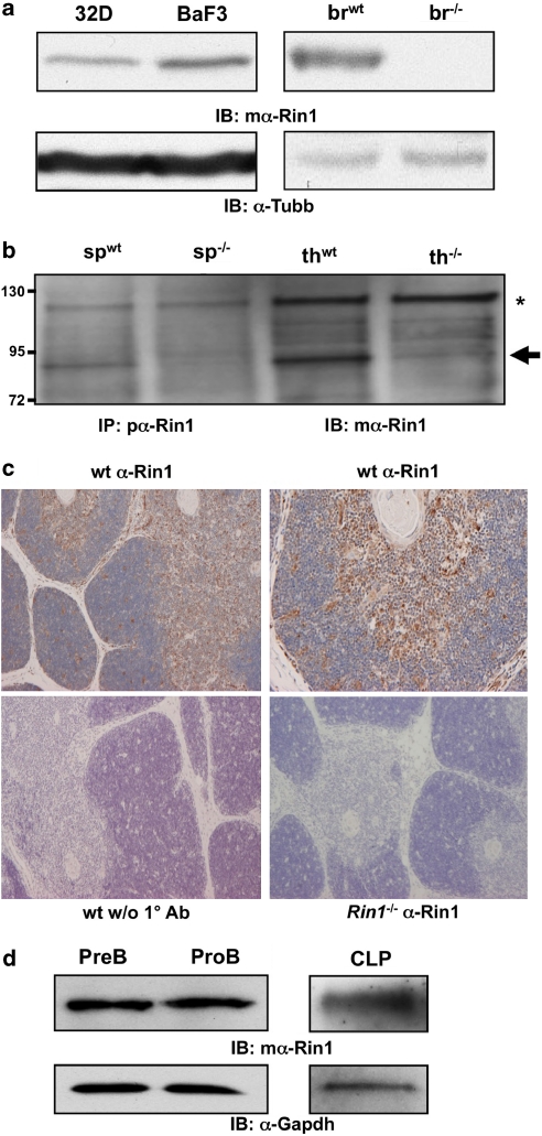
Endogenous mouse Rin1 expression. (a) Left: Immunoblot (IB) of Rin1 in 32D and BaF3 cells. Right: IB of wild-type (wt) and Rin1−/− (−/−) brain (br) tissue confirming antibody specificity. β-tubulin (Tubb) control below each blot. (b) IB of immunoprecipitated (IP) material from spleen (sp) and thymus (th) tissue of wt and Rin1−/− mice. Arrow indicates Rin1. Asterisk marks background band. mα, monoclonal antibody; pα, polyclonal antibody. (c) Immunohistochemical stain of Rin1 in mouse thymus. Top two panels show wild-type thymus probed with anti-Rin1 and counter stained with hematoxylin (left, × 4; right, × 10). Bottom left panel shows wild-type control without anti-Rin1. Bottom right panel shows Rin1−/− thymus control. (d) IB of Rin1 in PreB, ProB and common lymphoid progenitor (CLP) cells. Gapdh control below.
Results
BCR-ABL1 activity is increased by RIN1
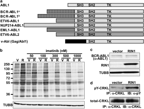
Because RIN1 stimulates tyrosine kinase activity by binding to the ABL SH3 and SH2 autoinhibitory domains,27, 28 and these domains are consistently present in human leukemogenic ABL fusion proteins (Figure 1a), we asked whether overexpression of RIN1 could enhance BCR-ABL1 kinase activity. K562 cells, which are derived from a BCR-ABL1-positive CML patient sample, were transduced with a RIN1 lentivirus or a control vector. The RIN1 overexpressing K562 cells had elevated levels of total cellular tyrosine phosphorylation, consistent with higher BCR-ABL1 kinase activity, when compared with vector control cells (Figure 1b, left two lanes). Note that RIN1, itself an ABL substrate, appears on the phosphotyrosine immunoblot near the 95 kDa marker. There was no change in the level of BCR-ABL1 protein in K562 cells overexpressing RIN1 (Figure 1c), indicating that activation of existing BCR-ABL1 was responsible for the observed increase in cellular tyrosine phosphorylation.
Figure 1.
BCR-ABL1 kinase activity in K562 cells is increased by RIN1. (a) Linear representation of ABL1 (top), translocation-derived human ABL1 and ABL2 fusion oncoproteins (middle six entries) and murine retroviral v-Abl (bottom). Src homology domains (SH2 and SH3) and tyrosine kinase (TK) domains are indicated. BCR-ABL1a represents the p190 isoform associated predominantly with ALL; BCR-ABL1b represents the p210 isoform associated predominantly with CML. (b) K562 cells transduced with vector (V) or RIN1 expression lentivirus (R) were treated with imatinib for 30 min at the indicated concentration and analyzed by immunoblot with anti-phosphotyrosine (MW markers in kDa at left). The ∼95 kDa band that intensifies in the R sample is most likely RIN1. β-tubulin (TUBB) immunoblot was used for normalization. (c) Levels of BCR-ABL1 (210 kDa) expression were evaluated by anti-ABL1 immunoblot of control (vector) and RIN1 overexpression (RIN1) K562 cells (ABL1 migrates below this region). RIN1 immunoblot showed ∼8- to 10-fold overexpression above endogenous levels. TUBB immunoblot was used to normalize extracts. (d) Tyrosine-phosphorylated endogenous CRKL (pY-CRKL) evaluated by immunoprecipitation with anti-CRKL and immunoblot with anti-phosphotyrosine (top) or anti-CRKL (bottom).
We next examined tyrosine phosphorylation of a specific BCR-ABL1 substrate. CRKL is a well-characterized target of ABL1, ABL2 and BCR-ABL1. Indeed, CRKL phosphorylation is used as an indicator of BCR-ABL1 activity in patient-derived CML cells.5 We observed an elevation of CRKL tyrosine phosphorylation levels in RIN1-transduced K562 cells compared with vector control cells (Figure 1d). These results emphasize that, although it is constitutively active relative to ABL1, the BCR-ABL1 oncogene is still responsive to the physiological regulator RIN1. Elevated kinase activity likely accounts, at least in part, for the enhancement of BCR-ABL1-mediated transformation and leukemogenesis by RIN1.
The ABL inhibitor imatinib works by binding to and stabilizing an inactive conformation of the kinase domain.35 We therefore tested whether RIN1, which our model predicts should induce an active conformation, might enhance catalytic activity even in the presence of this kinase inhibitor drug. The levels of total cellular phosphotyrosine remained elevated in RIN1 overexpression cells compared with control cells across a wide range of imatinib concentrations (Figure 1b), suggesting that enhancement of endogenous BCR-ABL1 kinase activity by RIN1 continues in the presence of imatinib. The persistence of higher tyrosine phosphorylation levels in the RIN1 overexpression K562 cells also correlated with resistance to long-term culture in imatinib (Supplementary Figure S1). This modest effect may reflect other changes, however, and its relevance to in vivo resistance has not been validated.
RIN1 is expressed in hematopoietic cells but is not needed for lineage development or engraftment
The mRin1 gene is expressed most highly not only in brain but also in other tissues.36 We detected Rin1 in murine hematopoietic cell lines 32D (myeloid) and BaF3 (lymphoid; pro-B) (Figure 2a), as well as in spleen and thymus, tissues that are rich in lymphoid cells (Figure 2b). Examination of thymus tissue showed Rin1 expression throughout the T-cell-rich medullary and cortical regions (Figure 2c). Rin1 was also observed in sorted populations of common lymphoid progenitor cells as well as in pro- and pre-B cells (Figure 2d). Rin1−/− mutant mice develop normally, however, with cell types and tissues that would otherwise express Rin1 appearing unchanged.37 When bone marrow cells from WT and matched Rin1−/− mice were examined in more detail using multimarker FACS analysis, no significant changes in lineage composition were uncovered (data not shown). This result suggests that, although some aspects of Ras signaling contribute to hematopoietic differentiation,38 the Ras effector Rin1 is not required for normal development in these lineages. Cultured Rin1−/− bone marrow cells also proliferated at the same rate as WT cells in response to interleukin 7 and macrophage colony-stimulating factor (data not shown), demonstrating that Rin1−/− bone marrow cells remain responsive to these physiological factors.
The surface marker and growth factor response analyses suggested that Rin1 is unnecessary for lineage development. However, alterations in hematopoietic stem or progenitor cells may have been undetectable with these methods. We next engrafted bone marrow from WT or Rin1−/− donors (CD45.2) into lethally irradiated recipients (CD45.1). At 34 and 105 days post transplantation, hematopoietic compartments (bone marrow, spleen and thymus) were isolated from recipient mice and examined for lineage composition. Similar profiles were observed for both genotypes at both time points (Table 1), suggesting that Rin1 is dispensable for the regeneration of a complete hematopoietic system.
Table 1. Engraftment of Rin1−/− bone marrow cells.
| Antibody (lineage) | Day 34 | Day 105 | ||
|---|---|---|---|---|
| Wild type (%) | Rin1−/− (%) | Wild type (%) | Rin1−/− (%) | |
| Bone marrow | ||||
| CD19 (B cell) | 18 | 15 | 26 | 25 |
| CD11b (mac/gran) | 26 | 25 | 42 | 41 |
| Ter119 (erythroid) | 14 | 16 | 22 | 27 |
| Spleen | ||||
| CD19 (B cell) | 50 | 52 | 73 | 72 |
| CD11b (mac/gran) | 4 | 4 | 5 | 4 |
| CD3e CD4 (helper T) | 6 | 4 | 7 | 15 |
| CD3e CD8 (cytotoxic T) | 3 | 2 | 8 | 6 |
| Thymus | ||||
| CD4+ CD8− (mature T) | 9 | 9 | 6 | 10 |
| CD4− CD8+ (mature T) | 3 | 3 | 7 | 6 |
| CD4+ CD8+ (immature T) | 80 | 81 | 82 | 82 |
| CD4− CD8− (immature T) | 2 | 2 | 5 | 3 |
CD45.2 wild type or Rin1−/− bone marrow samples were transplanted into lethally irradiated CD45.1 mice. After 34 or 105 days, flow cytometric analysis was performed on bone marrow, spleen and thymus cell suspensions. Data are presented as the percentage of single live donor cells (CD45.2+ 7AAD excluded).
Previous studies showed that Rin1−/− neurons and epithelial cells exhibit conditional (for example, stimulation and stress dependent) phenotypes.28, 37 We therefore considered whether Rin1−/− bone marrow cells might be compromised in their response to oncogenic forms of ABL1.
RIN1 is required for transformation by BCR-ABL1 and ETV6-ABL1
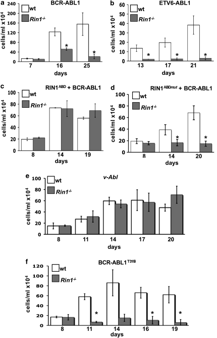
BCR-ABL1 transforms primary bone marrow cells, and this leads to measurable growth factor-independent proliferation in vitro.31 To test whether RIN1 is required for transformation, the p210 isoform of BCR-ABL1, which is common in CML, was introduced by retroviral transduction into primary bone marrow cells from WT or Rin1−/− mutant mice. Cells were then cultured in the absence of growth factors and periodically counted. Rin1−/− cells showed much less expansion than WT cells in these assays (Figure 3a), but grew equally well in the presence of growth factors (data not shown). This suggested that the growth factor independence normally conferred by BCR-ABL131 is to a large degree dependent on the presence of RIN1. Similar results were seen when using the ETV6-ABL1 oncogene (Figure 3b), indicating that the need for RIN1 is associated with the ABL tyrosine kinase rather than with the upstream fusion partner.
Figure 3.
Rin1 is required for transformation of primary bone marrow (BM) cells to growth factor independence. (a) BM cells from wild-type or Rin1−/− mice were infected with a BCR-ABL1 (p210) retrovirus, cultured without growth factors and counted at indicated times. (b) BM cell transformation as in ‘a', except using ETV6-ABL1 (a.k.a. TEL-ABL) retrovirus. (c) BM cell transformation as in ‘a', except using virus expressing both BCR-ABL1 and RIN1ABD. (d) BM cell transformation as in ‘c', except using a mutant (RIN1ABDmut) that does not bind ABL1. (e) BM cell transformation as in ‘a', except using v-Abl retrovirus. (f) BM cell transformation as in ‘a', except using the multidrug-resistant mutant BCR-ABL1T315I. All results are for duplicate samples counted in triplicate. Bars show standard deviation; * indicates P<0.05 between wt and Rin1−/−. Panel f shows 14-day sample P=0.06 between wt and Rin1−/−.
To determine whether RIN1 is required cell autonomously for ABL-mediated transformation, we transduced bone marrow cells with BCR-ABL1 and RIN1. For these experiments, we used the ABL-binding domain of RIN1 (RIN1ABD). This fragment, similar to full-length RIN1, can enhance cell transformation by BCR-ABL1.29 Reconstitution of RIN1 expression restored BCR-ABL1-mediated transformation of Rin1−/− cells (Figure 3c). RIN1 and RIN1ABD have no transforming activity on their own in this assay;29 hence, the recovery of transforming potential can be attributed to the restored collaboration of RIN1 and BCR-ABL1. As a further control, we used a RIN1 mutant with multiple tyrosine-to-phenylalanine substitutions that severely compromise ABL binding and activation.28, 30 The mutant was unable to rescue Rin1−/− bone marrow cells for transformation by BCR-ABL1 (Figure 3d). Taken together, these results support a direct and obligate role for RIN1 in the transformation of primary bone marrow cells by BCR-ABL1.
BCR-ABL1-transduced bone marrow cells from WT and Rin1−/− donors were also examined in transplantation experiments. We observed no striking differences in the penetrance or intensity of disease in models of lymphoid29 or myeloid39 leukemia (Supplementary Figure S2). The absence of a dramatic reduction in leukemogenesis comparable to that seen in growth factor independence assays of cultured bone marrow cells may reflect a richer growth environment and longer time frame for the in vivo experiments.
v-Abl does not require derepression by RIN1 for cell transformation
The murine v-Abl oncogene arises from the fusion of a retroviral Gag gene and cellular Abl1 sequences. Unlike human ABL fusion oncoproteins, v-Abl does not include the autoinhibitory SH3 domain40 (Figure 1a). As a consequence, v-Abl is a more active tyrosine kinase and a more potent transforming gene than BCR-ABL1.26 Rin1−/− bone marrow cells were transformed to growth factor independence by v-Abl as efficiently as WT bone marrow cells (Figure 3e), indicating that an ABL oncoprotein unhindered by SH3 domain autoinhibition does not require RIN1 binding. Although v-Abl differs from BCR-ABL1 in other ways, this result is consistent with RIN1 functioning through derepression of the ABL SH3 domain to promote transformation.
The kinase inhibitor-resistant BCR-ABL1T315I mutant still requires RIN1
We next asked whether RIN1 was required by a multidrug-resistant BCR-ABL1 mutation observed in CML patients. An otherwise normal (non-fusion) ABL1 kinase with the T315I kinase domain ‘gatekeeper' mutation is still responsive to RIN1,27 suggesting that this mutation does not alter RIN1 binding and subsequent kinase derepression. Indeed, although WT bone marrow cells transduced with BCR-ABL1T315I proliferated well, equivalent Rin1−/− bone marrow cells transduced with the same BCR-ABL1T315I virus were not transformed (Figure 3f). This provides further evidence that BCR-ABL1 is reliant on RIN1 for relief of autoinhibition and perhaps for additional contributions to signal-transduction pathways required for transformation. This result also demonstrates that regulation by RIN1 is independent of ABL active site alterations conferring resistance to kinase inhibitors, and strongly suggests that drug-resistant BCR-ABL1 mutants may be susceptible to blockade of RIN1 association.
RIN1 directly regulates drug response in human leukemia cells
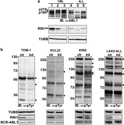
We next turned to cell lines originating from human leukemias with BCR-ABL1 oncogenes for additional insight into the role of RIN1. RIN1 was detected in multiple BCR-ABL1-positive myeloid leukemia lines (KCL22, JURL-MK1 and K562) and B-cell lineage leukemia cell lines (BV173, TOM-1 and SUP-B15) (Figure 4a). RIN1 was not detected by immunoblot in three other CML lines (LAMA-84, KU-812 and KY01). We found a suggestive association between RIN1 expression and imatinib sensitivity (Supplementary Figure S3); the three lines with undetectable RIN1 were the most sensitive to imatinib (Supplementary Figures S3A–C and H), although KCL22 had the highest RIN1 level and was the least sensitive (Supplementary Figures S3G and H). Further analysis with more leukemia cell samples and more precise drug response data will be needed to validate the significance of this correlation, which does not account for factors such as drug efflux rates.
Figure 4.
Analysis of human leukemia cells. (a) Panel of CML and ALL cell lines immunoblotted with anti-ABL1 (top), anti-RIN1 (middle) or anti-TUBB (bottom). CML cells (1=KCL22; 2=JURL-MK1; 3=K562) and a B lymphoid CML blast crisis cell line (4=BV173) express the p210 form of BCR-ABL, whereas ALL-derived cells (5=TOM-1; 6=SUP-B15) express the p190 form (arrowheads). Full-length RIN1 is marked with an arrow (faster migrating bands may be alternately spliced isoforms). (b) TOM-1, KCL22, K562 and primary pre-B-ALL cells infected with control (ctr) or RIN1-directed (kd) shRNA were analyzed by immunoblot with anti-phosphotyrosine. MW (kDa) markers are at left, arrowheads mark bands most clearly reduced by RIN1 silencing. Lower panels show immunoblots for TUBB, RIN1 and BCR-ABL1.
Given the unique nature of each established leukemia cell line, we chose to directly evaluate the contribution of RIN1 to BCR-ABL1 function in several lines. Constitutive knockdown of RIN1 expression using shRNA altered the total phosphotyrosine patterns in TOM-1, KCL22 and K562 cells (Figure 4b). Although many phosphoproteins appeared unaffected, numerous bands were weaker in the RIN1 knockdown cell lines. No change was detected in the amount of BCR-ABL1, consistent with a reduction in enzyme activity as an explanation for the primarily diminished cellular phosphotyrosine profile. The few band intensity increases in K562 cells may reflect enhanced kinases or repressed phosphatases downstream of BCR-ABL1.
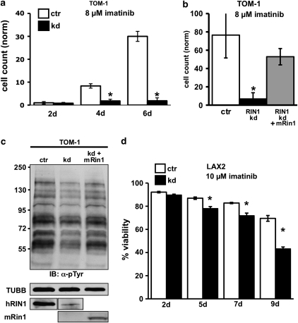
We reasoned that the loss of RIN1 might reduce the stability of the active BCR-ABL1 kinase conformation, and render these leukemia cells more susceptible to an ABL kinase inhibitor that preferentially binds the inactive conformation. Silencing RIN1 expression in TOM-141 cells markedly increased sensitivity to 8 μ imatinib (Figure 5a), a concentration selected as moderately cytotoxic to this ALL line. This result suggested that the loss of RIN1 can synergize with standard kinase inhibitors to block oncogenic ABL kinases. The viability and proliferation rate of TOM-1 cells with the RIN1-targeted shRNA showed no significant difference from TOM-1 cells with the scrambled shRNA control (Supplementary Figure S4A), perhaps because silencing was less than complete or because these established leukemia cell lines have been selected for additional mutations that promote vigorous growth in culture. To rule out off-target effects, the stable RIN1 knockdown TOM-1 cell line was transduced with a murine Rin1 cDNA that is resistant to human RIN1-targeted shRNA (data not shown). Ectopic Rin1 rescued imatinib resistance (Figure 5b). Consistent with restored BCR-ABL1 activity, expression of murine Rin1 also reversed most of the phosphotyrosine signal suppression in RIN1 kd TOM-1 cells (Figure 5c).
Figure 5.
RIN1 silencing sensitizes ALL cells to imatinib. (a) Control (ctr) and RIN1-silenced (kd) TOM-1 cells (1 × 10⁴/ml) were cultured in 8 μ imatinib for the indicated time. Cell counts normalized to 2d-ctr. (b) Control (ctr), RIN1 knockdown (RIN1 kd) and knockdown rescued with mouse Rin1 (RIN1 kd+mRin1) TOM-1 cells were cultured in 8 μ imatinib for 9 days. (c) Control, RIN1 kd and RIN1 kd+mRin1 TOM-1 cells were immunoblotted with anti-phosphotyrosine. TUBB and RIN1 immunoblots are shown below. Murine Rin1 and human RIN1 were detected using different antibodies. Note: hRIN1 bands are from the same exposure of a single immunoblot. (d) Control (ctr) and RIN1-silenced (kd) B-ALL cells were cultured in 10 μ imatinib for the indicated time. Cell viability was determined by propidium iodide stain and flow cytometry. Panels a, b and d: s.d. from triplicate samples counted in duplicate; * indicates P<0.05 between control and knockdown.
Primary pre-B ALL cells from the bone marrow biopsy of a 38-year-old male patient were used next to evaluate the translational relevance of RIN1-targeted silencing. These cells were collected from a relapse leukemia and carry the p210 form of BCR-ABL1 with a T315I mutation, which confers imatinib resistance. RIN1 knockdown reduced the intensity of the cellular phosphotyrosine signal (Figure 4b) and enhanced imatinib sensitivity (Figure 5d), without altering the baseline proliferation rate of the cells (Supplementary Figure S4C). These findings support and extend the conclusion that a direct ABL activator, RIN1, controls the set point for imatinib sensitivity, even in cells expressing the normally refractory BCR-ABL1T315I allele. They also imply that RIN1 silencing or inhibition might collaborate with a direct kinase inhibitor to overcome the leukemogenic properties of ABL fusion proteins.
Discussion
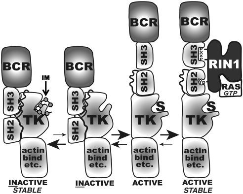
Translocations that create BCR-ABL1 and other human leukemogenic ABL gene fusions consistently retain the ABL SH3 and SH2 domains, despite the fact that biochemical and structure data have established that these domains restrain kinase activity. Our data suggest that RIN1 binding to ABL SH3 and SH2 domains stabilizes the active conformation of the kinase, enhancing the already constitutive activity of the fusion protein (Figure 6). The result is increased cellular tyrosine phosphorylation and transforming activity in the presence of RIN1, which comports with the observed overexpression of RIN1 in some leukemias42 and lymphomas.43 RIN1 loss greatly reduced the ability of ABL1 fusions to establish a transformed phenotype in bone marrow cells, consistent with a cell autonomous requirement for RIN1 to unleash the full potential of ABL tyrosine kinases. Even BCR-ABLT315I, a drug-resistant mutant with increased transformation potency,44 was dependent on RIN1 for bone marrow cell transformation. However, we did not detect any significant effects of Rin1 deletion on leukemogenesis by BCR-ABL1-transduced bone marrow cells in mouse transplantation model systems. These data suggest an important supporting role for RIN1 in BCR-ABL1-mediated transformation, but do not fully resolve the contribution of RIN1 in spontaneous leukemias.
Figure 6.
Model of RIN1 effect on BCR-ABL1 activity and imatinib sensitivity. BCR-ABL1 equilibrates between active and inactive conformations, favoring the active form (open to substrate (S)) relative to ABL1. Right: RIN1 binds to ABL1 SH3 and SH2 domains, alleviating residual autoinhibition and stabilizing a high activity conformation. RIN1 is also a RAS effector.28, 56 Left: Imatinib (IM) preferentially binds and stabilizes the inactive conformation of the ABL1 catalytic site.35 RIN1 overexpression shifts equilibrium to right; RIN1 silencing shifts equilibrium to left.
The absence of an SH3 domain in murine v-Abl (Gag-Abl1) increases its constitutive kinase activity and makes it a highly potent oncogene. Deletion of the autoinhibitory SH3 domain is likely to be more strongly activating than derepression by binding to endogenous RIN1. However, although an SH3 deletion mutant of BCR-ABL1 does potently transform cells in vitro and produces myeloproliferative disorders in a xenograft model,45 it does not possess full leukemogenic potential.46 There are also significant differences in the types of leukemias induced by v-Abl versus BCR-ABL1.47 Together with the fact that oncogenic fusions disrupting the ABL1 SH3 domain are extremely rare in human leukemias,24 it seems likely that the ABL SH3 domain is retained through selective pressure because it contributes to leukemogenesis and/or to leukemia progression in vivo. Additional evidence for this model comes from studies demonstrating that transphosphorylation of BCR-ABL1 SH3 and SH2 domain tyrosines modulates transforming activity.48 RIN1 likely functions, in part, to elevate ABL tyrosine kinase activity above a transformation threshold level while allowing the SH3 domain to enhance leukemia progression through other mechanisms. RIN1 might additionally contribute to oncogenic ABL signaling by influencing substrate specificity, directing subcellular distribution of ABL fusion proteins or recruiting signaling partners that promote expansion of the leukemic cell population. An alternative explanation for consistent SH3 domain retention is strong preference for recombination in the first ABL1 intron. This is unlikely given the evidence cited and because all oncogenic ABL2 fusions show the same breakpoint (in this case between exons 2 and 3).
As BCR-ABL1 is required to maintain cell transformation, a phenomenon described as ‘oncogene addiction', RIN1 silencing alone might be expected to impair tumor cell proliferation. Although reduced RIN1 expression did not arrest TOM-1 or LAX2 cells in our experiments, it did significantly heighten the sensitivity of these cells to imatinib. This enhanced drug responsiveness may result from a shift toward the inactive ABL1 kinase conformation that imatinib preferentially binds and stabilizes,35 and away from the active kinase conformation that RIN1 preferentially binds and stabilizes27 (Figure 6). Conformation stabilization does not imply protein stabilization, and we observed no indication that altered RIN1 levels affect BCR-ABL1 protein levels. The drug-sensitizing effect of RIN1 silencing suggests that disruption of RIN1 binding might synergize with kinase inhibitors to significantly expand the range of leukemias that respond to these drugs. We note that some BCR-ABL1-positive leukemia cell lines had relatively low RIN1 levels. RIN1 may nevertheless be important in those cells for BCR-ABL1 activity and transformation. Alternatively, low RIN1 expression may indicate additional genetic alterations, occurring in vivo or in culture, that confer a degree of independence from RIN1.
In epithelial cells, RIN1 has a role in plasma membrane receptor internalization and cell motility through the coordinated activation of RAB5 GTPases that regulate endocytosis and ABL tyrosine kinases that regulate actin remodeling.28, 49 In solid tumors, RIN1 is a tumor suppressor in breast cancer,50 but a tumor enhancer in non-small-cell lung cancer.51 In both cases, the main contribution of RIN1 appears to be through RAB5 activation and growth factor receptor trafficking. The rescue of Rin1−/− bone marrow cell transformation by a RIN1 amino terminal fragment with no RAB5-GEF domain, however, demonstrates that RAB5 activation is not required for collaboration with BCR-ABL1. The apparently contradictory roles for RIN1 as a myeloproliferative disorder enhancer and epithelial carcinoma suppressor (in breast cancer) are perhaps not surprising given the differences between these diseases and their cells of origin. Neither is it unprecedented; NOTCH1 gain of function is associated with T-cell leukemias,52 whereas NOTCH1 silencing contributes to the progression of cervical cancers.53
RIN1 silencing reduced BCR-ABL1 activity while enhancing imatinib sensitivity in established and primary leukemia cells, possibly the result of a shift toward the inactive ABL conformation. Mutations in the SH3-SH2 domains of BCR-ABL1 can also reduce imatinib sensitivity,54, 55 perhaps by overriding the dependence on RIN1 binding. Importantly, the only such mutations validated to confer imatinib sensitivity in patient samples never occurred simultaneously with kinase domain mutations.55 This suggests the potential value of small molecule inhibitors that target the functional interaction of RIN1 and BCR-ABL1 through direct binding blockade or allosteric interference. Such drugs might be combined with kinase active site-directed inhibitors for a first-line therapy that reduces relapse rates by requiring the simultaneous acquisition of independent resistance mutations.
Acknowledgments
The authors thank the following persons for their generosity in providing reagents and advice: Brian Druker, Kara Johnson, Kathleen Sakamoto, Charles Sawyers, Lee Goodglick, Lily Zhang, Gustavo Miranda-Carboni and Timothy Lane. This work was supported by NIH grants RO1 NS046787 (JC) and CA136699 (JC), and by the UCLA Jonsson Cancer Center Foundation. ONW is an Investigator of the Howard Hughes Medical Institute and MM is a Scholar of the Leukemia and Lymphoma Society.
Authorship contributions
Creation of expression constructs and lentivirus stocks, as well as immunostaining, immunoprecipitation and immunoblotting, was performed by Minh Thai. Collection, culturing and transduction of primary cells and cell lines, bone marrow transplantation experiments and FACS analyses were performed by Minh Thai, Pamela Ting, Jami McLaughlin and Donghui Cheng. Markus Müschen developed the primary ALL cell (LAX2) culture system. Owen Witte directed the design and analysis of mouse model experiments. John Colicelli oversaw the research project. All authors participated in the preparation of this manuscript.
The authors declare no conflict of interests.
Footnotes
Supplementary Information accompanies the paper on the Leukemia website (http://www.nature.com/leu)
Supplementary Material
References
- Wong S, Witte ON. The BCR-ABL story: bench to bedside and back. Annu Rev Immunol. 2004;22:247–306. doi: 10.1146/annurev.immunol.22.012703.104753. [DOI] [PubMed] [Google Scholar]
- Papadopoulos P, Ridge SA, Boucher CA, Stocking C, Wiedemann LM. The novel activation of ABL by fusion to an ets-related gene, TEL. Cancer Res. 1995;55:34–38. [PubMed] [Google Scholar]
- Iijima Y, Ito T, Oikawa T, Eguchi M, Eguchi-Ishimae M, Kamada N, et al. A new ETV6/TEL partner gene, ARG (ABL-related gene or ABL2), identified in an AML-M3 cell line with a t(1;12)(q25;p13) translocation. Blood. 2000;95:2126–2131. [PubMed] [Google Scholar]
- Deininger M, Buchdunger E, Druker BJ. The development of imatinib as a therapeutic agent for chronic myeloid leukemia. Blood. 2005;105:2640–2653. doi: 10.1182/blood-2004-08-3097. [DOI] [PubMed] [Google Scholar]
- Gorre ME, Mohammed M, Ellwood K, Hsu N, Paquette R, Rao PN, et al. Clinical resistance to STI-571 cancer therapy caused by BCR-ABL gene mutation or amplification. Science (NY) 2001;293:876–880. doi: 10.1126/science.1062538. [DOI] [PubMed] [Google Scholar]
- O'Hare T, Shakespeare WC, Zhu X, Eide CA, Rivera VM, Wang F, et al. AP24534, a pan-BCR-ABL inhibitor for chronic myeloid leukemia, potently inhibits the T315I mutant and overcomes mutation-based resistance. Cancer Cell. 2009;16:401–412. doi: 10.1016/j.ccr.2009.09.028. [DOI] [PMC free article] [PubMed] [Google Scholar]
- Shah NP, Skaggs BJ, Branford S, Hughes TP, Nicoll JM, Paquette RL, et al. Sequential ABL kinase inhibitor therapy selects for compound drug-resistant BCR-ABL mutations with altered oncogenic potency. J Clin Invest. 2007;117:2562–2569. doi: 10.1172/JCI30890. [DOI] [PMC free article] [PubMed] [Google Scholar]
- Bueno MJ, Perez de Castro I, Gomez de Cedron M, Santos J, Calin GA, Cigudosa JC, et al. Genetic and epigenetic silencing of microRNA-203 enhances ABL1 and BCR-ABL1 oncogene expression. Cancer Cell. 2008;13:496–506. doi: 10.1016/j.ccr.2008.04.018. [DOI] [PubMed] [Google Scholar]
- Chen R, Gandhi V, Plunkett W. A sequential blockade strategy for the design of combination therapies to overcome oncogene addiction in chronic myelogenous leukemia. Cancer Res. 2006;66:10959–10966. doi: 10.1158/0008-5472.CAN-06-1216. [DOI] [PubMed] [Google Scholar]
- Kawano T, Ito M, Raina D, Wu Z, Rosenblatt J, Avigan D, et al. MUC1 oncoprotein regulates Bcr-Abl stability and pathogenesis in chronic myelogenous leukemia cells. Cancer Res. 2007;67:11576–11584. doi: 10.1158/0008-5472.CAN-07-2756. [DOI] [PMC free article] [PubMed] [Google Scholar]
- Wu LX, Xu JH, Zhang KZ, Lin Q, Huang XW, Wen CX, et al. Disruption of the Bcr-Abl/Hsp90 protein complex: a possible mechanism to inhibit Bcr-Abl-positive human leukemic blasts by novobiocin. Leukemia. 2008;22:1402–1409. doi: 10.1038/leu.2008.89. [DOI] [PubMed] [Google Scholar]
- Dierks C, Beigi R, Guo GR, Zirlik K, Stegert MR, Manley P, et al. Expansion of Bcr-Abl-positive leukemic stem cells is dependent on Hedgehog pathway activation. Cancer Cell. 2008;14:238–249. doi: 10.1016/j.ccr.2008.08.003. [DOI] [PubMed] [Google Scholar]
- Gregory MA, Phang TL, Neviani P, Alvarez-Calderon F, Eide CA, O'Hare T, et al. Wnt/Ca2+/NFAT signaling maintains survival of Ph+ leukemia cells upon inhibition of Bcr-Abl. Cancer Cell. 2010;18:74–87. doi: 10.1016/j.ccr.2010.04.025. [DOI] [PMC free article] [PubMed] [Google Scholar]
- Hess P, Pihan G, Sawyers CL, Flavell RA, Davis RJ. Survival signaling mediated by c-Jun NH(2)-terminal kinase in transformed B lymphoblasts. Nat Genet. 2002;32:201–205. doi: 10.1038/ng946. [DOI] [PubMed] [Google Scholar]
- Zhao C, Chen A, Jamieson CH, Fereshteh M, Abrahamsson A, Blum J, et al. Hedgehog signalling is essential for maintenance of cancer stem cells in myeloid leukaemia. Nature. 2009;458:776–779. doi: 10.1038/nature07737. [DOI] [PMC free article] [PubMed] [Google Scholar]
- Nagar B, Hantschel O, Young MA, Scheffzek K, Veach D, Bornmann W, et al. Structural basis for the autoinhibition of c-Abl tyrosine kinase. Cell. 2003;112:859–871. doi: 10.1016/s0092-8674(03)00194-6. [DOI] [PubMed] [Google Scholar]
- Pluk H, Dorey K, Superti-Furga G. Autoinhibition of c-Abl. Cell. 2002;108:247–259. doi: 10.1016/s0092-8674(02)00623-2. [DOI] [PubMed] [Google Scholar]
- Nagar B, Hantschel O, Seeliger M, Davies JM, Weis WI, Superti-Furga G, et al. Organization of the SH3-SH2 unit in active and inactive forms of the c-Abl tyrosine kinase. Mol Cell. 2006;21:787–798. doi: 10.1016/j.molcel.2006.01.035. [DOI] [PubMed] [Google Scholar]
- Brasher BB, Van Etten RA. c-Abl has high intrinsic tyrosine kinase activity that is stimulated by mutation of the Src homology 3 domain and by autophosphorylation at two distinct regulatory tyrosines. J Biol Chem. 2000;275:35631–35637. doi: 10.1074/jbc.M005401200. [DOI] [PubMed] [Google Scholar]
- Dorey K, Engen JR, Kretzschmar J, Wilm M, Neubauer G, Schindler T, et al. Phosphorylation and structure-based functional studies reveal a positive and a negative role for the activation loop of the c-Abl tyrosine kinase. Oncogene. 2001;20:8075–8084. doi: 10.1038/sj.onc.1205017. [DOI] [PubMed] [Google Scholar]
- Plattner R, Kadlec L, DeMali KA, Kazlauskas A, Pendergast AM. c-Abl is activated by growth factors and Src family kinases and has a role in the cellular response to PDGF. Genes Dev. 1999;13:2400–2411. doi: 10.1101/gad.13.18.2400. [DOI] [PMC free article] [PubMed] [Google Scholar]
- Tanis KQ, Veach D, Duewel HS, Bornmann WG, Koleske AJ. Two distinct phosphorylation pathways have additive effects on Abl family kinase activation. Mol Cell Biol. 2003;23:3884–3896. doi: 10.1128/MCB.23.11.3884-3896.2003. [DOI] [PMC free article] [PubMed] [Google Scholar]
- Golub TR, Goga A, Barker GF, Afar DE, McLaughlin J, Bohlander SK, et al. Oligomerization of the ABL tyrosine kinase by the Ets protein TEL in human leukemia. Mol Cell Biol. 1996;16:4107–4116. doi: 10.1128/mcb.16.8.4107. [DOI] [PMC free article] [PubMed] [Google Scholar]
- Deininger MW, Goldman JM, Melo JV. The molecular biology of chronic myeloid leukemia. Blood. 2000;96:3343–3356. [PubMed] [Google Scholar]
- Smith KM, Yacobi R, Van Etten RA. Autoinhibition of Bcr-Abl through its SH3 domain. Mol Cell. 2003;12:27–37. doi: 10.1016/S1097-2765(03)00274-0. [DOI] [PMC free article] [PubMed] [Google Scholar]
- Kelliher MA, McLaughlin J, Witte ON, Rosenberg N. Induction of a chronic myelogenous leukemia-like syndrome in mice with v-abl and BCR/ABL. Proc Natl Acad Sci USA. 1990;87:6649–6653. doi: 10.1073/pnas.87.17.6649. [DOI] [PMC free article] [PubMed] [Google Scholar]
- Cao X, Tanis KQ, Koleske AJ, Colicelli J. Enhancement of ABL kinase catalytic efficiency by a direct binding regulator is independent of other regulatory mechanisms. J Biol Chem. 2008;283:31401–31407. doi: 10.1074/jbc.M804002200. [DOI] [PMC free article] [PubMed] [Google Scholar]
- Hu H, Bliss JM, Wang Y, Colicelli J. RIN1 is an ABL tyrosine kinase activator and a regulator of epithelial-cell adhesion and migration. Curr Biol. 2005;15:815–823. doi: 10.1016/j.cub.2005.03.049. [DOI] [PubMed] [Google Scholar]
- Afar DE, Han L, McLaughlin J, Wong S, Dhaka A, Parmar K, et al. Regulation of the oncogenic activity of BCR-ABL by a tightly bound substrate protein RIN1. Immunity. 1997;6:773–782. doi: 10.1016/s1074-7613(00)80452-5. [DOI] [PubMed] [Google Scholar]
- Hu H, Milstein M, Bliss JM, Thai M, Malhotra G, Huynh LC, et al. Integration of transforming growth factor beta and RAS signaling silences a RAB5 guanine nucleotide exchange factor and enhances growth factor-directed cell migration. Mol Cell Biol. 2008;28:1573–1583. doi: 10.1128/MCB.01087-07. [DOI] [PMC free article] [PubMed] [Google Scholar]
- McLaughlin J, Chianese E, Witte ON. In vitro transformation of immature hematopoietic cells by the P210 BCR/ABL oncogene product of the Philadelphia chromosome. Proc Natl Acad Sci USA. 1987;84:6558–6562. doi: 10.1073/pnas.84.18.6558. [DOI] [PMC free article] [PubMed] [Google Scholar]
- Hardy RR, Hayakawa K. B-lineage differentiation stages resolved by multiparameter flow cytometry. Ann N Y Acad Sci. 1995;764:19–24. doi: 10.1111/j.1749-6632.1995.tb55800.x. [DOI] [PubMed] [Google Scholar]
- Klemm L, Duy C, Iacobucci I, Kuchen S, von Levetzow G, Feldhahn N, et al. The B cell mutator AID promotes B lymphoid blast crisis and drug resistance in chronic myeloid leukemia. Cancer Cell. 2009;16:232–245. doi: 10.1016/j.ccr.2009.07.030. [DOI] [PMC free article] [PubMed] [Google Scholar]
- Mardiney M, III, Malech HL. Enhanced engraftment of hematopoietic progenitor cells in mice treated with granulocyte colony-stimulating factor before low-dose irradiation: implications for gene therapy. Blood. 1996;87:4049–4056. [PubMed] [Google Scholar]
- Schindler T, Bornmann W, Pellicena P, Miller WT, Clarkson B, Kuriyan J. Structural mechanism for STI-571 inhibition of abelson tyrosine kinase. Science (NY) 2000;289:1938–1942. doi: 10.1126/science.289.5486.1938. [DOI] [PubMed] [Google Scholar]
- Dzudzor B, Huynh L, Thai M, Bliss JM, Nagaoka Y, Wang Y, et al. Regulated expression of the Ras effector Rin1 in forebrain neurons. Mol Cell Neurosci. 2010;43:108–116. doi: 10.1016/j.mcn.2009.09.012. [DOI] [PMC free article] [PubMed] [Google Scholar]
- Dhaka A, Costa RM, Hu H, Irvin DK, Patel A, Kornblum HI, et al. The RAS effector RIN1 modulates the formation of aversive memories. J Neurosci. 2003;23:748–757. doi: 10.1523/JNEUROSCI.23-03-00748.2003. [DOI] [PMC free article] [PubMed] [Google Scholar]
- Miranda MB, Johnson DE. Signal transduction pathways that contribute to myeloid differentiation. Leukemia. 2007;21:1363–1377. doi: 10.1038/sj.leu.2404690. [DOI] [PubMed] [Google Scholar]
- Wong S, McLaughlin J, Cheng D, Shannon K, Robb L, Witte ON. IL-3 receptor signaling is dispensable for BCR-ABL-induced myeloproliferative disease. Proc Natl Acad Sci USA. 2003;100:11630–11635. doi: 10.1073/pnas.2035020100. [DOI] [PMC free article] [PubMed] [Google Scholar]
- Wang JY, Ledley F, Goff S, Lee R, Groner Y, Baltimore D. The mouse c-abl locus: molecular cloning and characterization. Cell. 1984;36:349–356. doi: 10.1016/0092-8674(84)90228-9. [DOI] [PubMed] [Google Scholar]
- Okabe M, Matsushima S, Morioka M, Kobayashi M, Abe S, Sakurada K, et al. Establishment and characterization of a cell line, TOM-1, derived from a patient with Philadelphia chromosome-positive acute lymphocytic leukemia. Blood. 1987;69:990–998. [PubMed] [Google Scholar]
- Morikawa J, Li H, Kim S, Nishi K, Ueno S, Suh E, et al. Identification of signature genes by microarray for acute myeloid leukemia without maturation and acute promyelocytic leukemia with t(15;17)(q22;q12)(PML/RARalpha) Int J Oncol. 2003;23:617–625. [PubMed] [Google Scholar]
- de Vos S, Hofmann WK, Grogan TM, Krug U, Schrage M, Miller TP, et al. Gene expression profile of serial samples of transformed B-cell lymphomas. Lab Invest. 2003;83:271–285. doi: 10.1097/01.lab.0000053913.85892.e9. [DOI] [PubMed] [Google Scholar]
- Skaggs BJ, Gorre ME, Ryvkin A, Burgess MR, Xie Y, Han Y, et al. Phosphorylation of the ATP-binding loop directs oncogenicity of drug-resistant BCR-ABL mutants. Proc Natl Acad Sci USA. 2006;103:19466–19471. doi: 10.1073/pnas.0609239103. [DOI] [PMC free article] [PubMed] [Google Scholar]
- Gross AW, Zhang X, Ren R. Bcr-Abl with an SH3 deletion retains the ability To induce a myeloproliferative disease in mice, yet c-Abl activated by an SH3 deletion induces only lymphoid malignancy. Mol Cell Biol. 1999;19:6918–6928. doi: 10.1128/mcb.19.10.6918. [DOI] [PMC free article] [PubMed] [Google Scholar]
- Skorski T, Nieborowska-Skorska M, Wlodarski P, Wasik M, Trotta R, Kanakaraj P, et al. The SH3 domain contributes to BCR/ABL-dependent leukemogenesis in vivo: role in adhesion, invasion, and homing. Blood. 1998;91:406–418. [PubMed] [Google Scholar]
- Clark SS, Chen E, Fizzotti M, Witte ON, Malkovska V. BCR-ABL and v-abl oncogenes induce distinct patterns of thymic lymphoma involving different lymphocyte subsets. J Virol. 1993;67:6033–6046. doi: 10.1128/jvi.67.10.6033-6046.1993. [DOI] [PMC free article] [PubMed] [Google Scholar]
- Chen S, O'Reilly LP, Smithgall TE, Engen JR. Tyrosine Phosphorylation in the SH3 Domain Disrupts Negative Regulatory Interactions within the c-Abl Kinase Core. J Mol Biol. 2008;383:414–423. doi: 10.1016/j.jmb.2008.08.040. [DOI] [PMC free article] [PubMed] [Google Scholar]
- Barbieri MA, Kong C, Chen PI, Horazdovsky BF, Stahl PD. The SRC homology 2 domain of Rin1 mediates its binding to the epidermal growth factor receptor and regulates receptor endocytosis. J Biol Chem. 2003;278:32027–32036. doi: 10.1074/jbc.M304324200. [DOI] [PubMed] [Google Scholar]
- Milstein M, Mooser CK, Hu H, Fejzo M, Slamon D, Goodglick L, et al. RIN1 is a breast tumor suppressor gene. Cancer Res. 2007;67:11510–11516. doi: 10.1158/0008-5472.CAN-07-1147. [DOI] [PMC free article] [PubMed] [Google Scholar]
- Tomshine JC, Severson SR, Wigle DA, Sun Z, Beleford DT, Shridhar V, et al. Cell proliferation and epidermal growth factor signaling in non-small cell lung adenocarcinoma cell lines is dependent on RIN1. J Biol Chem. 2009;284:26331–26339. doi: 10.1074/jbc.M109.033514. [DOI] [PMC free article] [PubMed] [Google Scholar]
- Weng AP, Ferrando AA, Lee W, Morris JPt, Silverman LB, Sanchez-Irizarry C, et al. Activating mutations of NOTCH1 in human T cell acute lymphoblastic leukemia. Science (NY) 2004;306:269–271. doi: 10.1126/science.1102160. [DOI] [PubMed] [Google Scholar]
- Talora C, Sgroi DC, Crum CP, Dotto GP. Specific down-modulation of Notch1 signaling in cervical cancer cells is required for sustained HPV-E6/E7 expression and late steps of malignant transformation. Genes Dev. 2002;16:2252–2263. doi: 10.1101/gad.988902. [DOI] [PMC free article] [PubMed] [Google Scholar]
- Azam M, Latek RR, Daley GQ. Mechanisms of autoinhibition and STI-571/imatinib resistance revealed by mutagenesis of BCR-ABL. Cell. 2003;112:831–843. doi: 10.1016/s0092-8674(03)00190-9. [DOI] [PubMed] [Google Scholar]
- Sherbenou DW, Hantschel O, Kaupe I, Willis S, Bumm T, Turaga LP, et al. BCR-ABL SH3-SH2 domain mutations in chronic myeloid leukemia patients on imatinib Blood 2010116E-pub ahead of print (PMID 20519627). [DOI] [PMC free article] [PubMed] [Google Scholar]
- Han L, Colicelli J. A human protein selected for interference with Ras function interacts directly with Ras and competes with Raf1. Mol Cell Biol. 1995;15:1318–1323. doi: 10.1128/mcb.15.3.1318. [DOI] [PMC free article] [PubMed] [Google Scholar]
Associated Data
This section collects any data citations, data availability statements, or supplementary materials included in this article.


