Abstract
Purpose
To determine the genetic and genomic alterations underlying classic aniridia in Saudi Arabia, a region with social preference for consanguineous marriage.
Methods
Prospective study of consecutive patients referred to a pediatric ophthalmologist in Saudi Arabia (2005–2009). All patients had paired box gene 6 (PAX6) analysis (sequencing and multiplex ligation-dependent probe amplification analysis if sequencing was normal). If PAX6 analysis was negative, the following were performed: candidate gene sequencing (forkhead box C1 [FOXC1], paired-like homeodomain transcription factor 2 [PITX2], cytochrome P450, family 1, subfamily B [CYP1B1], paired-like homeodomain transcription factor 3 [PITX3], and v-maf avian musculoaponeurotic fibrosarcoma oncogene homolog [MAF]) and molecular karyotyping by array competitive genomic hybridization (250K single nucleotide polymorphism (SNP) arrays).
Results
All 12 probands (4 months – 25 years of age; four boys and eight girls) had lens opacity and foveal hypoplasia in addition to no grossly visible iris. Four cases were familial. All cases were products of consanguineous unions except for three, one of which was endogamous. Heterozygous PAX6 mutations (including two novel mutations) were detectable in all but two cases, both of which were sporadic. In one of these two cases, the phenotype segregated with homozygosity for a previously-reported pathogenic missense FOXC1 variant (p.P297S) when homozygosity for chromosome 11q24.2 deletion (chr11:125,001,547–125,215,177 [rs114259885; rs112291840]) was also present. In the other, no genetic or genomic abnormalities were found.
Conclusions
The classic aniridia phenotype in Saudi Arabia is typically caused by heterozygous PAX6 mutations, even in the setting of enhanced homozygosity from recent shared parental ancestry. For PAX6-negative cases, interaction between missense variation in an anterior segment developmental gene and copy number variation elsewhere in the genome may be a potential mechanism for the phenotype.
Introduction
Classic aniridia (OMIM 106210) is a panocular disorder which, in addition to lack of grossly visible iris, is characterized by keratopathy, lens opacity, juvenile-onset glaucoma, foveal hypoplasia, and optic nerve hypoplasia [1]. This classic phenotype is caused by heterozygous mutation in the ocular developmental gene paired box gene 6 (PAX6; OMIM *607108; 11p13), typically through haploinsufficiency [2]. For cases in which the contiguous gene Wilms tumor 1 (WT1; OMIM 194070; 11p13) is also disrupted, affected children are typically syndromic and at risk for juvenile Wilms tumor of the kidney (WAGR syndrome, OMIM 194072) [3]. Previous studies have shown that dominant PAX6 mutations are the only known cause of classic aniridia. However, even with comprehensive analysis of PAX6 including copy number analysis, up to 10% of classic anirida cases have no identifiable mutation [2,4].
Congenital iris abnormality in general can also be due to heterozygous PAX6 mutation but is both phenotypically and genotypically more heterogenous. Heterozygous mutation in the ocular developmental gene forkhead box C1 (FOXC1; OMIM *601090; 6p25) or paired-like homeodomain transcription factor 2 (PITX2; OMIM *601542; 4q25-q26) causes the Axenfeld-Rieger spectrum (OMIM 602482, 180500, 137600), an anterior segment dysgenesis which can include mild iris stromal hypoplasia, corectopia, polycoria, iris pigment border hyperplasia (“ectropion uvea”), and/or virtually complete lack of iris [5]. FOXC1 duplication can cause the phenotype as well [6,7]. Affected children have a propensity for congenital or juvenile glaucoma, can have neural crest-related non-ocular findings (e.g., maxillary hypoplasia, dental anomalies), and can show significant intrafamilial phenotypic variation for the same mutation [5,8,9]. Congenital iris abnormality can also occur in the setting of congenital glaucoma from homozygous or compound heterozygous mutations in cytochrome P450, family 1, subfamily B (CYP1B1; OMIM *601771; 2p22-p21) [10], in the setting of anterior segment mesenchymal dysgenesis due to heterozygous mutation in paired-like homeodomain transcription factor 3 (PITX3; OMIM +602669; 10q25) [11], and in the setting of anterior segment dysgenesis due to heterozygous mutation in v-maf avian musculoaponeurotic fibrosarcoma oncogene homolog (MAF; OMIM *177075; 16q22-q23) [12]. The variation in iris phenotype for a given mutation that has been observed in FOXC1-related disease as well as in other genetic causes of congenital iris abnormality is likely related to the influence of yet unidentified modifiers.
Although a small percentage of aniridia cases have no detectable PAX6 mutation, to the best of our knowledge no other genotype has been associated with the classic aniridia phenotype. We propose two potential causes for PAX6-negative classic anirida: 1) genomic alteration that affect cis or trans regulatory elements of PAX6, or 2) point mutations or deletions in other genes known to be involved in the pathogenesis of congenital iris abnormalities. Genome-wide copy number analysis of classic aniridia cases with negative PAX6 sequencing can investigate the first possibility. In addition to interrogation for genomic alteration in a genome-wide fashion, this would also further characterize PAX6 deletions. Regarding the second possibility, i.e., mutations in other known genes, gross deletions would also be uncovered by genome-wide copy number analysis while sequencing variants can be investigated by direct sequencing of a panel of genes known to cause congenital iris abnormalities when mutated. Moreover, if recessive mutation in other known genes can cause classic aniridia, this would most likely be uncovered by the study of affected consanguineous families, for whom a recessive cause is more likely to be found if a recessive cause for the phenotype exists [13]. In the current study, we analyze the underlying genotype of classic aniridia in mostly consanguineous patients by PAX6 analysis and, for PAX6-negative cases, by candidate gene sequencing and array genomic copy number variation analysis.
Methods
Institutional review board approval was obtained for this study (KFSHRC IRB #2070023
KKESH IRB #?????). Consecutive patients with classic aniridia referred to the pediatric ophthalmology service of one of the authors (A.O.K.) from 2005 to 2009 were prospectively enrolled in the study. Classic anirida was defined as no grossly visible iris in addition to keratopathy, lens opacity, and/or foveal hypoplasia [1]. Patients underwent complete ophthalmic examination and venous blood sampling for analysis. The strategy for genetic and genomic analysis was as follows: If direct sequencing did not reveal PAX6 mutation, multiplex ligation-dependent probe amplification (MLPA) was performed to assess for PAX6 deletions. If this was normal, the candidate genes FOXC1, PITX2, CYP1B1, PITX3, and MAF were sequenced. In addition, genomic molecular karyotyping was performed via array-based comparative genomic hybridization (array CGH) in all cases that lacked an identifiable PAX6 mutation. When available and appropriate, relatives underwent ophthalmic examination and venous blood sampling for confirmatory genetic analysis.
DNA extraction
Genomic DNA was extracted from whole blood anti-coagulated with EDTA using the Purgene Gentra DNA Extraction Kit (Cat. # D-5000; Gentra Systems, Minneapolis, MN) according to the manufacturer’s instructions. The DNA was quantified spectrophotometrically and stored in aliquots at −20 °C until required.
PCR amplification and DNA sequencing
PCR amplification was performed on a thermocycler (DNA Engine Tetrad, MJResearch, Inc., Hercules, CA) in a total volume of 25 µl, containing 10 ng DNA, 50 mM KCl, 10 mM Tris-HCl (pH 9.0), 1.5 mM MgCl2, 0.1% Triton X-100, 0.25m M of each dNTP, 0.8 µM of each primer and 0.5 Units of Taq polymerase (D-40724; QIAGEN, Hilden, Germany). For PCR, an initial denaturation step at 95 °C for 10 min was followed by 40 cycles of denaturation at 95 °C for 30 s, annealing at 59 °C for 30 s and extension at 72 °C for 30 s followed by a final extension step of 72 °C for 10 min. Genomic DNA of each patient was screened for coding regions and boundary site variants by sequencing reactions using BigDye® Terminator v 3.0 (Applied Biosystems, Inc., Foster City, CA). Following the manufacturer’s instructions, sequencing reactions were desalted and unincorporated nucleotides removed using ethanol precipitation and re-suspended in a deionized distilled water for injection on a Applied Biosystems 3730xl DNA Analyzer (Applied Biosystems, Inc.). Sequence analysis was performed using the SeqManII module of the Lasergene (DNA Star Inc., Madison, WI) software package and was compared to the relevant reference sequence.
MLPA analysis
MLPA analysis of PAX6 was performed using the commercial Kit P219 PAX6 (MCR Holland, Amsterdam, The Netherlands). The manufacturer’s instructions were followed and data was analyzed using Coffalyser software (MCR Holland).
Molecular karyotyping
Affymetrix Genechip Human Mapping 250K SNP arrays (Affymetrix, Santa Clara, CA) were used. All procedures were performed according to Affymetrix standard protocols. The SNP call rate and sample mismatch report was determined with GTYPE software (Affymetrix). Two algorithms, the Affymetrix® Genotyping Console™ and Copy Number Analyzer for GeneChip® arrays (CNAG) Version 3.021 software were used to infer copy number variation among affected individuals based on the hybridization intensity signal of the probes.
Results
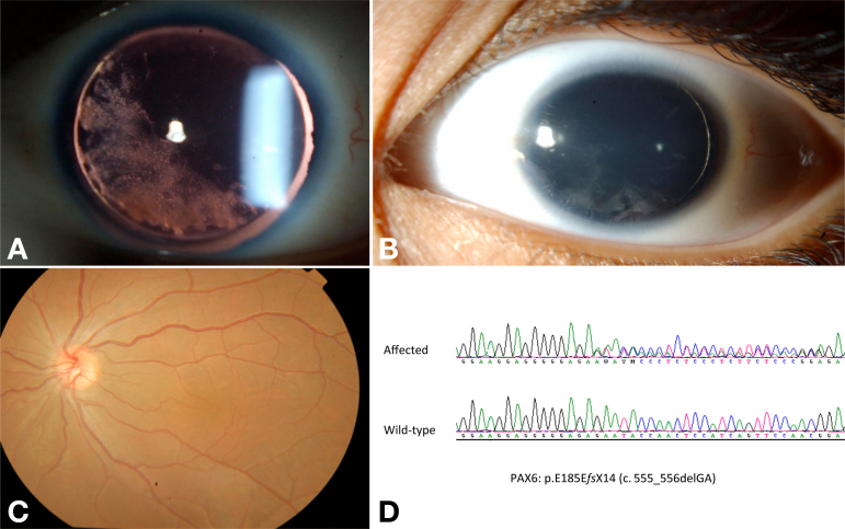
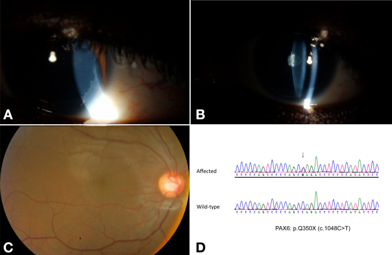
Twelve probands (four familial, eight sporadic) were included in the study – four boys and eight girls who ranged from 4 months to 25 years of age. Two of the familial cases were previously-reported [14]. All patients were products of consanguineous unions except for three cases, one of which was endogamous. None had associated non-ocular congenital malformation or complicated birth history and all had had comprehensive physical examination by a pediatrician. All probands had lens opacity, foveal hypoplasia, and gross lack of iris; in addition, all probands had keratopathy except for two sporadic cases (patients 7 and 12 in Table 1). All four familial cases and six of the eight sporadic cases had PAX6 mutation, including two novel PAX6 mutations (Table 1, Figure 1, and Figure 2). The two sporadic cases without detectable PAX6 mutation (patients 7 and 9 in Table 1), both from consanguineous families, are discussed further below.
Table 1. Summary of the results of molecular analysis of patients with classic aniridia.
| ID | Inbred? | Age | Sex | # | PAX6 mutation | Gene analysis | Copy number variation | Comment |
|---|---|---|---|---|---|---|---|---|
| 1 |
Consang |
6 |
F |
9 |
p.Arg240X (c.1195C>T) |
N/A |
N/A |
Family from reference [14] |
| 2 |
No |
10 |
F |
4 |
**p.E185EfsX14 (c.555_556delGA) |
N/A |
N/A |
|
| 3 |
Consang |
9 |
F |
3 |
p.Pro39ArgfsX14 (c.112del1) |
N/A |
N/A |
|
| 4 |
Consang |
8 |
F |
4 |
p.Asn273IlefsX91 (c.delA1294) |
N/A |
N/A |
Family from reference [14] |
| 5 |
Consang |
25 |
M |
1 |
p.Arg240X (c.718C>T) |
N/A |
N/A |
|
| 6 |
Consang |
9 |
M |
1 |
** p.Gln350X (c.1048C>T) |
N/A |
N/A |
developed juvenile glaucoma |
| 7 |
Consang |
8 |
F |
1 |
none |
no mutation found |
none found |
no keratopathy |
| 8 |
No |
7 |
M |
1 |
p.Ala37ProfsX16 (c.109del1) |
N/A |
N/A |
accommodative esotropia |
| 9 |
Consang |
7 |
F |
1 |
none |
homozygous p.Pro297Ser FOXC1 (c.889C>Tr) |
chr11q24.2:125,001,547-125,215,177 (rs114259885;rs112291840) |
|
| 10 |
Consang |
3 |
M |
1 |
p.Ser167X (c.500C>A) |
N/A |
N/A |
optic nerve hypoplasia |
| 11 |
Consang |
1 |
F |
1 |
PAX6 gene deletion (see copy number variation column) |
N/A |
chr11:30,877,006-32,440,841 (1,563,836 bp) |
|
| 12 | Endogom | 4/12 | F | 1 | PAX6 and WT1 gene deletion (see copy number variation column) | N/A | chr11:27,206,264-42,280,976 (15,074,713 bp) | Non-consanguineous but endogamous; no keratopathy |
#denotes number of affected individuals), N/A: not applicable; **denotes novel mutation. Consang: consanguineous; Endogom: endogamous.
Figure 1.
Patient 2, without pharmacologic mydriasis. A: Retroillumination shows lack of iris and the lenticular changes (left eye shown). B: Diffuse illumination reveals surface keratopathy (left eye shown). C: There was no defined fovea by indirect ophthalmoscopy (left eye shown). D: sequencing revealed a novel PAX6 mutation (p.E185EfsX14).
Figure 2.
Patient 6, without pharmacologic mydriasis. A: Slit illumination revealed surface limbal keratopathy (right eye shown). B: Slit illumination shows lack of iris and a posterior lenticular opacity (right eye shown). C: The fovea is not well defined (right eye shown). D: sequencing revealed a novel PAX6 mutation (p.Q350X).
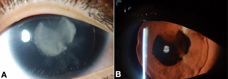
Patient 7 (Figure 3) had classic aniridia in that in addition to lack of iris she had lens opacity and foveal hypoplasia. She had normal genetic and genomic analysis; no underlying cause was found for the ocular phenotype.
Figure 3.
Patient 7, without pharmacologic mydriasis. A, B: In addition to lack of iris, this patient without detectable PAX6 mutation had lenticular opacity and foveal hypoplasia (not shown). Further genetic and genomic analyses were unremarkable. The right eye is shown.
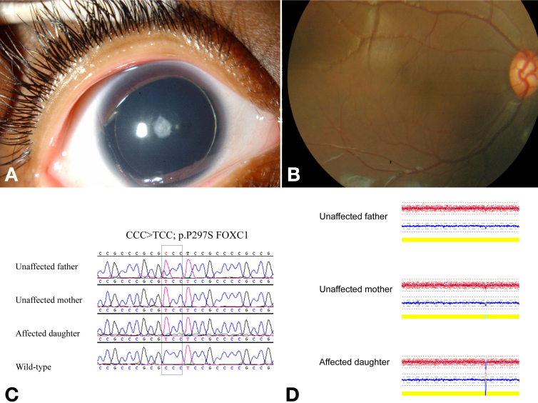
Patient 9 (Figure 4) had classic aniridia in that in addition to lack of iris she had keratopathy, lens opacity, and foveal hypoplasia. She was homozygous for a missense variant in FOXC1 (p.P297S) that was previously been reported as pathogenic and as a cause for dominant anterior segment dysgenesis in two unrelated individuals [15]. However, her father was heterozygous for the variant while her mother was homozygous and neither parent had significant ophthalmic findings despite careful clinical ophthalmic examination with attention to the anterior segment and fovea. Molecular karyotyping revealed homozygosity (nullizygosity) for a chromosome 11q24.2 deletion (chr11:125,001,547–125,215,177 [rs114259885 and rs112291840]) present in the child and heterozygosity (hemizygosity) for the deletion in both parents. Thus the child's phenotype segregated with homozygosity for the FOXC1 missense variant in conjunction with homozygosity for the deletion (Figure 4). The region of this deletion contains five genes (CHK1 checkpoint homolog [CHEK1], acrosomal vesicle protein 1 [ACRV1], prostate and testis expressed 4 [PATE4], prostate and testis expressed 2 [C11ORF38], and FLJ41047), none of which has known ocular function. Neither the FOXC1 variant nor the deletion was found in 100 ethnically-matched controls.
Figure 4.
Patient 9, without pharmacologic mydriasis. A, B: In addition to lack of iris, this patient without detectable PAX6 mutation had anterior lens opacity, limbal keratopathy (not shown), and foveal hypoplasia. C: Further genetic and genomic analyses revealed homozygosity for both a previously-described heterozygous FOXC1 mutation (p.P279S) and a for chromosome 11q24.2 deletion (D) while neither unaffected parent was homozygous for both.
Discussion
In this series of classic aniridia in mostly consanguineous families from Saudi Arabia, the heterozygous PAX6 mutation was detected in 4/4 familial cases and 6/8 sporadic cases. One PAX6-negative case harbored homozygosity for both a previously-reported pathogenic missense FOXC1 variant and for a deletion on chromosome 11q24.2; the unaffected parents were heterozygous or homozygous for the FOXC1 variant and both were heterozygous for the deletion. The other PAX6-negative case remained idiopathic despite candidate gene sequencing and whole genome copy number analysis.
Studies of consanguineous families are more likely to uncover a recessive cause for a given phenotype if a recessive cause exists because of parental shared recent ancestry. Every individual is a heterozygous carrier for mutated alleles that would potentially cause recessive disease in the homozygous (or compound heterozygous) state. Consanguineous marriage increases the expression of rare recessive disease because unless carriers are related, they are unlikely to marry a partner who carries the same disorder [16]. In the current study of classic aniridia of mostly inbred families, 3/3 familial cases from consanguineous families and 6/8 sporadic cases from consanguineous or endogamous families harbored heterozgyous PAX6 mutation. Thus even in the setting of enhanced homozygosity from recent shared parental ancestry, heterozgyous PAX6 mutation typically underlies the phenotype of classic aniridia.
Two patients, both from consanguineous families, had no detectable PAX6 mutation. Although no genetic or genomic cause for classic aniridia was found in one (Patient 7, Figure 3), in the other (Patient 9, Figure 4) there was a homozygous FOXC1 missense variant previously reported as responsible for anterior segment dysgenesis in the heterozygous state [15]. However, both unaffected and otherwise normal parents of the patient harbored this variant – in the heterozygous state in the father and in the homozygous state in the mother – and both unaffected parents had no significant ophthalmic findings. Additional analysis by array CGH revealed a homozygous deletion in the child on chromosome 11q24.2 that was present in the heterozygous state in both parents. One can speculate that either the homozygous 11q24.2 deletion alone or in concert with the homozygous FOXC1 missense variant was associated with the classic aniridia phenotype. The latter seems more plausible as the genes affected by the deletion do not have known ocular function while p.P297S FOXC1 has been previously described as pathogenic [15]. Functional work suggests that p.P297S FOXC1 alters interaction with other yet unidentified factors involved in FOXC1 transactivation and degradation, thus extending the half-life of the protein and causing an increased dosage effect akin to the mechanism of FOXC1 duplication [15]. Consistent with this, cases associated with p.P297S FOXC1 as well as those associated with FOXC1 duplication have been reported with phenotypes of iridogoniodysgeneis that can resemble the iris appearance of patients with classic anirida [15,17]. While the genes contained within this 11q24.2 deletion are not known to be involved in eye development, such a role has not been excluded. In addition, we cannot exclude the possibility that non-coding regulatory elements for PAX6 may exist within this region. Either of these two hypothesized causal links could be either sufficient or dependent on the FOXC1 mutation. Patient 9 also had developmental delay, which may or may not have been related to the deletion alone or in combination with p.P297S FOXC1.
For a particular mutation in a given gene, differences in a given individual's background genotype and environmental exposure cause intrafamilial phenotypic variability. Intrafamilial phenotypic variability has been well documented for FOXC1-related disease [5,9]. Background genomic copy number variation – both deletion and repeats – can increase susceptibility to ocular disease and contribute to phenotypic variability [18,19]. In patient 9 from this study (Figure 4), the homozygous deletion may have been responsible for the expression of a p.P279S FOXC1-related anterior segment phenotype, i.e., the homozygous deletion is a genomic explanation for the intrafamilial phenotypic variability and the observed classic aniridia in patient 9. Although there have been reports of lack of visible iris in patients with heterozygous FOXC1 [20] as well as heterozygous PITX2 [21] mutations, these previously-reported patients would not be considered classic aniridia as they did not have documented keratopathy, lens opacity, or foveal hypoplasia. In addition, the previously-reported patient [20] with lack of iris and heterozgyous FOXC1 mutation also had obvious newborn glaucoma, which is not part of classic aniridia [10]. There is a previous report of gross lack of iris and concurrent keratopathy associated with heterozygous FOXC1 mutation; this was in a baby boy who also had severe newborn glaucoma [22]. Again, his phenotype would not be considered classic aniridia because of newborn glaucoma [10].
Our finding of a homozygous deletion of a previously unreported copy number variation in a patient with this phenotype could be interpreted as a rare recessive form. However, this case has to be viewed in the context of the FOXC1 mutation and lack of other siblings in whom we can verify the segregation pattern of this deletion with respect to the classic aniridia phenotype. In other words, we caution against the over-interpretation of this finding as an example of a recessive form of aniridia at this time. It is hoped that our ongoing analysis of more classic aniridia cases in our highly consanguineous population will address this possibility. Future studies with even higher resolution microarrays are needed to further investigate the potential role of genomic copy number variation in classic aniridia patients without PAX6 mutations.
Acknowledgments
This study was funded in part by a KACST grant (08-MED497–20) to F.S.A. The corresponding author had access to all data, confirms the integrity of all data, and confirms the accuracy of data analysis.
References
- 1.Nelson LB, Spaeth GL, Nowinski TS, Margo CE, Jackson L. Aniridia. A review. Surv Ophthalmol. 1984;28:621–42. doi: 10.1016/0039-6257(84)90184-x. [DOI] [PubMed] [Google Scholar]
- 2.Prosser J, van Heyningen V. PAX6 mutations reviewed. Hum Mutat. 1998;11:93–108. doi: 10.1002/(SICI)1098-1004(1998)11:2<93::AID-HUMU1>3.0.CO;2-M. [DOI] [PubMed] [Google Scholar]
- 3.van Heyningen V, Hoovers JM, de Kraker J, Crolla JA. Raised risk of Wilms tumour in patients with aniridia and submicroscopic WT1 deletion. J Med Genet. 2007;44:787–90. doi: 10.1136/jmg.2007.051318. [DOI] [PMC free article] [PubMed] [Google Scholar]
- 4.Lee H, Khan R, O'Keefe M. Aniridia: Current pathology and management. Acta Ophthalmol. 2008;86:708–15. doi: 10.1111/j.1755-3768.2008.01427.x. [DOI] [PubMed] [Google Scholar]
- 5.Alward WL. Axenfeld-Rieger syndrome in the age of molecular genetics. Am J Ophthalmol. 2000;130:107–15. doi: 10.1016/s0002-9394(00)00525-0. [DOI] [PubMed] [Google Scholar]
- 6.Nishimura DY, Searby CC, Alward WL, Walton D, Craig JE, Mackey DA, Kawase K, Kanis AB, Patil SR, Stone EM, Sheffield VC. A spectrum of FOXC1 mutations suggests gene dosage as a mechanism for developmental defects of the anterior chamber of the eye. Am J Hum Genet. 2001;68:364–72. doi: 10.1086/318183. [DOI] [PMC free article] [PubMed] [Google Scholar]
- 7.Lehmann OJ, Ebenezer ND, Jordan T, Fox M, Ocaka L, Payne A, Leroy BP, Clark BJ, Hitchings RA, Povey S, Khaw PT, Bhattacharya SS. Chromosomal duplication involving the forkhead transcription factor gene FOXC1 causes iris hypoplasia and glaucoma. Am J Hum Genet. 2000;67:1129–35. doi: 10.1016/s0002-9297(07)62943-7. [DOI] [PMC free article] [PubMed] [Google Scholar]
- 8.Shields MB, Buckley E, Klintworth GK, Thresher R. Axenfeld-Rieger syndrome. A spectrum of developmental disorders. Surv Ophthalmol. 1985;29:387–409. doi: 10.1016/0039-6257(85)90205-x. [DOI] [PubMed] [Google Scholar]
- 9.Honkanen RA, Nishimura DY, Swiderski RE, Bennett SR, Hong S, Kwon YH, Stone EM, Sheffield VC, Alward WL. A family with axenfeld-rieger syndrome and peters anomaly caused by a point mutation (Phe112Ser) in the FOXC1 gene. Am J Ophthalmol. 2003;135:368–75. doi: 10.1016/s0002-9394(02)02061-5. [DOI] [PubMed] [Google Scholar]
- 10.Khan AO, Aldahmesh MA, Al-Abdi L, Mohamed JY, Hashem M, Al-Ghamdi I, Alkuraya FS. Molecular characterization of newborn glaucoma including a distinct aniridic phenotype. Ophthalmic Genet. 2011 doi: 10.3109/13816810.2010.544365. [DOI] [PubMed] [Google Scholar]
- 11.Semina EV, Ferrell RE, Mintz-Hittner HA, Bitoun P, Alward WL, Reiter RS, Funkhauser C, Daack-Hirsch S, Murray JC. A novel homeobox gene PITX3 is mutated in families with autosomal-dominant cataracts and ASMD. Nat Genet. 1998;19:167–70. doi: 10.1038/527. [DOI] [PubMed] [Google Scholar]
- 12.Jamieson RV, Perveen R, Kerr B, Carette M, Yardley J, Heon E, Wirth MG, van Heyningen V, Donnai D, Munier F, Black GC. Domain disruption and mutation of the bZIP transcription factor, MAF, associated with cataract, ocular anterior segment dysgenesis and coloboma. Hum Mol Genet. 2002;11:33–42. doi: 10.1093/hmg/11.1.33. [DOI] [PubMed] [Google Scholar]
- 13.Alkuraya FS. Autozygome Decoded. Genet Med. 2010;12:765–71. doi: 10.1097/GIM.0b013e3181fbfcc4. [DOI] [PubMed] [Google Scholar]
- 14.Khan AO, Aldahmesh MA. PAX6 analysis of two unrelated families from the Arabian Peninsula with classic hereditary aniridia. Ophthalmic Genet. 2008;29:145–8. doi: 10.1080/13816810802078195. [DOI] [PubMed] [Google Scholar]
- 15.Fetterman CD, Mirzayans F, Walter MA. Characterization of a novel FOXC1 mutation, P297S, identified in two individuals with anterior segment dysgenesis. Clin Genet. 2009;76:296–9. doi: 10.1111/j.1399-0004.2009.01210.x. [DOI] [PubMed] [Google Scholar]
- 16.Modell B, Darr A. Science and society: Genetic counselling and customary consanguineous marriage. Nat Rev Genet. 2002;3:225–9. doi: 10.1038/nrg754. [DOI] [PubMed] [Google Scholar]
- 17.Strungaru MH, Dinu I, Walter MA. Genotype-phenotype correlations in Axenfeld-Rieger malformation and glaucoma patients with FOXC1 and PITX2 mutations. Invest Ophthalmol Vis Sci. 2007;48:228–37. doi: 10.1167/iovs.06-0472. [DOI] [PubMed] [Google Scholar]
- 18.Zhou J, Hu J, Guan H. The association between copy number variations in glutathione S-transferase M1 and T1 and age-related cataract in a Han Chinese population. Invest Ophthalmol Vis Sci. 2010;51:3924–8. doi: 10.1167/iovs.10-5240. [DOI] [PubMed] [Google Scholar]
- 19.Schmid-Kubista KE, Tosakulwong N, Wu Y, Ryu E, Hecker LA, Baratz KH, Brown WL, Edwards AO. Contribution of copy number variation in the regulation of complement activation locus to development of age-related macular degeneration. Invest Ophthalmol Vis Sci. 2009;50:5070–9. doi: 10.1167/iovs.09-3975. [DOI] [PubMed] [Google Scholar]
- 20.Khan AO, Aldahmesh MA, Al-Amri A. Heterozygous FOXC1 mutation (M161K) associated with congenital glaucoma and aniridia in an infant and a milder phenotype in her mother. Ophthalmic Genet. 2008;29:67–71. doi: 10.1080/13816810801908152. [DOI] [PubMed] [Google Scholar]
- 21.Perveen R, Lloyd IC, Clayton-Smith J, Churchill A, van Heyningen V, Hanson I, Taylor D, McKeown C, Super M, Kerr B, Winter R, Black GC. Phenotypic variability and asymmetry of rieger syndrome associated with PITX2 mutations. Invest Ophthalmol Vis Sci. 2000;41:2456–60. [PubMed] [Google Scholar]
- 22.Ito YA, Footz TK, Berry FB, Mirzayans F, Yu M, Khan AO, Walter MA. Severe molecular defects of a novel FOXC1 W152G mutation result in aniridia. Invest Ophthalmol Vis Sci. 2009;50:3573–9. doi: 10.1167/iovs.08-3032. [DOI] [PubMed] [Google Scholar]


