

Abstract
RNA localization is a common mechanism for regulating cell structure and function. Localized RNAs in Xenopus oocytes are critical for early development, including germline specification by the germ plasm. Despite the importance of these localized RNAs, only approximately 25 have been identified and fewer are functionally characterized. Using microarrays, we identified a large set of localized RNAs from the vegetal cortex. Overall, our results indicate a minimum of 275 localized RNAs in oocytes, or 2–3% of maternal transcripts, which are in general agreement with previous findings. We further validated vegetal localization for 24 candidates and further characterized three genes expressed in the germ plasm. We identified novel germ plasm expression for reticulon 3.1, exd2 (a novel exonuclease-domain encoding gene), and a putative noncoding RNA. Further analysis of these and other localized RNAs will likely identify new functions of germ plasm and facilitate the identification of cis-acting RNA localization elements.
Keywords: Xenopus, germ plasm, vegetal cortex, localized RNAs, microarray, primordial germ cells
INTRODUCTION
Adult germ cells arise during embryogenesis from a set of pluripotent precursor cells, the primordial germ cells (PGCs). In many organisms, including most invertebrates, fish, Anuran amphibians, and birds, PGC differentiation requires the inheritance of germ plasm from the egg (Eddy, 1975). Germ plasm is morphologically similar across species, typically forming a distinct assembly of mitochondria, and ribonucleoprotein-rich germinal granules within an electrondense fibrillar material (Beams and Kessel, 1974). Mammalian and urodele embryos generate PGCs through inductive signaling (Nieuwkoop and Sutasurya, 1976) and lack maternally inherited germ plasm during early development. Germ plasm does, however, accumulate during the meiotic stages of PGC development in these organisms. More recent molecular characterization of the germ plasm has identified numerous RNA-binding proteins, implicating regulation of RNA metabolism, transport, and translation in PGC development (reviewed in Houston and King, 2000a). Other germ plasm localized molecules have been identified that mediate transcriptional repression, regulate migration, and control mitotic divisions in PGCs (reviewed in Seydoux and Braun, 2006). Additionally, germ plasm-specific proteins such as Vasa (Tanaka et al., 2000) and Tudor (Chuma et al., 2006) have conserved roles in PGC formation and germ plasm assembly from flies to mammals, suggesting that germ plasm is universally important for germline development. Despite its importance, a comprehensive molecular picture of germ plasm is only beginning to emerge as the functions of its individual components are elucidated.
Genetic screens in Drosophila and Caenorhabditis elegans have identified numerous molecules important for germ plasm function (reviewed in Wylie, 1999). In vertebrates, where such screens are not practical, germ plasm-localized RNAs have been readily cloned from Xenopus oocytes and embryos, taking advantage of their large size, amenability to micromanipulation, and abundant maternal RNA stores. Germ plasm-localized RNAs are one main class of localized RNAs in Xenopus, the other being the so-called late pathway, which operates beginning around stage IV of oogenesis and results in RNAs becoming broadly localized throughout the cortex of the vegetal hemisphere (reviewed in King et al., 2005).
The first vertebrate germ plasm mRNA identified was the Nanos-like gene, xcat2/nanos1 (nos1), which was discovered as a cytoskeleton-associated transcript (Mosquera et al., 1993). The localization pattern of nos1 mirrors that of the germ plasm itself, first becoming localized to the mitochondrial cloud/Balbiani Body of previtellogenic (stage I) oocytes (Forristall et al., 1995), the source of the germ plasm (Heasman et al., 1984). The nos1 mRNA is translocated to the vegetal pole along with the cloud material during the early vitellogenic stages of oogenesis and is colocalized with germ plasm throughout early embryogenesis (Forristall et al., 1995). Additional germ plasm-localized RNAs include dazl, deadsouth, dead end (dnd), and hermes, which encode RNA-binding proteins; and xpat, germes, and fatvg/adipophilin (adfp), which encode novel proteins and a lipid droplet-associated protein, respectively (reviewed in King et al., 2005). Functional studies, using antisense-mediated depletion of maternal mRNAs, identified a role for dazl in the migration and differentiation of PGCs (Houston and King, 2000b). Similar migration defects have been found in embryos depleted of Dnd (both fish and frog; Weidinger et al., 2003, Horvay et al., 2006), Nos1 (fish; Köprunner et al., 2001), and Grip2.1 (frog; Tarbashevich et al., 2007), suggesting that germ plasm is involved in regulation of the migratory phenotype, potentially through the regulation of germ cell-specific RNA metabolism.
Recently, maternal depletion studies have shown that a subset of germ plasm-localized mRNAs adfp (Chan et al., 2007), wnt11b (Tao et al., 2005), and trim36 (Cuykendall and Houston, 2009) are required for dorsal axis formation. It is possible that germ plasm has additional roles in controlling patterning of the early embryo, roughly analogous to the role of pole plasm in Drosophila posterior fate specification. In this work, we describe a genomics approach to identify vegetally localized RNAs using RNA from dissected Xenopus vegetal cortices to probe microarrays. Among the RNAs identified by this approach, we report several new RNAs that localize to the germ plasm, including a novel exonuclease with potential roles in RNA interference and an mRNA encoding an endoplasmic reticulum component. We further describe the expression of a putative noncoding RNA enriched in the germ plasm. Functional and bioinformatics analyses of these RNAs should lead to more comprehensive models of germ plasm function and RNA localization mechanisms.
RESULTS AND DISCUSSION
Microarray Analysis of mRNAs Enriched in the Vegetal Cortex
To profile the expression of localized mRNAs in Xenopus, we used microarray analysis to identify genes with enriched expression in the cortex of the full-grown oocyte. To obtain a highly enriched population of localized mRNAs, we manually dissected vegetal cortices from stage VI oocytes and extracted total RNA. The mass of total cortical RNA recovered was approximately 66 ng per cortex, which is in line with previous estimates of 100 ng per cortex (Elinson et al., 1993). To verify that these samples were enriched in localized RNAs, we compared the expression of two known localized mRNAs, vegt and wnt11, by reverse transcriptase-polymerase chain reaction (RT-PCR) using cDNA from vegetal cortex and whole oocyte samples. Both genes were found more abundantly in the cortex than in the whole oocyte (data not shown), whereas a ubiquitous RNA, ornithine decarboxylase (odc), was less abundant in the cortex sample.
RNAs from two biological replicates of cortices and intact oocytes were obtained from different females and used to generate labeled complementary RNA by a two-step linear amplification method. These probes were used to hybridize Affymetrix microarrays (Xenopus Genome Array), which are printed with sense probe sets to 14,400 transcripts present in Xenopus laevis expressed sequence tag (EST) databases, including previously characterized genes as well as uncharacterized genes. Data were analyzed by GeneSifter software and the results sorted according to the ratio of increase in the vegetal cortex versus whole oocyte. We identified 430 sequences with statistically significant elevation in the cortex (t-test; P < 0.05), all of which were enriched by at least 1.5-fold. Of these, 282 sequences were present in equal or greater abundance than the least expressed known localized mRNA, hermes (UniGene ID Xl. 449), which was enriched 2.92-fold. These sequences are thus likely to represent bona fide localized mRNAs. In addition to hermes, we also found significant enrichment of all other known localized RNAs that were present on the array (Table 1), thus validating this approach for identifying localized mRNAs. Because some genes have multiple probes sets represented on the array (e.g., xoo1, camk2g), we estimate the minimum number of localized RNAs at approximately 275. This represents approximately 2–3% of the estimated number of maternally expressed genes (~10,000). Of interest, these percentages are consistent with the estimates obtained from analytical hybridization experiments of vegetally enriched poly(A)+ RNAs of around 3–5% of the total mRNA population (Carpenter and Klein, 1982).
TABLE 1.
Summary of Microarray Data for Known Localized RNAsa
| Ratio | Gene Identifier | Probe ID | Gene Name | UniGene Cluster | Gene ID [ref] |
|---|---|---|---|---|---|
| 18.56 | U31427 | Xl.302.1.S1_at | Ephrin-B1 (EPH family ligand) | Xl.302 | efnb1 [1] |
| 16.04 | BF048890 | Xl.1145.1.S1_at | Xcat-2 protein | Xl.1145 | xcat2 [2] |
| 14.56 | AY172320 | Xl.7252.1.S1_at | Germes | Xl.7252 | germes [3] |
| 10.57 | BM180550 | Xl.14891.1.S1_at | Glutamate receptor interacting protein 2 | Xl.64243 | xgrip2 [4, 5] |
| 9.24 | AF190623 | Xl.670.1.S1_at | DEADSouth RNA helicase | Xl.670 | dead south [6] |
| 8.66 | BC046740 | Xl.7122.1.S1_at | Similar to fatty acid Coenzyme A ligase | Xl.7122 | acsl1 [7] |
| 8.44 | AF224746 | Xl.641.1.S1_at | Bicaudal-C | Xl.641 | bic-C [8] |
| 8.43 | L23542 | Xl.1073.1.S1_at | Maternal protein | Xl.24008 | wnt11 [9,10] |
| 7.53 | BE189094 | Xl.12195.1.A1_at | Transcribed locus | Xl.71390 | arhbeta [11] |
| 7.32 | AF017778 | Xl.311.1.S1_at | DAZ-like protein | Xl.311 | dazl [12] |
| 7.18 | AY029294 | Xl.781.1.S1_at | Transcription factor Otx1, orthodenticle homolog 1 | Xl.781 | otx1 [13] |
| 6.93 | AJ002384 | Xl.38.1.S1_at | Xpat protein | Xl.38 | xpat [14] |
| 6.37 | BI448233 | Xl.18931.1.A1_at | NIF protein | Xl.18931 | nif [15] |
| 6.30 | AF061982 | Xl.476.1.S1_at | CCCH zinc finger protein C3H-3 | Xl.476 | c3h-3 [1] |
| 5.06 | BE026894 | Xl.25471.1.A1_at | Dead end protein | Xl.29785 | dead end [16] |
| 4.61 | BM191810 | Xl.12012.2.A1_at | Beta-transducin repeat containing | Xl.968 | betatrcp [17] |
| 4.11 | AF184090 | Xl.576.1.S1_at | Fatvg protein | Xl.29614 | fatvg [18] |
| 3.77 | BG016938 | Xl.25780.1.S1_at | Transforming growth factor-beta Vg1 gene | Xl.25780 | vg1 [19, 20] |
| 3.72 | BM262613 | Xl.3767.1.A1_at | weakly similar to (Asp-Glu-Ala-Asp) box polypeptide 43 | Xl.3767 | centroid [21] |
| 3.56 | U89707 | Xl.1775.1.S1_at | Brachyury- and Tbx-related protein | Xl.1775 | vegt [22] |
| 2.89 | AF107889 | Xl.449.1.S1_at | RRM-type RNA-binding protein hermes | Xl.449 | hermes [23] |
RNAs with previously described vegetal localization ranked by the ratio of mean signal in cortex samples compared with oocyte samples. Significance was assessed by t-test, using the Benjamini and Hochberg correction. Other known vegetal cortex RNAs, xlsirt 13.2 (Kloc et al., 1993) and xvelo1 (Claussen and Pieler, 2004) do not have antisense targets on the array. Probe ID is the Affymetrix probe set number.
References:
Germ plasm-localized transcripts are shaded.
A list of the top 120 enriched sequences is shown in Supp. Table S1, which is available online (enriched greater than six-fold). The raw data from our experiments were deposited in the GEO database under accession number GSE17713 (Supp. Table S1).
We assessed the predicted molecular function of these cortex-enriched transcripts by using BLAST and Gene Ontology and analyses. Over 70% of the genes are uncharacterized or non-annotated, encoding either a transcribed locus or hypothetical protein. Of the remaining genes, we identified six RNA-binding proteins, 15 zinc finger proteins, and nine transcription factors. We also identified localized RNAs for growth factor receptors not previously implicated in maternal signaling events, including fgfr2 (Xl.16135), igfr1b (Xl.269), and alk2 (Xl. 722). In addition to alk2, other members of the bone morphogenetic protein (BMP) signaling pathway were found in the set of localized genes, including tob2 (Xl.12100) and madh2/smad7 (Xl.1228), suggesting a possible role for maternal BMP signaling in oogenesis or early development. Numerous genes for metabolic proteins and those linked to endoplasmic reticulum function were also enriched in the vegetal cortex, highlighting this region as one of high metabolic activity.
Identification of Novel Localized mRNAs
To confirm the cortical localization of genes identified through the array, we synthesized primers against 25 top-scoring and uncharacterized sequences that were linked to full-length cDNAs, and verified their enrichment by means of RT-PCR. We initially focused on full-length cDNAs so we could readily obtain functional constructs for gain-of-function analysis in embryos. Of this set, 23 sequences were enriched in the cortex, one was apparently not enriched in the cortex (Xl.13266.1) and one clone was not detected (Fig. 1). Interestingly, the putatively unlocalized gene encoded arhβ, which is known to encode both localized and unlocalized transcripts (Zhou et al., 2004). Analysis of our primer sequences showed that they recognized predominantly the unlocalized form, whereas the sequences on the array recognized the localized form (not shown).
Fig. 1.

Confirmation of vegetal cortex enrichment of selected genes. Reverse transcriptase-polymerase chain reaction (RT-PCR) analysis of novel cortex-enriched transcript expression in whole oocyte (Oo) and vegetal cortex (VgCx) samples. The −RT lane is an oocyte sample processed in the absence of reverse transcriptase. ornithine decarboxylase (odc) is shown as a loading control.
Because RT-PCR analysis is unable to distinguish the spatial expression patterns of localized mRNAs within the vegetal cortex, we next used in situ hybridization to identify any germ plasm-expressed mRNAs in our set of candidates. We obtained cDNA clones for the 23 genes described above, plus the clone corresponding to the top hit overall. Four of these genes were expressed in the germ plasm (see below): trim36 (Xl. 6926), rtn3.1 (Xl.57382), transcribed locus (Xl.29596), and exd2 (Xl.6566). To date, approximately 12 of the ~25 known localized RNAs show germ plasm localization (Table 1), whereas ~17% of the genes from our set of initial candidates were in the germ plasm. This could be due to chance, or could suggest that germ plasm-localized RNAs were isolated more readily using molecular cloning methods. Alternatively, because we chose full-length cDNAs to characterize, germ plasm RNAs might be less well represented in full-length libraries. A more complete characterization of the expression patterns of localized RNAs will be necessary to determine the true contribution of germ plasm-localized RNAs to the overall localized RNA population.
We recently described and characterized trim36, showing its role regulating cortical rotation and dorsal axis formation (Cuykendall and Houston, 2009); the others we describe in more detail in the latter part of this work. The remaining 20 candidate genes were broadly localized throughout the cortex of the vegetal hemisphere of stage VI oocytes (Fig. 2), and expressed uniformly in early stage oocytes (data not shown). We also determined the zygotic expression for these genes to identify any tissue-specific or otherwise interesting patterns. We examined gastrula (stage 11), neurula (stage 18) and tail bud (stage 30) stages, in addition to stage VI oocytes. Shown in Figure 2 are the results for the 16 of these candidate genes, excluding those that have been described to date (igfr1b, Xl.269; Richard-Parpaillion et al., 2002; and acyl coa synthetase long chain 1 [acsl1], Xl.7122), and those that are the subject of ongoing functional studies in our lab (sim to borg4, Xl.15910, and trim69, Xl.14571). Few of these genes had highly tissue-specific expression, with many genes showing ubiquitous expression or enrichment in the neural plate. Some of the notable examples are described: (1) serca3 (Xl. 1572) is expressed in a scattered population of cells in the epidermis which do not co-stain for tubulin (data not shown). These cells likely represent scattered small cells derived from the deep ectodermal layer, a population of cells recently characterized by Hayes et al. (2007), the function of which is not known. (2) milk fat globule-egf factor 8 (mfge8, Xl.5125) shows enriched expression in the tail bud fin epidermis. (3) sestrin 1 (sesn1, Xl.6911) is expressed throughout the gastrula and becomes highly expressed in the notochord and epidermis of the neurula. Interestingly, by the tail bud, sesn1 becomes weakly expressed in the foregut endoderm. (4) zinc finger, an1-type domain 3 (zfand3, Xl.26137) is broadly expressed in the anterior neural plate at stage 18 and then becomes localized to the forebrain and the midbrain–hindbrain boundary, as well as showing expression in the eye and spinal cord. (5) calcium binding protein 39 (cab39, Xl.5662) and trio and f-actin binding protein (triobp, Xl.12480) are expressed in the developing pronephros in tail bud embryos. cab39 is highly expressed throughout the embryo, whereas triobp also shows a highly enriched staining in differentiating neurons flanking the otic vesicle. In summary, although these genes are all localized to the vegetal cortex during oogenesis, their zygotic expression patterns are quite dissimilar. In the remainder of this work, we focus on the genes expressed in the germ plasm.
Fig. 2.
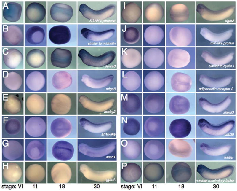
Expression of non-germ plasm localized RNAs. In situ hybridizations for each mRNA, each set of four columns shows stage VI oocytes (left panel), stage 11 embryos (second panel), stage 18 embryos (third panel), and stage 30 embryos (right hand panel). Oocytes and stage 11 embryos are vegetal views; stage 18 embryos are dorsal views, anterior to the left; and stage 30 embryos are lateral views, anterior to the left.
Germ Plasm Expression of Previously Identified Genes, rtn3.1 and a Putative Noncoding RNA
We identified a sequence representing reticulon 3.1 (rtn3.1) in the set of vegetal cortex-enriched transcripts, which was increased ~8.6-fold compared with the whole oocyte (data not shown). Proteins of the reticulon family are thought to associate with the endoplasmic reticulum and may have roles in regulating apoptosis in neurons (Park et al., 2005). rtn3.1 is the only family member to be maternally expressed in Xenopus, and the embryonic expression and sequence analysis of rtn3.1 has been previously reported (Park et al., 2005), although its expression in the germ plasm was not noted. We used a full-length rtn3.1 clone as a probe and analyzed its expression at different stages by whole-mount in situ hybridization, focusing on oocyte and early embryo stages. Our in situ results generally agreed with the Park et al. (2005) study, including evidence of expression in the animal pole of the blastula, neural plate, branchial arches, and epidermis (Fig. 3). In addition, we identified expression in germ plasm islands at the vegetal apex of full-grown oocytes and in clustered germ plasm at the cleavage stages (Fig. 3A,B). Also, rtn3.1 expression was evident in cells at the vegetal pole of some early gastrula stage embryos (Fig. 3C). This is consistent with the position of germ plasm-containing cells at this stage (Whitington and Dixon, 1975; Hudson and Woodland, 1998), and most likely represents a small number of PGCs that have not yet been fully internalized into the endoderm. We also observed this expression pattern for the other germ plasm RNAs described in this study. Although rtn3.1 is enriched in germ plasm, it is also expressed at a lower level ubiquitously at all stages examined.
Fig. 3.

Expression and germ plasm localization of rtn3.1. In situ hybridization against rtn3.1. A: Stage VI oocyte, vegetal view. B: Two-cell embryo, vegetal view. C: Stage 10, vegetal view. The arrows in A–C indicate rtn3.1 expression in germ plasm. D: Stage 18, anterior view, showing ubiquitous expression and enrichment in the neural plate. E: Stage 24, lateral view, showing enrichment of rtn3.1 in the eye (ey) and branchial arches (ba).
The most highly enriched sequence in our analysis was described only as transcribed locus (Xl.29596; 45-fold enriched). Examination of the Gen-Bank record (AB331172; Kataoka, K. and Orii, H. 2007, Direct Submission) indicated a designation of noncoding RNA enriched in germ plasm. This sequence was originally identified in another screen for genes expressed in the vegetal hemisphere (Kataoka et al., 2005), but its expression pattern was not presented in detail. We obtained a slightly longer clone commercially (BQ383794) and performed whole-mount in situ hybridization to determine its expression pattern. Consistent with the previous report (Kataoka et al., 2005), we detected robust expression in the germ plasm of oocytes and early embryos through the early gastrula stage (Fig. 4). Later, this transcript was detected in the intersomitic boundaries and in the branchial arches. Expression in the germ plasm and PGCs was undetectable after gastrulation, suggesting the gene is absent from migrating PGCs (Fig. 4). For this work, we have designated this gene germ plasm transcribed locus 1 (gptl1).
Fig. 4.

Expression and germ plasm localization of gptl1. In situ hybridization against gptl1. A: Early stage oocytes (stage I–II, left panel; stage III–IV, right panel). Roman numerals indicate oocyte stages; mc, mitochondrial cloud. B: Stage VI oocyte, vegetal view. C: Four-cell embryo, vegetal view. D: Stage 10.5, vegetal view. E: Stage 30 embryo, cleared in Murray’s clear, showing expression in branchial arches and intersomitic boundaries and the absence of staining in primordial germ cells (PGCs). Inset: in situ for xpat showing positive PGC staining in a cleared embryo.
To try to verify whether gptl1 was indeed an untranslatable noncoding RNA, we performed basic sequence analysis and database searches. The transcript has four open reading frames (ORF), ranging from 35 to 64 amino acids, all encoding novel proteins (data not shown). Database searches of nonredundant nucleotide and protein databases yielded no significant homologies to any of the predicted protein sequences. The start codons of these ORFs also have indications of functional initiation codons (not shown). The GenBank description of the original clone indicated that the 5′ end was identical to the 5′ end of U2 snRNA. The 5′ end of the clone we obtained differs from the reported sequence, reducing the similarity to U2 snRNA to 93% over 29 nucleotides, an insignificant E value (0.003; data not shown). It is unclear that these sequence data alone can accurately predict a noncoding RNA function for gptl1. Arguments such as predicted nontranslatability and lack of homologous sequences were used to predict that polar granule component (pgc) acts as a noncoding RNA (Nakamura et al., 1996), although recent data suggest that pgc is translated into a small, Drosophila spp.-specific polypeptide, which functions in transcriptional silencing (Hanyu-Nakamura et al., 2008). Functional data will likely also be needed to determine the role of the gptl1 transcript in Xenopus development.
Sequence and Expression Analysis of a Novel Germ Plasm RNA, exonuclease 3′-5′ domain containing 2 (exd2)
Analysis of the full-length exd2 EST clone identified a novel ~2500 n.t. cDNA, including a 1928 n.t. coding region (CDS), preceded by an in-frame stop codon. The translated sequence of the cDNA encodes a putative protein of 642 amino acids. BLAST searches revealed conserved predicted proteins encoded in sequenced mammalian genomes, including human and mouse. Exd2 proteins contain a conserved 3′–5′ exonuclease domain of the DnaQ-like exonuclease superfamily, which includes the proofreading domains of DNA polymerases and the exonuclease RNase D. Specifically, this domain in Exd2 is within the WRN-exo family of 3′–5′ exonuclease domains, related to the exonuclease domain found in the Werner Syndrome protein. This domain contains three conserved motifs harboring acidic residues involved in magnesium binding and a conserved tyrosine required for catalysis (DEDDy). The protein sequences surrounding these residues are conserved in the mouse and human Exd2 proteins (Fig. 5). In addition, the carboxy-terminus of the Exd2 protein is well conserved, although there are no discernable functional motifs in that region (Fig. 5). We identified a single orthologue for exd2 in the Xenopus tropicalis genome, which was >95% identical to the X. laevis gene at the nucleic acid level (data not shown). Shared synteny comparisons across species using the Metazome database (www.metazome.com) showed that Xenopus tropicalis exd2 occupies a conserved relative position throughout vertebrate genomes.
Fig. 5.

Amino acid sequence alignment of Exd2 proteins. Alignment of Xenopus laevis Exd2 with human and mouse Exd2 proteins (hsaEXD2 and mmuExd2, respectively). Identical residues are shown in black, similar residues in gray. Motifs conserved in 3′–5′ exonucleases are circled and indicated to the right (ExoI, ExoII, and ExoIII); acidic residues involved in magnesium ion coordination are labeled with asterisks and the catalytic tyrosine residue is indicated by an arrow.
Temporal analysis of exd2 expression by RT-PCR showed abundant maternal transcripts, which declined steadily throughout development (data not shown). Spatial analysis by in situ hybridization showed a pattern of expression consistent with that of the germ plasm. exd2 mRNA is localized to the mitochondrial cloud of stage I previtellogenic oocytes (Fig. 6). In fully grown stage VI oocytes, exd2 is localized in small islands within a compact vegetal expression domain, also consistent with germ plasm. exd2 is also expressed in the germ plasm of fertilized eggs and cleavage stage embryos, clustering in yolk-free islands along vegetal cleavage furrows. Furthermore, transcripts for exd2 remain associated with germ plasm through gastrulation, including the transition to a perinuclear position, a characteristic of germ plasm at this stage (Fig. 6). Expression is absent from PGCs after gastrulation and from migrating PGCs (data not shown). exd2 is not expressed outside the early germline and was not detectable in later neurula and tail bud stage embryos. Interestingly, exd2 expression was robust in spermatogenic cells of the adult testis (Fig. 6), suggesting possible additional roles in male germ cell development or more generally in meiosis.
Fig. 6.

Expression and germ plasm localization of exd2. In situ hybridization against exd2. A: High magnification view of stage I oocytes; mc, mitochondrial cloud. B: Stage IV (left) and stage VI (right) oocytes, vegetal views. C: Four-cell embryos, vegetal view. D: Stage 18, showing lack of expression. E: Stage 7 sagittal section. F: Stage 11 sagittal section. Insets show low power views in E, F. G: Section of adult testis, exd2 antisense probe. H: Sense control on testis.
Although the function of Exd2 is not known, a related C. elegans gene, mut-7, is required for transposon silencing and RNA interference (Ketting et al., 1999). Very little is known about transposon silencing in Xenopus. The Piwi proteins, which have been implicated in multiple aspects of germ cell specification and transposon silencing (Houwing et al., 2007), are expressed in Xenopus eggs (Wilczynska et al., 2009) and a Xenopus Piwi homolog (Xiwi) is enriched in the germ plasm (Lau et al., 2009). Also, the small RNAs bound by Piwi proteins (piRNAs) represent the major class of small RNA in Xenopus oocytes (Lau et al., 2009). It is tempting to speculate that Exd2 is required for transposon silencing either by regulating piRNA biogenesis or activity. It is also possible that Exd2 is required for other aspects of RNA metabolism, because numerous other proteins that control RNA splicing, capping, and translation are also enriched in the germ plasm (reviewed in Seydoux and Braun, 2006).
Analysis of Putative Germ Plasm RNA Localization Elements
Although we have focused on identifying interesting candidate genes for functional analysis, we anticipate that this listing of putative localized RNAs will be useful in identifying cis-acting RNA localization elements. These elements are typically found in the 3′-untranslated regions (UTRs) of localized messages and are characterized by clusters of TTCAC motifs (UUCAC for the RNA sequence), which are recognized by the Vera/Vg1RBP RNA binding protein (King et al., 2005, for review). Additionally, a mitochondrial cloud localization element (MCLS; Chang et al., 2004) has been identified in germ plasm localized RNAs and is characterized by repeats of GCAC. Of interest, clustering of repeats containing CAC and/or Vera binding sites are general to RNA localization mechanisms in species ranging from ascidians to humans (Betley et al., 2002).
To determine the extent that clusters of CAC-containing motifs occur in trim36, rtn3.1, gptl1, and exd2, we analyzed their 3′UTRs using REPFIND, a program developed to identify clustered, nonrandom short repeats in a nucleotide sequence (Betley et al., 2002). We first screened for all significant repeats (P ≤ 1 × 10⁻⁴; Fig. 7A–D), a strategy that has been used to predict functional localization elements (Betley et al., 2002; Andken et al, 2007). trim36 and rtn3.1 showed significant clustering of non–CAC-containing motifs; however, these were not common among the RNAs and did not correspond to known localization motifs (Fig. 7A,B). The exd2 3′UTR lacked significant clusters of repeats, although the X. tropicalis exd2 homologue does have a significant cluster of ATTT (Fig. 7C; P = 2 × 10⁻⁵). Only the gptl1 3′UTR contained significant clusters of CAC-containing motifs. For gptl1, the top scoring cluster of repeats was GCAC (Fig. 7D; P = 7.23 × 10⁻⁹), a sequence critical for mitochondrial cloud localization (Chang et al., 2004).
Fig. 7.

REPFIND analysis of localized RNA 3′-untranslated region (UTR) sequences. A: Upper panel, significant repeat clusters in the 3′UTR from trim36. Only those with P values < 1 × 10⁻⁵ are shown. Lower panel, clusters of putative Vera binding elements in the trim36 3′UTR (P < 0.01). B: Upper panel, significant repeat clusters (P ≤ 0.0001) in the 3′UTR from rtn3.1. Lower panel, clusters of putative CAC-containing elements in the rtn3.1 3′UTR (P < 0.01). C: Upper panel, significant repeat clusters (P ≤ 0.0001) in the 3′UTR from exd2 (X. tropicalis). Lower panel, cluster of putative GCAC-containing elements in the exd2 3′UTR (X. laevis; P < 0.01). D: Upper panel, significant repeat clusters in the 3′UTR from gptl1. Only those with P values < 1 × 10⁻⁶ are shown. Lower panel, clusters of putative Vera binding elements in the gptl1 3′UTR (P < 0.01). The box indicates a region containing clusters of CAC, GCAC, and TTCAC motifs. In all panels, cDNA sequences are shown, and nucleotide positions are indicated across the top, starting at the first nucleotide in the 3′UTR following the stop codon. Colored bars indicate the strongest cluster; individual repeats are separated by different colors.
We next determined specifically the incidence of known motifs, TTCAC and GCAC, using a higher P-value cutoff (0.01). These motifs can found in experimentally defined localization elements with P values between 0.0001 and 0.01 (Betley et al., 2002). We identified two putative Vera binding elements in the 3′UTR of trim36 (P = 0.009) and two putative MCLSs in exd2 (P = 0.0028) (Fig. 7A,C, lower panels). rtn3.1 lacked significant clusters of TTCAC and GCAC, but did have several clusters of different CAC-containing motifs (Fig. 7B, lower panel). gptl1 showed a cluster of two Vera binding sites (P = 0.00125) adjacent to one of its GCAC clusters (see above), suggesting a likely region for the gptl1 localization element (Fig. 7D, lower panel).
Our analysis of putative localization elements in these genes shows a varying degree of prevalence of canonical localization motifs. gptl1 contains tight clusters of well-characterized motifs, enabling a reasonable guess as to where the minimal element might be. By contrast, exd2 contains very few significant repeats and only one cluster of two GCAC elements. rtn3.1 appears to lack Vera binding sites and MCLSs, but does have a few CAC-containing repeats. This observation is consistent with our data showing ubiquitous rtn3.1 expression in addition to localized transcripts. Experimental identification of localization elements in these RNAs should prove useful for comparing the relative roles of existing elements in mediating localization. Also, a more sophisticated bioinformatics analysis of the ~400 localized RNAs we have identified in this study may facilitate the identification of novel cis-localization elements.
EXPERIMENTAL PROCEDURES
Oocytes and Embryos
Xenopus laevis ovary tissue was surgically removed from anesthetized females, divided into small segments and stored at 18°C. Oocytes were manually defolliculated in modified oocyte culture medium (OCM; 70% L-15 media, 0.04% bovine serum albumin, 1 mM Glutamax supplement, 1.0 μg/ml gentimicin, pH 7.6–7.8; Heasman et al., 1991) and cultured at 18°C. For cortex isolations and in situ hybridization, oocytes were isolated by collagenase treatment. Ovary pieces (10–20 oocytes) were rinsed in 0.5× phosphate buffered saline (PBS), calcium/magnesium-free (Delbecco’s PBS; D-PBS), and incubated with 0.2% collagenase A (Roche Applied Science) in 0.5× D-PBS for 90 min. This was followed by extensive washing in 0.5× D-PBS, and then by a final wash in OCM. Females were stimulated to ovulate by injection of hCG (1,000 U; Sigma). Eggs were fertilized using a sperm suspension and the embryos were reared in 0.1× MMR.
Cortex Isolation
Manual isolation of vegetal cortices was performed using the methods of Elinson et al. (1993), with modifications. Briefly, collagenased stage VI oocytes were transferred to 1× MEM (100 mM MOPS pH 7.4, 2 mM ethyleneglycoltetraacetic acid, 1 mM MgSO4,) in a clean glass dish and bisected into animal and vegetal halves using a new scalpel blade. The vitelline membrane was removed, and the vegetal halves were placed cortex side down onto a nitrocellulose filter. A cover slip was used to compress away the yolky cytoplasm. Afterward, gentle agitation was used to free the cortex from the membrane and to dislodge any additional yolk. Isolated cortices were rinsed in fresh buffer and frozen in batches of 10 on dry ice. Intact stage VI oocytes from the same female were frozen as controls.
RNA Isolation and Microarray Screening
RNA was isolated from oocyte and cortex samples using a proteinase K digestion buffer followed by phenol: chloroform extraction and isopropanol precipitation. The phenolic phases of cortex samples were back-extracted with lysis buffer, and the aqueous phases combined. RNAs were re-suspended in nuclease-free water, and RNA quality and quantity were assessed by means of automated electrophoresis (Experion System, Bio-Rad Laboratories). For each sample, 50 ng of RNA was used to synthesize cDNA and the enrichment of cortical RNAs was verified by RT-PCR as described (Houston and Wylie, 2005). Oocyte and cortex total RNAs were then used in two-cycle target labeling reactions using the GeneChip 3′ IVT Express Kit, according to the manufacturer’s instructions (Affymetrix, Inc.). Briefly, 100 ng of total mRNA was reverse transcribed using oligo(dT) primers containing a T7 promoter site. Following second-strand cDNA synthesis, labeled RNA was generated by in vitro transcription using T7 polymerase and biotin-labeled NTPs. The resulting labeled antisense RNAs were used to probe Xenopus laevis Genome Arrays, according to the manufacturer’s instructions (Affymetrix, Inc.). Data were analyzed by Genespring v.7.3 and Genesifter software.
Whole-Mount In Situ Hybridization
Plasmids containing full-length clones were obtained from Open Bio-systems and plasmid insert sizes were confirmed by restriction digestion. Whole-mount in situ hybridization was performed essentially as described (Sive et al., 2000). Templates were prepared by digestion with SalI or EcoRI, followed by proteinase K treatment. Digoxigenin-labeled RNA probes were synthesized using T7 RNA polymerase and reaction buffers from Promega. RNAs were precipitated with ethanol and re-suspended in 30 μl of nuclease-free H2O and used at 1.0 μg/ml in hybridization buffer. Oocyte and embryo samples were fixed in MEMFA (1× MEM salts containing 3.7% formaldehyde) for up to 2 hr at room temperature. Samples were dehydrated in methanol and stored at −20°C. In situs were performed as described (Sive et al., 2000; Kerr et al., 2008).
REPFIND Analysis
Analyses of the 3′UTRs was performed using the REPFIND Web server (http://zlab.bu.edu/repfind/form.html) with the following parameters: Minimum repeat length, 3; Maximum repeat length, infinity; Low complexity filter, on; Statistical background, Xenopus 3′UTR database; Order of background Markov model, 1. The P value cutoffs were either 0.0001 or 0.01.
Supplementary Material
Acknowledgments
The authors thank The University of Iowa DNA Facility for performing the RNA amplification, labeling, and array hybridization. D.W.H. was funded by the NIH.
Grant sponsor: NIH; Grant number: 5R01 GM083999-02.
Footnotes
Additional Supporting Information may be found in the online version of this article.
References
- Andken BB, Lim I, Benson G, Vincent JJ, Ferenc MT, Heinrich B, Jarzylo LA, Man HY, Deshler JO. 3′-UTR SIRF: a database for identifying clusters of short interspersed repeats in 3′ untranslated regions. BMC Bioinformatics. 2007;8:274–287. doi: 10.1186/1471-2105-8-274. [DOI] [PMC free article] [PubMed] [Google Scholar]
- Beams HW, Kessel RG. The problem of germ cell determinants. Int Rev Cytol. 1974;39:413–479. doi: 10.1016/s0074-7696(08)60944-4. [DOI] [PubMed] [Google Scholar]
- Berekelya LA, Ponomarev MB, Luchinskaya NN, Belyavsky AV. Xenopus Germes encodes a novel germ plasm-associated transcript. Gene Expr Patterns. 2003;3:521–524. doi: 10.1016/s1567-133x(03)00055-3. [DOI] [PubMed] [Google Scholar]
- Betley JN, Frith MC, Graber JH, Choo S, Deshler JO. A ubiquitous and conserved signal for RNA localization in chordates. Curr Biol. 2002;12:1756–1761. doi: 10.1016/s0960-9822(02)01220-4. [DOI] [PubMed] [Google Scholar]
- Carpenter CD, Klein WH. A gradient of poly(A)+ RNA sequences in Xenopus laevis eggs and embryos. Dev Biol. 1982;91:43–49. doi: 10.1016/0012-1606(82)90006-9. [DOI] [PubMed] [Google Scholar]
- Chan AP, Kloc M, Bilinski S, Etkin LD. The vegetally localized mRNA fatvg is associated with the germ plasm in the early embryo and is later expressed in the fat body. Mech Dev. 2001;100:137–140. doi: 10.1016/s0925-4773(00)00517-7. [DOI] [PubMed] [Google Scholar]
- Chan AP, Kloc M, Larabell CA, LeGros M, Etkin LD. The maternally localized RNA fatvg is required for cortical rotation and germ cell formation. Mech Dev. 2007;124:350–363. doi: 10.1016/j.mod.2007.02.001. [DOI] [PMC free article] [PubMed] [Google Scholar]
- Chang P, Torres J, Lewis RA, Mowry KL, Houliston E, King ML. Localization of RNAs to the mitochondrial cloud in Xenopus oocytes through entrapment and association with endoplasmic reticulum. Mol Biol Cell. 2004;15:4669–4681. doi: 10.1091/mbc.E04-03-0265. [DOI] [PMC free article] [PubMed] [Google Scholar]
- Chuma S, Hosokawa M, Kitamura K, Kasai S, Fujioka M, Hiyoshi M, Takamune K, Noce T, Nakatsuji N. Tdrd1/Mtr-1, a tudor-related gene, is essential for male germ-cell differentiation and nuage/germinal granule formation in mice. Proc Natl Acad Sci U S A. 2006;103:15894–15899. doi: 10.1073/pnas.0601878103. [DOI] [PMC free article] [PubMed] [Google Scholar]
- Claussen M, Pieler T. Xvelo1 uses a novel 75-nucleotide signal sequence that drives vegetal localization along the late pathway in Xenopus oocytes. Dev Biol. 2004;266:270–284. doi: 10.1016/j.ydbio.2003.09.043. [DOI] [PubMed] [Google Scholar]
- Cuykendall TN, Houston D. Vegetally localized Xenopus trim36 regulates cortical rotation and dorsal axis formation. Development. 2009;136:3057–3065. doi: 10.1242/dev.036855. [DOI] [PMC free article] [PubMed] [Google Scholar]
- Eddy EM. Germ plasm and the differentiation of the germ cell line. Int Rev Cytol. 1975;43:229–280. doi: 10.1016/s0074-7696(08)60070-4. [DOI] [PubMed] [Google Scholar]
- Elinson RP, King ML, Forristall C. Isolated vegetal cortex from Xenopus oocytes selectively retains localized mRNAs. Dev Biol. 1993;160:554–562. doi: 10.1006/dbio.1993.1329. [DOI] [PubMed] [Google Scholar]
- Forristall C, Pondel M, Chen L, King ML. Patterns of localization and cytoskeletal association of two vegetally localized RNAs, Vg1 and Xcat-2. Development. 1995;121:201–208. doi: 10.1242/dev.121.1.201. [DOI] [PubMed] [Google Scholar]
- Hanyu-Nakamura K, Sonobe-Nojima H, Tanigawa A, Lasko P, Nakamura A. Drosophila Pgc protein inhibits P-TEFb recruitment to chromatin in primordial germ cells. Nature. 2008;451:730–733. doi: 10.1038/nature06498. [DOI] [PMC free article] [PubMed] [Google Scholar]
- Hayes JM, Kim SK, Abitua PB, Park TJ, Herrington ER, Kitayama A, Grow MW, Ueno N, Wallingford JB. Identification of novel ciliogenesis factors using a new in vivo model for mucociliary epithelial development. Dev Biol. 2007;315:115–130. doi: 10.1016/j.ydbio.2007.09.01. [DOI] [PMC free article] [PubMed] [Google Scholar]
- Heasman J, Quarmby J, Wylie CC. The mitochondrial cloud of Xenopus oocytes: the source of germinal granule material. Dev Biol. 1984;105:458–469. doi: 10.1016/0012-1606(84)90303-8. [DOI] [PubMed] [Google Scholar]
- Heasman J, Holwill S, Wylie CC. Fertilization of cultured Xenopus oocytes and use in studies of maternally inherited molecules. Methods Cell Biol. 1991;36:213–230. doi: 10.1016/s0091-679x(08)60279-4. [DOI] [PubMed] [Google Scholar]
- Horvay K, Claussen M, Katzer M, Landgrebe J, Pieler T. Xenopus Dead end mRNA is a localized maternal determinant that serves a conserved function in germ cell development. Dev Biol. 2006;291:1–11. doi: 10.1016/j.ydbio.2005.06.013. [DOI] [PubMed] [Google Scholar]
- Houston DW, King ML. A critical role for Xdazl, a germ plasm-localized RNA, in the differentiation of primordial germ cells in Xenopus. Development. 2000a;127:447–456. doi: 10.1242/dev.127.3.447. [DOI] [PubMed] [Google Scholar]
- Houston DW, King ML. Germ plasm and molecular determinants of germ cell fate. Curr Top Dev Biol. 2000b;50:155–181. doi: 10.1016/s0070-2153(00)50008-8. [DOI] [PubMed] [Google Scholar]
- Houston DW, Wylie C. Maternal Xenopus Zic2 negatively regulates Nodal-related gene expression during anteroposterior patterning. Development. 2005;132:4845–4855. doi: 10.1242/dev.02066. [DOI] [PubMed] [Google Scholar]
- Houston DW, Zhang J, Maines JZ, Wasserman SA, King ML. A Xenopus DAZ-like gene encodes an RNA component of germ plasm and is a functional homologue of Drosophila boule. Development. 1998;125:171–180. doi: 10.1242/dev.125.2.171. [DOI] [PubMed] [Google Scholar]
- Houwing S, Kamminga LM, Berezikov E, Cronembold D, Girard A, van den Elst H, Filippov DV, Blaser H, Raz E, Moens CB, Plasterk RH, Hannon GJ, Draper BW, Ketting RF. A role for Piwi and piRNAs in germ cell maintenance and transposon silencing in Zebrafish. Cell. 2007;129:69–82. doi: 10.1016/j.cell.2007.03.026. [DOI] [PubMed] [Google Scholar]
- Hudson C, Woodland HR. Xpat, a gene expressed specifically in germ plasm and primordial germ cells of Xenopus laevis. Mech Dev. 1998;73:159–168. doi: 10.1016/s0925-4773(98)00047-1. [DOI] [PubMed] [Google Scholar]
- Hudson JW, Alarcón VB, Elinson RP. Identification of new localized RNAs in the Xenopus oocyte by differential display PCR. Dev Genet. 1996;19:190–198. doi: 10.1002/(SICI)1520-6408(1996)19:3<190::AID-DVG2>3.0.CO;2-4. [DOI] [PubMed] [Google Scholar]
- Kaneshiro K, Miyauchi M, Tanigawa Y, Ikenishi K, Komiya T. The mRNA coding for Xenopus glutamate receptor interacting protein 2 (XGRIP2) is maternally transcribed, transported through the late pathway and localized to the germ plasm. Biochem Biophys Res Commun. 2007;355:902–906. doi: 10.1016/j.bbrc.2007.02.059. [DOI] [PubMed] [Google Scholar]
- Kataoka K, Tazaki A, Kitayama A, Ueno N, Watanabe K, Mochii M. Identification of asymmetrically localized transcripts along the animal-vegetal axis of the Xenopus egg. Dev Growth Differ. 2005;47:511–521. doi: 10.1111/j.1440-169X.2005.00826.x. [DOI] [PubMed] [Google Scholar]
- Kerr T, Cuykendall T, Luettjohann L, Houston D. Maternal Tgif1 regulates nodal gene expression in Xenopus. Dev Dyn. 2008;237:2862–2873. doi: 10.1002/dvdy.21707. [DOI] [PubMed] [Google Scholar]
- Ketting RF, Haverkamp TH, van Luenen HG, Plasterk RH. Mut-7 of C. elegans, required for transposon silencing and RNA interference, is a homolog of Werner syndrome helicase and RNaseD. Cell. 1999;99:133–141. doi: 10.1016/s0092-8674(00)81645-1. [DOI] [PubMed] [Google Scholar]
- King ML, Messitt TJ, Mowry KL. Putting RNAs in the right place at the right time: RNA localization in the frog oocyte. Biol Cell. 2005;97:19–33. doi: 10.1042/BC20040067. [DOI] [PubMed] [Google Scholar]
- Kloc M, Chan AP. Centroid, a novel putative DEAD-box RNA helicase maternal mRNA, is localized in the mitochondrial cloud in Xenopus laevis oocytes. Int J Dev Biol. 2007;51:701–706. doi: 10.1387/ijdb.072293mk. [DOI] [PubMed] [Google Scholar]
- Kloc M, Spohr G, Etkin LD. Translocation of repetitive RNA sequences with the germ plasm in Xenopus oocytes. Science. 1993;262:1712–1714. doi: 10.1126/science.7505061. [DOI] [PubMed] [Google Scholar]
- Kloc M, Larabell C, Chan AP, Etkin LD. Contribution of METRO pathway localized molecules to the organization of the germ cell lineage. Mech Dev. 1998;75:81–93. doi: 10.1016/s0925-4773(98)00086-0. [DOI] [PubMed] [Google Scholar]
- Köprunner M, Thisse C, Thisse B, Raz E. A zebrafish nanos-related gene is essential for the development of primordial germ cells. Genes Dev. 2001;15:2877–2885. doi: 10.1101/gad.212401. [DOI] [PMC free article] [PubMed] [Google Scholar]
- Ku M, Melton DA. Xwnt-11: a maternally expressed Xenopus wnt gene. Development. 1993;119:1161–1173. doi: 10.1242/dev.119.4.1161. [DOI] [PubMed] [Google Scholar]
- Lau N, Ohsumi T, Borowsky M, Kingston R, Blower M. Systematic and single cell analysis of Xenopus Piwi-interacting RNAs and Xiwi. EMBO J. 2009;28:2945–2958. doi: 10.1038/emboj.2009.237. [DOI] [PMC free article] [PubMed] [Google Scholar]
- MacArthur H, Houston DW, Bubunenko M, Mosquera L, King ML. DEAD-South is a germ plasm specific DEAD-box RNA helicase in Xenopus related to eIF4A. Mech Dev. 2000;95:291–295. doi: 10.1016/s0925-4773(00)00357-9. [DOI] [PubMed] [Google Scholar]
- Mosquera L, Forristall C, Zhou Y, King ML. A mRNA localized to the vegetal cortex of Xenopus oocytes encodes a protein with a nanos-like zinc finger domain. Development. 1993;117:377–386. doi: 10.1242/dev.117.1.377. [DOI] [PubMed] [Google Scholar]
- Nakamura A, Amikura R, Mukai M, Kobayashi S, Lasko PF. Requirement for a noncoding RNA in Drosophila polar granules for germ cell establishment. Science. 1996;274:2075–2079. doi: 10.1126/science.274.5295.2075. [DOI] [PubMed] [Google Scholar]
- Nieuwkoop PD, Sutasurya LA. Embryological evidence for a possible polyphyletic origin of the recent amphibians. J Embryol Exp Morphol. 1976;35:159–167. [PubMed] [Google Scholar]
- Pannese M, Cagliani R, Pardini CL, Boncinelli E. Xotx1 maternal transcripts are vegetally localized in Xenopus laevis oocytes. Mech Dev. 2000;90:111–114. doi: 10.1016/s0925-4773(99)00228-2. [DOI] [PubMed] [Google Scholar]
- Park EC, Shim S, Han JK. Identification and expression of XRTN2 and XRTN3 during Xenopus development. Dev Dyn. 2005;233:240–247. doi: 10.1002/dvdy.20327. [DOI] [PubMed] [Google Scholar]
- Rebagliati MR, Weeks DL, Harvey RP, Melton DA. Identification and cloning of localized maternal RNAs from Xenopus eggs. Cell. 1985;42:769–777. doi: 10.1016/0092-8674(85)90273-9. [DOI] [PubMed] [Google Scholar]
- Richard-Parpaillon L, Héligon C, Chesnel F, Boujard D, Philpott A. The IGF pathway regulates head formation by inhibiting Wnt signaling in Xenopus. Dev Biol. 2002;244:407–417. doi: 10.1006/dbio.2002.0605. [DOI] [PubMed] [Google Scholar]
- Seydoux G, Braun RE. Pathway to totipotency: lessons from germ cells. Cell. 2006;127:891–904. doi: 10.1016/j.cell.2006.11.016. [DOI] [PubMed] [Google Scholar]
- Sive H, Grainger RM, Harland RM. Early development of Xenopus laevis: a laboratory manual. New York: Cold Spring Harbor Laboratory Press; 2000. pp. 249–297. [Google Scholar]
- Tanaka SS, Toyooka Y, Akasu R, Katoh-Fukui Y, Nakahara Y, Suzuki R, Yokoyama M, Noce T. The mouse homolog of Drosophila Vasa is required for the development of male germ cells. Genes Dev. 2000;14:841–853. [PMC free article] [PubMed] [Google Scholar]
- Tao Q, Yokota C, Puck H, Kofron M, Birsoy B, Yan D, Asashima M, Wylie CC, Lin X, Heasman J. Maternal wnt11 activates the canonical wnt signaling pathway required for axis formation in Xenopus embryos. Cell. 2005;120:857–871. doi: 10.1016/j.cell.2005.01.013. [DOI] [PubMed] [Google Scholar]
- Tarbashevich K, Koebernick K, Pieler T. XGRIP2.1 is encoded by a vegetally localizing, maternal mRNA and functions in germ cell development and anteroposterior PGC positioning in Xenopus laevis. Dev Biol. 2007;311:554–565. doi: 10.1016/j.ydbio.2007.09.012. [DOI] [PubMed] [Google Scholar]
- Weeks DL, Melton DA. A maternal mRNA localized to the vegetal hemisphere in Xenopus eggs codes for a growth factor related to TGF-beta. Cell. 1987;51:861–867. doi: 10.1016/0092-8674(87)90109-7. [DOI] [PubMed] [Google Scholar]
- Weidinger G, Stebler J, Slanchev K, Dumstrei K, Wise C, Lovell-Badge R, Thisse C, Thisse B, Raz E. dead end, a novel vertebrate germ plasm component, is required for zebrafish primordial germ cell migration and survival. Curr Biol. 2003;13:1429–1434. doi: 10.1016/s0960-9822(03)00537-2. [DOI] [PubMed] [Google Scholar]
- Wessely O, De Robertis EM. The Xenopus homologue of Bicaudal-C is a localized maternal mRNA that can induce endoderm formation. Development. 2000;127:2053–2062. doi: 10.1242/dev.127.10.2053. [DOI] [PMC free article] [PubMed] [Google Scholar]
- Whitington PM, Dixon KE. Quantitative studies of germ plasm and germ cells during early embryogenesis of Xenopus laevis. J Embryol Exp Morphol. 1975;33:57–74. [PubMed] [Google Scholar]
- Wilczynska A, Minshall N, Armisen J, Miska EA, Standart N. Two Piwi proteins, Xiwi and Xili, are expressed in the Xenopus female germline. RNA. 2009;15:337–345. doi: 10.1261/rna.1422509. [DOI] [PMC free article] [PubMed] [Google Scholar]
- Wylie C. Germ cells. Cell. 1999;96:165–174. doi: 10.1016/s0092-8674(00)80557-7. [DOI] [PubMed] [Google Scholar]
- Zearfoss NR, Chan AP, Wu CF, Kloc M, Etkin LD. Hermes is a localized factor regulating cleavage of vegetal blastomeres in Xenopus laevis. Dev Biol. 2004;267:60–71. doi: 10.1016/j.ydbio.2003.10.032. [DOI] [PubMed] [Google Scholar]
- Zhang J, King ML. Xenopus VegT RNA is localized to the vegetal cortex during oogenesis and encodes a novel T-box transcription factor involved in mesodermal patterning. Development. 1996;122:4119–4129. doi: 10.1242/dev.122.12.4119. [DOI] [PubMed] [Google Scholar]
- Zhou Y, Zhang J, King ML. Polarized distribution of mRNAs encoding a putative LDL receptor adaptor protein, xARH (autosomal recessive hypercholesterolemia) in Xenopus oocytes. Mech Dev. 2004;121:1249–1258. doi: 10.1016/j.mod.2004.05.008. [DOI] [PubMed] [Google Scholar]
Associated Data
This section collects any data citations, data availability statements, or supplementary materials included in this article.