Abstract
Two alternative promoter trap libraries, based on the green fluorescence protein (gfp) reporter and on the chloramphenicol acetyltransferase (cat) cassette, were constructed for isolation of potent Francisella tularensis promoters. Of the 26,000 F. tularensis strain LVS gfp library clones, only 3 exhibited visible fluorescence following UV illumination and all appeared to carry the bacterioferritin promoter (Pbfr). Out of a total of 2,000 chloramphenicol-resistant LVS clones isolated from the cat promoter library, we arbitrarily selected 40 for further analysis. Over 80% of these clones carry unique F. tularensis DNA sequences which appear to drive a wide range of protein expression, as determined by specific chloramphenicol acetyltransferase (CAT) Western dot blot and enzymatic assays. The DNA sequence information for the 33 unique and novel F. tularensis promoters reported here, along with the results of in silico and primer extension analyses, suggest that F. tularensis possesses classical Escherichia coli σ70-related promoter motifs. These motifs include the −10 (TATAAT) and −35 [TTGA(C/T)A] domains and an AT-rich region upstream from −35, reminiscent of but distinct from the E. coli upstream region that is termed the UP element. The most efficient promoter identified (Pbfr) appears to be about 10 times more potent than the F. tularensis groEL promoter and is probably among the strongest promoters in F. tularensis. The battery of promoters identified in this work will be useful, among other things, for genetic manipulation in the background of F. tularensis intended to gain better understanding of the mechanisms involved in pathogenesis and virulence, as well as for vaccine development studies.
The bacterium Francisella tularensis is a Gram-negative, facultative intracellular human pathogen which was recognized as the etiological agent of tularemia at the beginning of the 20th century (39). As of today, the disease is relatively rare in the Western world and is efficiently treated by prompt antibiotic administration (40). Yet, owing to the low bacterial dose necessary for the onset of inhalatory infection and the potential airborne route of dissemination, F. tularensis was recently classified by the Centers for Disease Control and Prevention as a category A biothreat select agent. This has led to a surge of studies of this human pathogen in an attempt to better understand the pathogenesis of the bacteria and to design novel approaches for diagnostics, prophylaxis, and treatment strategies. Such studies strongly depend on the availability of genetic tools that enable the examination of individual bacterial proteins in a variety of experimental approaches (e.g., directed disruption of genes and/or controlled expression of heterologous proteins), and the paucity of these tools severely limited F. tularensis research for many years (10). We therefore decided to search for, isolate, and characterize different F. tularensis promoters to increase the number of genetic tools that will allow the modulation of gene expression in the background of F. tularensis.
To date, a small number of functional Francisella promoters have been adapted for such purposes, among which is the groEL promoter (9) that has been widely used for gene expression both in vivo and in vitro (16, 24, 27, 31, 35, 36). Other promoters include the acpA promoter (34), which was used to drive the expression of green fluorescent protein (GFP) in F. tularensis strain LVS during a murine macrophage infection (27), and the FTN_1451 promoter (11), which was used to express the kanamycin resistance gene in the process of adapting the Targeton system for use in F. tularensis (35, 36). Promoter trap studies were previously conducted in F. tularensis LVS, resulting in the identification of several promoters that were active in vivo, but to the best of our knowledge, their identity was not disclosed (18, 33). In addition, an LVS promoter trap library constructed and screened in Escherichia coli resulted in the characterization of the FTL_0580 glucose-repressible promoter (15).
The relatively limited repertoire of F. tularensis promoters available for genetic and recombinant DNA manipulations (such as allelic exchange and complementation experiments) may stem from the fact that F. tularensis RNA polymerase possesses two distinct and unique α subunits (6, 20). Indeed, some studies suggested that the expression of heterologous genes is more efficient in F. tularensis when their transcription is driven by endogenous rather than heterologous promoters. For example, the transformation efficiency of the F. tularensis Schu S4 strain with a plasmid carrying the aphA kanamycin resistance gene was significantly lower when the aphA gene was transcribed from its native promoter than when aphA was transcribed from the F. tularensis groEL promoter (24). In another study, it was observed that a transposon mutant F. tularensis subsp. novicida library exhibits a significant insert orientation bias in favor of the direction of the gene residing upstream from the insertion site. Such orientation probably enabled the expression of the antibiotic resistance gene from promoters of the genes residing upstream from the insertion sites, overcoming the poor expression of the kanamycin resistance marker from its native promoter (11).
In the present study, we describe the use of two alternative promoter trap screening procedures in order to identify Francisella promoters. The first procedure, which is a nonselective method, relies on the expression of the GFP gene as a reporter gene, while the other is dependent on the selection of chloramphenicol-resistant (Cmr) colonies due to expression of the cat gene. The screening and selection procedures resulted in the identification of numerous novel promoters, representing different intergenic chromosomal loci, which exhibit a wide dynamic range of heterologous gene expression in the background of F. tularensis. Inspection of the sequences of 33 promoters, as well as primer extension analyses of some of them, provides new insight into the architecture of F. tularensis promoters which could serve as new tools for genetic manipulation of F. tularensis genes.
MATERIALS AND METHODS
Bacterial strains, media, and growth conditions.
The bacterial strains used in this study are listed in Table 1. Escherichia coli DH5α was grown in Luria-Bertani (LB) medium containing 100 μg/ml of ampicillin or 10 μg/ml tetracycline. F. tularensis LVS wild-type and recombinant strains were grown in TSBC broth (0.1% l-cysteine, 3% tryptic soy broth) or CHA agar (1% hemoglobin, 5.1% Bacto heart infusion) supplemented with 2 μg/ml chloramphenicol (Cm) or 10 μg/ml tetracycline when they contained the pTRAP or the pKK214 vector, respectively. In experiments in which the possible enrichment of highly potent promoters was evaluated, the bacteria were cultured in medium supplemented with up to 80 μg/ml Cm. Cultures were grown to exponential or stationary phase at 37°C under vigorous agitation (200 rpm) for 12 to 16 h to an optical density at 600 nm (OD600) of 0.4 or >1.0. F. tularensis agar plates were typically incubated for 72 h at 37°C.
TABLE 1.
Bacterial strains, plasmids, and primers used in this study
| Strain or plasmid | Description or sequencea | Source or reference |
|---|---|---|
| Strains | ||
| F. tularensis LVS | F. tularensis subsp. holarctica live vaccine strain | ATCC 29684 |
| E. coli DH5α | endA1 recA1 | Clontech |
| Plasmids | ||
| pRIT5 | Apr in E. coli, Cmr in Gram-positive organisms, pC194 origin of replication | Pharmacia (29) |
| pTE | pRIT5 vector in which the protein A gene was deleted; E. coli-F. tularensis shuttle vector | This study |
| pWH1012 | Souce of the gfp+ gene | 37 |
| pTRAP | Promoterless gfp+ gene from pWH1012; inserted as an EcoRI-PstI fragment in pTE | This work |
| pASC-1 | E. coli-Bacillus expression vector, carrying the B. anthracis pagA gene | 8 |
| pKK214 | Tetr in E. coli and F. tularensis; p15A origin of replication in E. coli; oriFT in F. tularensis; promoterless cat gene | 18 |
| pKK202 | Tetr in E. coli and F. tularensis; p15A origin of replication in E. coli; oriFT in F. tularensis | 30 |
| pTRAP (groEL-gfp) | pTRAP containing the groEL promoter upstream from the gfp gene | This work |
| pKK (bfr-cat) | pKK214 containing the bfr promoter upstream from the cat gene | This work |
| Primers | ||
| GFP-F | gtcgatcatcgaattctctagagatcttacaataaggagtacgtatATGGCTAGCAAAGGAGAAG | |
| GFP-R | tgaccatagcctgcagtggtaccccgggTTATTTGTAGAGCTCATCCATGC | |
| GFP-seq | TTTGTGCCCATTAACATCACC | |
| CAT-seqF | CCAAAACGATCTCAAGAAGATC | |
| CAT-seqR | GATGCCATTGGGATATATCAACGG | |
| PE P2Ab | GACGAATGTTCATAACAATCTTACTCC | |
| PE P2B | GCACGACGAACTAATACTCTATCTTG | |
| PE P3A | CCACAGATACCTAAAATATGAATATG | |
| PE P3B | CTAATACTGCTAAAGAACCCATAAAAG | |
| PE P39A | GCTCAACTATTATATGGTTAACTCTAG | |
| PE P39B | CTTCTGGCTTAAGATCTTCTTC | |
| PE PbfrA | CTTGTTTATTTTCTAATTTAAGTTCC | |
| PE PbfrB | CGAGTTCTAAGATTTTATTTAATTG | |
| PE P18A | GTTTGATTTTTTTCATTGTATTGC | |
| PE P18B | CTAACTAGGGTAAGTGTAGCTAATG | |
| CAT-F | ATGGACAACTTCTTCGCCCC | |
| CAT-R | CAAACGGCATGATGAACCTG | |
| Tet-F | CTAACAATGCGCTCATCGTCA | |
| Tet-R | CCGGCAGTACCGGCATAAC | |
| Det-P2F | TGGTAATGCTCAAGAGAAACCTAG | |
| Det-P2R | CTTGTTTTGCAGCCATTTTTAATTTC | |
| Det-P39F | CTACAGATATGACTGATAAACTAACTG | |
| Det-P39R | TTCTTCTTTAACGCCTAATTGCTC | |
| Det-P29F | CTAGATAAGATTGAAAATTAAACTC | |
| Det-P29R | CTGTTAACATAAATTTACTCC | |
| Det-PbfrF | GCATAATATCATTTTTATTAAAATATC | |
| Det-PbfrR | TGTTTATTTTCTAATTTAAGTTC | |
| groEL-F | GCCAAAAAACagatctCCTATTGTATGGATTAGTCGAGC | |
| groEL-R | CGAATGTTCtacgtaATCTTACTCCTTTG | |
| bfr-F | caatactgcatctagaGATCCATACCCATGATGGTTAC | |
| bfr-R | cataagaattcctgcagGATCAATAATTTCTTGTTTATTTTC |
The region of homology to the coding sequence is in uppercase letters. The restriction sites, used for cloning of the corresponding PCR fragments, are underlined.
PE primers were used for primer extension analyses of the various promoters as indicated by their numbers.
Electroporation of F. tularensis.
Plasmids were introduced into F. tularensis by electroporation as previously reported (2). Briefly, F. tularensis LVS TSBC cultures (150 ml) were grown to an OD600 of 0.2 to 0.4, washed twice with wash solution (0.5 M sucrose, 15% glycerol) and resuspended in 250 μl of the same solution. Plasmid DNA was mixed with 200 μl of electrocompetent cells, and the mixture was pulsed in a 0.2-cm-gap cuvette (Bio-Rad) at 2.5 kV, 600 Ω, and 25 μF. Immediately after being pulsed, the cells were resuspended in 2 ml of TSBC and incubated for 4 h (37°C, 150 rpm) prior to selection on CHA plates.
Construction of a gfp promoter trap library and related clones.
An E. coli-F. tularensis shuttle vector, pTE, was constructed by SalI digestion and self-ligation of the pASC-1 plasmid. A promoterless version of the gfp+ reporter gene was amplified by PCR from plasmid pWH1012 using primers GFP-F and GFP-R. The PCR product was ligated into a linearized pTE vector as an EcoRI-PstI fragment, and the resulting plasmid was designated pTRAP. Chromosomal DNA from F. tularensis subsp. holarctica LVS was isolated by the method of Marmur (28). The purified DNA was partially digested with Sau3AI and size fractionated by agarose gel electrophoresis. DNA fragments ranging from 0.3 to 2.0 kb were purified and ligated into the BglII site of pTRAP. The ligation mixture was introduced into E. coli DH5α cells, and transformants were plated on LB agar containing 100 μg/ml ampicillin, resulting in about 9,000 colonies. Visualization of fluorescent colonies expressing the gfp+ gene was carried out at a wavelength of 365 nm using a UV illuminator. Estimation of the number of insert-containing colonies was carried out by PCR analysis using primers GFP-F and GFP-seq. The extent of genome coverage of the library was calculated using the equation N = ln(1 − P)/ln(1 − a/b), where N is the number of library clones needed for full genome coverage at the desired probability (P), a represents the average insert size (bp), and b is the complete genome size. One microgram of plasmid DNA prepared from a pool of the E. coli clones was electroporated into competent LVS cells and plated on large (10- by 10-cm) CHA agar plates containing 2 μg/ml Cm. Screening of the resulting colonies was performed using UV light. Fluorescent colonies were isolated and stored for further analysis.
The pTRAP (groEL-gfp) vector was constructed by PCR amplification of the groEL promoter region using primers groEL-F and groEL-R and subsequent ligation into the BglII-SnaBI linearized pTRAP vector. Similarly, a PCR fragment containing the bfr promoter region was amplified using the bfr-F and bfr-R primers and ligated to an XbaI-PstI linearized pKK214 vector to obtain the pKK (bfr-cat) plasmid.
Analysis of GFP fluorescence.
Quantification of fluorescence in cultures of the pTRAP(groEL-gfp) and pTRAP(bfr-gfp) clones was performed with a Spectrafluor Plus (Tecan) fluorimetric microplate reader, using a 485-nm filter for excitation and a 510-nm filter for emission. Culture samples were diluted to an OD600 of 1.0 with TSBC broth prior to analysis. Flow cytometry data were obtained from exponential-phase bacteria that were washed twice in phosphate-buffered saline (PBS) and resuspended in PBS supplemented with 1% bovine serum albumin (BSA) at a concentration of 107 CFU/ml. Flow cytometry analysis was performed with a FACSCalibur (Becton-Dickinson Immunocytometry Systems, San Jose, CA). Bacterial labeling for fluorescence-activated cell sorter (FACS) analysis was performed with the total Alexa 647-conjugated IgG collected from hyperimmune antiserum prepared by immunization with formalin-killed LVS cells as previously described (1). Labeling was carried out at a concentration of 1 μg/ml for 30 min in 4°C. Data were analyzed using the FlowJo software.
Construction of cat-based promoter trap library.
The F. tularensis subsp. holarctica LVS genomic DNA preparation was partially digested with AluI, and DNA fragments ranging from 50 to 1,500 bp were purified and ligated into the SmaI-linearized pKK214 plasmid. The ligation products were used to transform E. coli DH5α cells as a means for plasmid amplification. The resulting recombinant colonies were plated on LB medium containing 10 μg/ml tetracycline. One hundred nanograms of plasmid DNA prepared from a pool of the E. coli colonies was electroporated into LVS cells and incubated in 2 ml of TSBC broth containing 5 μg/ml Cm at 37°C under agitation (150 rpm) for 3 h, diluted 1:20 into fresh medium, and incubated under the same conditions for 2 additional hours. The same procedure was then repeated by a 1:20 dilution of the propagated bacteria in fresh medium supplemented with 10 μg/ml Cm. One hundred-microliter fractions of the 2-ml bacterial culture originating from the last liquid medium propagation were then plated on CHA plates containing 10 μg/ml Cm, and the plates were incubated at 37°C for 3 days. Forty of 2,000 Cmr colonies obtained were arbitrarily isolated for further characterization.
PCR analysis for specific promoters in the promoter trap libraries.
In order to identify specific cat-isolated promoters in the original gfp library, a plasmid DNA preparation (50 ng) made from the pool of gfp clones served as a template for PCR analysis using primers Det-P2F and Det-P2R, Det-P39F and Det-P39R, and Det-P29F and Det-P29R (Table 1) for identification of the PgroEL, P39, and P29 promoters, respectively. Similarly, the presence of the bfr promoter in the original cat library was verified by PCR analysis of the plasmid pool using primers Det-PbfrF and Det-PbfrR (Table 1).
Qualitative evaluation of expression levels of CAT by immunoblot analysis.
For determination of intracellular chloramphenicol acetyltransferase (CAT) levels, individual LVS Cmr library clones were cultured in a TSBC broth supplemented with 10 μg/ml Cm to an OD600 of 0.5. Cell pellets originating from 1-ml cultures were harvested by centrifugation and resuspended in SDS sample buffer (50 mM Tris-Cl, pH 6.8, 100 mM dithiothreitol [DTT], 2% SDS, 0.1% bromophenol blue, 10% glycerol) to an OD600 of 10.0 and boiled for 10 min. Western blot analysis was performed according to standard procedures. For dot blot analysis, 0.5- to 1-μl amounts of samples were spotted onto a dry Hybond ECL nitrocellulose membrane (Amersham Biosciences) and handled as described for the Western blots. Immunoblotting was performed using commercial anti-CAT antibodies (Sigma) and developed with horseradish peroxidase (HRP)-labeled donkey anti-rabbit immunoglobulin G (Sigma) according to standard procedures.
Quantification of CAT activity by an enzymatic analysis.
A mid-log-phase bacterial cell culture of each of the 40 recombinant Cmr LVS library clones grown in TSBC broth at 37°C under aeration was centrifuged, and cell pellets were adjusted to a calculated turbidity of 0.2 OD600/ml. Pellets were subjected to a single freeze-thaw cycle, resuspended in 500 μl of cold (4°C) disruption buffer (50 mM Tris, pH 7.8, 30 μM DTT), vigorously vortexed for 30 s, and centrifuged, and the protein concentration in the soluble fraction was determined using the Bradford assay (3). Equal amounts of proteins from the supernatant fractions were used for the spectrophotometric assay of CAT activity according to the method of Shaw (38). Briefly, total protein (1 μg) from each sample was diluted 1:10 into a 100-μl CAT reaction buffer {0.4 mg/ml DTNB [5,5′-dithiobis(2-nitrobenzoic acid)], 0.1 mM acetyl-coenzyme A, and 0.1 M Tris buffer, pH 8.0}. CAT activity was determined at 25°C by monitoring the changes in absorbance at 405 nm under conditions of excess substrate.
Characterization of chromosomal inserts in selected Cmr and GFP-expressing clones.
Cmr LVS colonies were analyzed by PCR using primers CAT-seqF and CAT-seqR for Cm library clones or GFP-F and GFP-seq for gfp library clones (Table 1). The PCR amplicons were electrophoretically analyzed to determine insert sizes. Plasmid DNA isolated from LVS was prepared using Qiagen miniprep spin columns. The insert fragments were sequenced by the dideoxy termination method with primers CAT-seqF and CAT-seqR (for Cm library clones) or GFP-F and GFP-seq (for gfp library clones) and analyzed with an automated sequencing system (Applied Biosystems). The insert sequences were analyzed by GenBank database searches using the National Center for Biotechnology Information BLAST web server (http://www.ncbi.nlm.nih.gov/BLAST).
Total RNA isolation, primer extension, and quantitative real-time PCR (qRT-PCR) analyses.
Total RNA from F. tularensis strain LVS grown to mid-log phase in TSBC broth was extracted using a RiboPure-bacteria kit (Ambion). Residual genomic DNA was removed by DNase I treatment according to the manufacturer's instructions. The RNA was quantified spectrophotometrically, and its integrity was examined by agarose gel electrophoresis. Primer extension (PE) reactions were carried out according to the method of Lloyd and colleagues (22). Briefly, for each reaction, the respective FAM (6-carboxyfluorescein)-labeled primer (Table 1) (10 nM) was added to 15 to 20 μg of total RNA, and first-strand cDNA synthesis was performed using the avian myeloblastosis virus (AMV) reverse transcriptase enzyme (Promega). FAM-labeled cDNAs were purified using a Performa DTR gel filtration cartridge (Edge Bio) according to the manufacturer's instructions and then air-dried with a heated Speed-Vac centrifuge and resuspended in 25 μl of UltraPure formamide (Invitrogen) with 1.0 μl MapMarker 1000 (BioVentures, Inc.). The mixture was heated to 90°C for 2 min, chilled on ice for 5 min, and then used for electrophoresis, using an ABI310 genetic analyzer (Applied Biosystems). The DNA fragments were sized using GeneScan Analysis software version 3.1 (Applied Biosystems).
For qRT-PCR, cDNA was generated with Omniscript reverse transcriptase (Qiagen), 10 μM random primers (BioLabs), and 250 ng of RNA prepared from each of the tested clones. A 1:1,000 dilution of each of the cDNA preparations was amplified in two separate 50-μl reaction mixtures using 500 nM cat-specific primers (CAT-F and CAT-R) or tetracycline (tet)-specific primers (Tet-F and Tet-R), 5 mM magnesium, 0.25 mM deoxynucleoside triphosphate, PCR buffer, 100 nM Super ROX (BioSearch Technologies), AmpliTaq Gold DNA polymerase (Applied Biosystems), and EVA green (Biotium, Inc.). The experiments were repeated at least twice for each clone tested and analyzed using a 7500 ABI real-time PCR system (Applied Biosystems). The pKK214 plasmid DNA (10 pg/ml to 10,000 pg/ml) served as a reference for cDNA quantification. The relative potency of each clone tested was determined by normalizing the amount of cat PCR products generated to the amount of common tet PCR products.
Antibiotic sensitivity tests (Etests).
Isolated individual LVS colonies, originating from a CHA agar plate, were suspended in TSBC broth to an OD660 of 0.15. A 100-μl aliquot of each suspension was plated to a lawn on a CHA agar plate and left to dry for 10 min, and an Etest strip (AB bioMérieux, Solna, Sweden) was placed on the plate. The plates were incubated at 37°C for 48 to 76 h, and MICs were determined following the clear emergence of the bacterial monolayer according to the manufacturer's instructions.
Use of chloramphenicol for direct selection of highly potent promoters from the cat-based promoter trap system.
The 2,000 Cmr colonies isolated from the cat-based promoter trap system were pooled and grown in the presence of 5 μg/ml Cm and then diluted into increasing concentrations of Cm (20, 40, and 80 μg/ml). Total plasmid DNA was isolated to determine variations in plasmid abundance from the bacteria cultured at the lowest (5 μg/ml) and the highest (80 μg/ml) antibiotic concentrations. The abundance of plasmids was analyzed by qRT-PCR using primers specific for six representative promoters of different strengths (P2, P3, P18, P36, P39, and Pbfr). All plasmids, regardless of the promoter potency, could be identified in cultures with both the low and the high concentration of Cm.
Computational analysis of promoter elements.
The sequences of all the unique promoter clone DNA inserts were subjected to regulatory element analysis using the BPROM bacterial promoter prediction program (Softberry). Multiple alignment of predicted promoter elements was carried out using the T-coffee algorithm (32) and visualized using the BOXSHADE viewer (http://bioweb.pasteur.fr/seqanal/interfaces/boxshade.html).
RESULTS
Screening an F. tularensis LVS promoter trap library using gfp as a reporter gene.
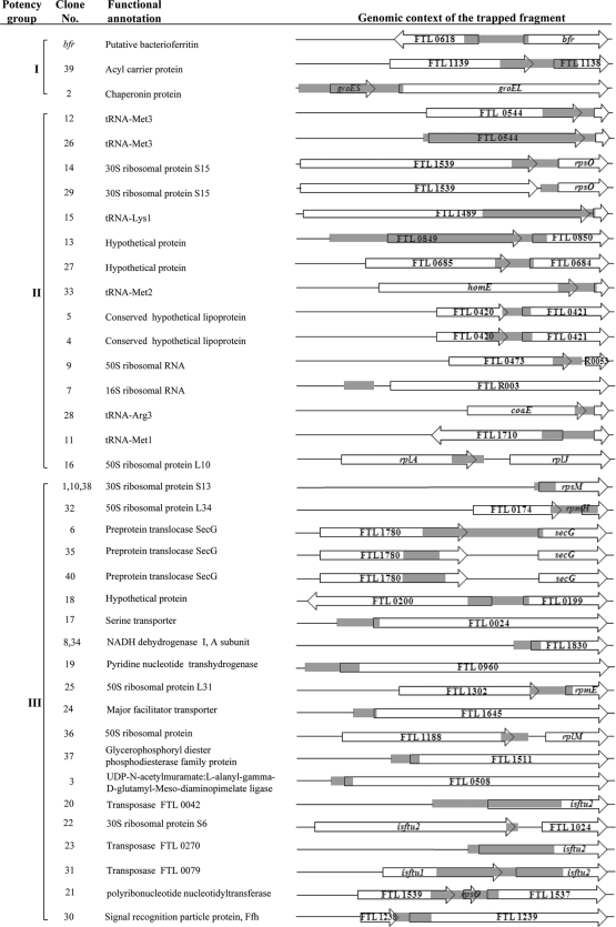
The initial attempt to identify F. tularensis promoters was based on screening of a gfp-based promoter trap system. For this purpose, a novel pTRAP E. coli-Francisella shuttle vector was constructed based on a derivative of the pRIT5 vector (29) (Pharmacia, Uppsala), which contains the promiscuous pC194 origin of replication (14) (see Materials and Methods). The GFP vector includes a promoterless copy of the gfp+ gene (37) located downstream from a synthetic ribosome binding site (RBS) which is separated from the gfp+ start codon by seven nucleotides that comprise a SnaBI restriction site. Sau3A-digested F. tularensis LVS chromosomal DNA fragments were inserted into the pTRAP plasmid vector. This ligation mixture was used to transform E. coli cells, resulting in about 9,000 colonies, many of which exhibited visible fluorescence. DNA restriction analysis performed on a sample of 50 colonies revealed that about 50% of the transformants contained chromosomal inserts, with an average size of 410 ± 50 bp (average ± standard deviation). Based on the F. tularensis genome size (1.9 Mbp), the calculated genome coverage achieved by the inserts in the library is about 50% (see Materials and Methods). Plasmid DNA isolated from the pool of E. coli transformants was used to transform F. tularensis LVS. Of the 2.6 × 10⁴ F. tularensis transformant colonies that were inspected by UV illumination, only three colonies exhibited visible fluorescence (in marked contrast to the many fluorescence-positive clones in the E. coli pool). DNA restriction and sequence analysis revealed that all three clones carry the same chromosomal fragment, which contains the bacterioferritin promoter (Pbfr) region upstream from the gfp reporter. The map of the cloned fragment present in all three of these clones is provided in the first line of the chromosomal locus tabulation in Fig. 1. As will be shown below, Pbfr is the most potent among all the promoters analyzed in this study.
FIG. 1.
Chromosomal gene arrangement and genomic context of the strain LVS promoters analyzed. The maps of the 38 clones compiled in this figure represent 33 unique F. tularensis promoters that were isolated from the gfp and the cat promoter trap libraries. Gray boxes represent the regions of the LVS cloned fragments. The chromosomal loci of the various promoters are depicted in the orientation that is compatible with expression of the reporter gene (cat or gfp). The genes adjacent to the cloned fragments are indicated as open arrows, and their locus tags (FTL numbers) according to the F. tularensis subsp. holarctica complete genome sequence in the NCBI database are indicated. Whenever available, the genes are designated by their name symbols. Promoters are arranged according to their relative potencies (Fig. 3B and text). Note that clones 1, 10, and 38 represent the same DNA fragments and thus appear together in their respective entries, as is the case for clones 8 and 34. For more details on the cloned fragments, please refer to the text and Table S1 in the supplemental material.
Use of a cat-based promoter trap library for the selection of F. tularensis LVS promoters.
In view of the limited results obtained with the gfp promoter trap system, we decided to employ a different screening approach that relies on cat as a reporter gene. The cat-based promoter library was constructed from partially AluI-digested F. tularensis LVS chromosomal DNA fragments that were cloned upstream from a promoterless cat gene in the E. coli-F. tularensis shuttle vector pKK214 (18). Since attempts to construct the library directly in the LVS background resulted in an insufficient number of colonies on chloramphenicol (Cm) selection plates, we first amplified the library in E. coli. Plasmids isolated from the pool of E. coli transformants were electroporated into LVS cells and resulted in a total of 2,000 Cmr LVS clones. Of these 2,000 Cmr clones, we arbitrarily selected 40 colonies for further characterization. Restriction analysis revealed that all of the clones contained LVS DNA fragments of various sizes (50 to 1,500 bp; average size, 350 ± 50 bp).
Identification of the chromosomal loci of the LVS promoters analyzed.
The cloned chromosomal DNA fragments from all of the 40 clones isolated from the cat library, as well as those of the 3 clones which exhibited fluorescence in the gfp library, were sequenced, and the genomic location of each was determined based on the sequence of the F. tularensis subsp. holarctica LVS genome in the NCBI databank (Fig. 1). As mentioned above, all three clones originating from the gfp library appeared to be identical and all were derived from the bfr promoter region; hence, they are represented as a single clone in the first line of Fig. 1. Of the clones isolated from the cat library, three, P1, P10, and P38, contained identical fragments, as did clones P8 and P34. Some chromosomal regions were represented in more than one clone, yet they clearly represent independent cloning events, since their respective inserts were not identical (P4 and P5; P12 and P26; P14 and P29; and P6, P35, and P40) (Fig. 1; also see Table S1 in the supplemental material). Most of the clones include inserts derived from intergenic fragments, consistent with their expected location in regulatory regions. Furthermore, the orientation of these inserts with respect to the cat reporter gene is in line with the orientation expected for transcription in the context of the genome. The vast majority of the trapped DNA regulatory fragments contained a putative ribosome binding site (RBS). In the case of the five inserts that did not contain an apparent RBS (P9, P7, P16, P22, and P36), it appears that the expression of the reporter gene exploited the existing RBS in the expression vector. Six clones, P7, P22, P27, P32, P33, and P36, were found to include DNA inserts that resulted from the ligation of two separate AluI genomic fragments, yet in each case, only one of the two ligated fragments was found to represent a promoter-like 5′ untranslated region of an open reading frame (ORF). We note that, apart from P2, which is located upstream of the previously described groEL operon (9), all of the sequenced clones represent novel F. tularensis promoters. In summary, of 43 analyzed clones originating from both the gfp and the cat libraries, 38 clones contained different genomic fragments representing 33 unique promoters.
In silico compilation and experimental analysis of F. tularensis promoters.
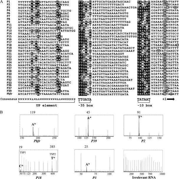
Computational analysis of the newly identified promoters was carried out in a search for regulatory elements, using the BPROM tool (Softberry). Consensus sequences were derived from multiple alignment of the 33 unique sequences (Fig. 2A). Accordingly, a putative −10 region matching the consensus hexamer of the σ70 family promoters (TATAAT) was identified 4 to 8 nucleotides upstream from the predicted transcription start point (TSP), and a putative −35 region highly similar to the classical TTGACA consensus sequence was found to be located 14 to 21 bp upstream from the −10 region (Fig. 2A). Inspection of the LVS promoter sequences located upstream from the −35 consensus (positions −38 to −59) revealed a typical AT-rich region that resembled the upstream region, termed the UP element, that is necessary for efficient transcription from some promoters in both Gram-positive and Gram-negative bacteria (12).
FIG. 2.
Sequence analysis of the cloned F. tularensis promoters. (A) Multiple alignment of all unique promoter sequences determined in this study. The promoters are arranged in ascending order. The TSP, −10, and −35 boxes were predicted using the BPROM tool and aligned using the T-coffee multiple-alignment tool. Nucleotides conserved among more than 50% of the sequences are indicated by black boxes. The predicted TSP nucleotide is boxed, while the 5 experimentally determined TSP nucleotides are circled. The consensus sequences of the −10, the −35, and the UP element are indicated at the bottom. The arrow on the bottom right indicates the TSP (nucleotide position +1). (B) Analysis of primer extension (PE) products specific for the indicated promoters. PE fragments were sized using GeneScan Analysis software (Applied Biosystems) (for detailed experimental conditions, see Materials and Methods). MapMaker 1000 internal lane standards are included in each electropherogram. The first 5′ nucleotide of the transcript is marked by an asterisk above the translation start diagnostic peak. The number above each panel refers to the length of the 5′ untranslated region of each promoter (i.e., the distance between the transcription starting point and the respective translation starting point). Note the absence of cDNA signal in the irrelevant-RNA negative-control panel.
In order to confirm that the predictions based on this in silico analysis reflect the actual TSPs, we further selected five promoters for primer extension analysis. Total RNA prepared from F. tularensis LVS bacteria grown to mid-logarithmic phase served as a template for a modified primer extension analysis (see Materials and Methods) using FAM-labeled primers as previously described (22). Two independent extension reactions, using two different primers complementary to the 5′ region of the relevant open reading frame, were carried out for each promoter inspected. As depicted in Fig. 2B, a single TSP was determined for each of the five clones except for P18, in which two TSPs were identified. All of the TSPs that were experimentally determined were located within a range of 1 to 5 nucleotides of the TSP predicted by the in silico analysis. The TSP of P2, which is located upstream of the groEL operon, was mapped to reside 91 bp upstream from the groES start codon, which is in excellent agreement with previously published results (9). The TSPs of the open reading frames regulated by the Pbfr, P39, and P3 promoters are located 119, 45, and 25 bp upstream from the respective ATG start codons. The first transcriptional initiation site of promoter 18 (TSP1) is located 19 bp upstream from the translational start codon of its putatively regulated gene, FTL_0199. The second transcription start point (TSP2) is located 385 bp upstream from the FTL_0199 translational start codon and is a part of the FTL_0200 ORF. However, TSP2 is not a part of the cloned cat library fragment, and therefore, cat transcription in clone 18 is driven by the proximal rather than the distal promoter.
It was of interest to determine to what extent the sequences of the promoters originating from the F. tularensis subsp. holarctica LVS strain are similar to or diverge from those of the virulent F. tularensis subsp. tularensis Schu S4 strain. It appears that of the 33 LVS promoter sequences, 26 are completely identical to their corresponding loci in the Schu S4 strain, while 7 promoters exhibited minor differences consisting of single-nucleotide replacement/deletion/insertion, all within the region spanning between the −10 and −35 elements. This region is known to be promiscuous with respect to both sequence composition and length.
Relative potencies of the analyzed cat-derived promoters.
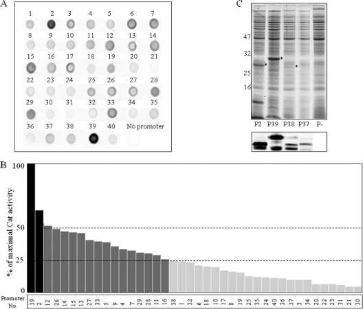
In order to assess the relative potencies of the promoters derived from the cat library, we conducted a dot blot Western analysis of cell extracts from each clone using specific anti-CAT antibodies (Fig. 3A). This analysis revealed that all the clones tested interact specifically with the anti-CAT antibodies, while control LVS cells carrying the promoterless cat cassette showed no Western blot signal. Furthermore, the tested clones exhibited various signal intensities, suggesting a wide dynamic range of CAT expression levels.
FIG. 3.
Expression level of chloramphenicol acetyltransferase (CAT) from selected clones isolated from the cat library. (A) Western dot blot analysis for determination of CAT levels in cytosolic extracts, using anti-CAT antibodies. (B) Relative CAT activity in cytosolic cell extracts determined by CAT enzymatic assays. Promoters are ordered according to descending levels of potency. Black, dark-gray, and light-gray bars denote strong, moderate, and weak promoters, respectively. (C) SDS-PAGE (Coomassie, top) and immunoblot (anti-CAT, bottom) analyses from four selected promoter clones. CAT protein is indicated by asterisks (the additional high-molecular-weight CAT form generated in the P39 clone probably stems from an alternative upstream translation initiation codon within the P39 insert). The control lane designated (P−) represents cells carrying the promoterless cat plasmid pKK214.
In order to better quantify the potencies of the different cloned promoters, an enzymatic assay was performed using equal amounts of cell extracts derived from each clone (see Materials and Methods). The CAT activity levels determined by the assay were consistent with the results derived from the dot blot analysis in most cases. Based on the enzymatic assay, the promoters could be arbitrarily classified into three potency groups (Fig. 3B). The relative potencies of selected promoters were further manifested by SDS-PAGE and Western blot (for example, see Fig. 3C). Both the Western and the enzymatic analyses established that 2 out of the 40 clones analyzed (5%) entail promoters of high potency, 15 clones (37%) represent promoters of moderate potency, and 23 clones (58%) include relatively weak promoters (it should be noted that in E-tests, even the weakest LVS promoter [e.g., promoter P30] provided at least a 5-fold-higher MIC value than that of pKK202 [30] in the LVS background). In view of the presence of clones containing plasmids with different promoter strengths, we tested the possibility of direct selection of highly potent promoters using increasing Cm concentrations. However, no enrichment of clones containing the more potent promoters was attained by this selection procedure (see Materials and Methods).
To directly assess the relative transcription initiation potencies of the promoters, the number of cat mRNA molecules generated under the control of various promoters was determined by qRT-PCR analysis. The pKK214 tet gene, which confers tetracycline resistance, was used as an internal control in the RT-PCR assays. The relative levels of cat amplicons in promoter clones P39, P2, and P3 were 100%, 67%, and 7%, respectively. These results confirm the potencies of the promoters inferred from the CAT assay and the Western analysis and demonstrate that these assays correctly reflect the promoter potency at the transcriptional level.
Potency of the bfr promoter and limitations of the GFP screening method.
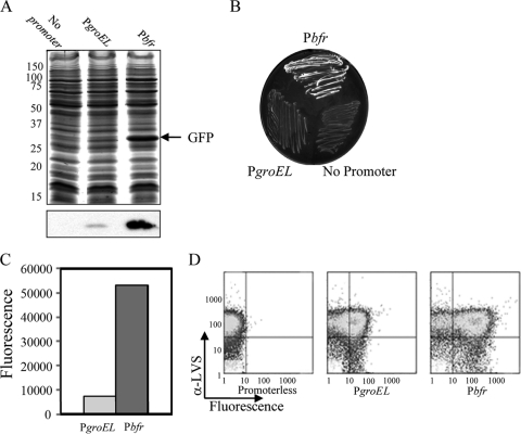
While the Cm-based selection allowed the isolation of numerous promoters, the gfp-based promoter trap system essentially generated only 3 clones, all representing the bfr promoter (Pbfr). In order to determine the reason for this extremely low prevalence of fluorescent LVS clones, we probed the original plasmid pool of the gfp library for the presence of 3 potent promoters (P39, P2, and P29) which were identified in the cat library (see Materials and Methods). The PCR analysis revealed that the DNA fragments of each of these promoters is present in the gfp library. This result strongly suggests that the GFP expression level driven by these 3 promoters was probably insufficient to allow visible fluorescence in the LVS background (unlike that driven by Pbfr), in spite of their relative strength. To further confirm this notion, we decided to compare the GFP expression levels in the cells carrying the bfr promoter to those generated by the well-characterized F. tularensis promoter groEL (identified as P2 by the Cm screen and classified as one of the two most potent promoters). Accordingly, the groEL (P2) promoter was cloned upstream from the promoterless gfp+ (in the pTRAP vector), and the level of GFP production in the LVS transformants was compared to that of the bfr-GFP cells. Indeed, the levels of GFP expressed by the LVS cells as analyzed by SDS-PAGE, as well as by inspection of the fluorescence emitted by colonies grown on solid medium, were significantly higher when the transcription was driven by the bfr promoter (Fig. 4A and B). Furthermore, quantitative fluorimetry, as well as FACS analysis of cell extracts, allowed us to estimate that the fluorescence level driven by Pbfr was nearly 10-fold higher than that driven by PgroEL (Fig. 4C and D).
FIG. 4.
Comparison of relative potencies of the bfr and groEL promoters determined by GFP expression. (A) SDS-PAGE (top) and Western blot (anti-GFP, bottom) analyses of F. tularensis LVS cell extracts expressing GFP from indicated promoters cloned in the pTRAP vector. Bacterial cultures were collected 12 h postinoculation. (B) Direct inspection of fluorescence emitted by cells expressing GFP from the indicated promoters. (C) Quantification by direct fluorimetry of GFP fluorescence levels emitted by cells expressing GFP from either the bfr or groEL promoter. Fluorescence quantification was carried out with cultures of identical densities. (D) FACS analysis for quantification of GFP fluorescence levels emitted by equal numbers of cells expressing GFP from either the bfr or groEL promoter.
While the bfr promoter was not identified among the 40 randomly selected cat colonies, its presence within the cat library plasmid pool was verified by PCR analysis (see Materials and Methods). To further directly evaluate the potency of the bfr promoter relative to the potencies of other promoters in the cat context, we cloned the bfr promoter fragment (isolated from the gfp library) upstream from the promoterless cat gene in the pKK214 vector and compared the CAT expression level to that driven by promoter 39 (the strongest promoter identified in the cat library). This assay showed that the activity of the CAT enzyme driven by the bfr promoter was higher than that driven by P39 (80 OD405/min/OD600 and 60 OD405/min/OD600, respectively [OD405 is the measurement for CAT enzyme activity and OD600 is the measurement for culture density]). Based on all these observations, we conclude that the bfr promoter is the most potent among all promoters identified in the present work and is probably one of the most potent promoters in F. tularensis.
DISCUSSION
The previously identified groEL, acpA, and FTN_1451 Francisella promoters provided essential genetic tools for studying and manipulating genes of F. tularensis. Nevertheless, the number of Francisella promoters available to date is rather limited, and those identified are still poorly characterized. In the current study, two alternative in vitro large-scale screening procedures were employed in an attempt to identify new potent Francisella promoters. The first approach relied on gfp as a reporter gene. This procedure allowed the identification of a highly potent promoter (the bfr promoter). In fact, this promoter was the only promoter that could drive the expression of gfp in an amount that enabled the visualization of the fluorescent clone on agar plates using UV light. This phenomenon is clearly not due to underrepresentation of insert-containing clones, since we estimated that 50% out of the initial 9,000 library clones contained LVS sequences. The fact that many LVS promoters were able to generate visible fluorescence in the background of E. coli but failed to do so in the LVS background may be due to one or a combination of the following reasons. (i) The visible GFP fluorescence elicited in LVS is somehow quenched, and thus, only very potent promoters can allow the detection of GFP-expressing colonies. Indeed, while GFP expression from the groEL promoter was confirmed by Western blot analysis, the fluorescence level of the groEL-gfp clone was hardly detectable in the LVS background (Fig. 4). Actually, detection of the fluorescence emitted by a gfp clone required that gfp expression be driven by a promoter 10 times stronger than the groEL promoter, such as the bfr promoter (Fig. 4A and B). We note that in another study, the expression of GFP from the F. tularensis rpsL promoter was noted to be poorly visible in strain LVS, while it was clearly visible in the E. coli background, as judged by confocal microscopy imaging (23). (ii) A certain proportion of the cloned chromosomal segments that promoted GFP fluorescence in E. coli may not represent authentic Francisella promoters. It is well established that the E. coli σ70-dependent promoters are capable of driving significant levels of transcription solely upon recognition of an AT-rich −10 consensus promoter element (TATAAT) together with a TGn −10 motif (4, 17). Since the F. tularensis genome is AT rich, it is likely that some F. tularensis chromosomal DNA regions may be recognized as promoters by the E. coli transcription machinery just by virtue of the high frequency of AT-rich regions. Similar observations were reported for the pKK214 vector, which contains a promoterless cat gene and still confers chloramphenicol resistance when introduced into E. coli (18). (iii) It is possible that a higher copy number of the pTRAP vector in E. coli could have contributed to the high prevalence of fluorescent E. coli clones.
The second screening approach that was implemented in this study relied on direct selection of Cmr colonies through expression from cat as a reporter gene. Many studies of bacterial pathogens, including F. tularensis, have used cat-based promoter trap systems in order to select for bacterial genes that are specifically induced during infection (7, 18, 25, 26). Here we used, for the first time in F. tularensis, an in vitro approach that took advantage of the strong Cm-mediated selective pressure in order to select Francisella promoters in a genomic library. Out of 2,000 Cmr LVS colonies, we arbitrarily selected 40 clones for further characterization. All of these clones appeared to contain inserts of various sizes upstream of the cat reporter gene, as attested by both restriction and sequence analyses. Sequence analysis revealed that 37 of the 40 cat library clones contain different genome fragments and 32 represent unique promoter sequences. The low level of redundancy of the cloned promoters in the population suggests that the 2,000 cat library clones provide good coverage of most if not all F. tularensis promoters.
The two screening approaches (gfp and cat cassette) yielded 33 different and unique promoters with various levels of potency. The 3 most potent promoters were found to belong to a putative bacterioferritn (Pbfr, FTL_0617), an acyl carrier protein (PacpP, FTL_1138), and a heat shock protein which belongs to the groESL operon (PgroEL, FTL_1715). The high potency exhibited by Pbfr and PgroEL is in line with previous reports documenting that Bfr and the 2 proteins residing on the groESL operon (i.e., GroES and GroEL) are among the 12 major proteins secreted by LVS cells (21). The side-by-side quantitative comparison of GFP expression driven by the bfr and the groEL promoter suggests that the bfr promoter is about 10-fold more potent than the groEL promoter. Interestingly, 16 of the 33 unique clones contain promoters that regulate genes belonging to components of the translational machinery (6 clones contain promoters that regulate the transcription of tRNAs, 2 clones include rRNA promoters, and the rest of the clones harbor various ribosomal protein promoters). This high prevalence of promoters related to the translational machinery is surprising since only 5.5% of the entire F. tularensis genome encodes such functions (2% encodes tRNAs [38 genes], 0.5% encodes rRNA [10 genes] and less than 2.7% encodes ribosomal proteins). We note in this context that the promoters of genes encoding rRNA, tRNA, and ribosomal proteins are among the most active in the bacterial cell (19).
Very little is known about the regulation of gene expression in Francisella or the architecture of the promoters. Apart from the groEL operon, in which the transcription start point was determined (9), no other Francisella promoters have been characterized at the molecular level. Multiple sequence alignment of the 33 newly identified promoters revealed a σ70-relevant −10 motif with a 6-nucleotide conserved sequence identical to the TATAAT consensus sequence from E. coli (13). This F. tularensis −10 motif is found at the expected location upstream of the predicted transcription start point (Fig. 2). Within the −35 region, we could determine a TTGA(C/T)A consensus sequence which is in good agreement with the E. coli TTGACA consensus sequence. The spacer DNA segment between the two conserved regions ranged from 17 to 21 bases in most cases, and the 2 most potent promoters (Pbfr and P39) exhibited a 17-bp spacer, in line with the architecture of strong promoters previously reported in E. coli (13). The results of primer extension analyses carried out for five selected promoter clones are in good agreement with the predicted location of the transcription initiation site and, thus, provide additional experimental support for the in silico-predicted consensus promoter elements of Francisella tularensis. In addition to the −10 and −35 regions, a 22-nucleotide upstream region (termed the UP element) exhibiting an AT-rich sequence mediating recognition by the α-subunit of RNA polymerase is often needed for efficient transcription (12). We observe that such an AT-rich region is present in all the promoters analyzed. And yet, no consensus sequence which resembles the E. coli UP element consensus sequence could be found in this region. Collectively, these results show that the two well-established promoter elements (the −10 and −35 regions) described in other microorganisms as σ70-binding sites can also be identified in F. tularensis. However, the inability to distinguish the canonical cis-acting UP element consensus among F. tularensis promoters may imply that recognition of this DNA element by the F. tularensis α-subunit is different from that described for E. coli.
The battery of promoters identified in the present work may serve in future studies to achieve controlled levels of expression of selected proteins in the genetic background of F. tularensis strains, as well as for various studies aimed at understanding the mechanisms of pathogenesis and virulence. Furthermore, F. tularensis strain LVS has been used as a vaccine for many years; however, its efficacy against the virulent Schu S4 strain has been questioned (5). Our study could provide the means for selected expression of specific LVS-/Schu S4-derived proteins in the background of LVS and, thus, may allow the development of a more efficacious F. tularensis vaccine.
Supplementary Material
Acknowledgments
We thank Anders Norqvist and Ake Forsberg for providing the pKK202 and pKK214 plasmids and Michael Niederweis for kindly providing the pWH1012 plasmid. We are grateful to Erez Bar-Haim for the FACS analysis. The excellent technical support of Gila Friedman is highly appreciated.
Footnotes
Published ahead of print on 30 December 2010.
Supplemental material for this article may be found at http://aem.asm.org/.
REFERENCES
- 1.Bar-Haim, E., et al. 2008. Interrelationship between dendritic cell trafficking and Francisella tularensis dissemination following airway infection. PLoS Pathog. 4:e1000211. [DOI] [PMC free article] [PubMed] [Google Scholar]
- 2.Baron, G. S., S. V. Myltseva, and F. E. Nano. 1995. Electroporation of Francisella tularensis. Methods Mol. Biol. 47:149-154. [DOI] [PubMed] [Google Scholar]
- 3.Bradford, M. M. 1976. A rapid and sensitive method for the quantitation of microgram quantities of protein utilizing the principle of protein-dye binding. Anal. Biochem. 72:248-254. [DOI] [PubMed] [Google Scholar]
- 4.Browning, D. F., and S. J. Busby. 2004. The regulation of bacterial transcription initiation. Nat. Rev. Microbiol. 2:57-65. [DOI] [PubMed] [Google Scholar]
- 5.Burke, D. S. 1977. Immunization against tularemia: analysis of the effectiveness of live Francisella tularensis vaccine in prevention of laboratory-acquired tularemia. J. Infect. Dis. 135:55-60. [DOI] [PubMed] [Google Scholar]
- 6.Charity, J. C., et al. 2007. Twin RNA polymerase-associated proteins control virulence gene expression in Francisella tularensis. PLoS Pathog. 3:e84. [DOI] [PMC free article] [PubMed] [Google Scholar]
- 7.Chiang, S. L., J. J. Mekalanos, and D. W. Holden. 1999. In vivo genetic analysis of bacterial virulence. Annu. Rev. Microbiol. 53:129-154. [DOI] [PubMed] [Google Scholar]
- 8.Cohen, S., et al. 2000. Attenuated nontoxinogenic and nonencapsulated recombinant Bacillus anthracis spore vaccines protect against anthrax. Infect. Immun. 68:4549-4558. [DOI] [PMC free article] [PubMed] [Google Scholar]
- 9.Ericsson, M., I. Golovliov, G. Sandstrom, A. Tarnvik, and A. Sjostedt. 1997. Characterization of the nucleotide sequence of the groE operon encoding heat shock proteins chaperone-60 and -10 of Francisella tularensis and determination of the T-cell response to the proteins in individuals vaccinated with F. tularensis. Infect. Immun. 65:1824-1829. [DOI] [PMC free article] [PubMed] [Google Scholar]
- 10.Frank, D. W., and T. C. Zahrt. 2007. Genetics and genetic manipulation in Francisella tularensis. Ann. N. Y. Acad. Sci. 1105:67-97. [DOI] [PubMed] [Google Scholar]
- 11.Gallagher, L. A., et al. 2007. A comprehensive transposon mutant library of Francisella novicida, a bioweapon surrogate. Proc. Natl. Acad. Sci. U. S. A. 104:1009-1014. [DOI] [PMC free article] [PubMed] [Google Scholar]
- 12.Gourse, R. L., W. Ross, and T. Gaal. 2000. UPs and downs in bacterial transcription initiation: the role of the alpha subunit of RNA polymerase in promoter recognition. Mol. Microbiol. 37:687-695. [DOI] [PubMed] [Google Scholar]
- 13.Hawley, D. K., and W. R. McClure. 1983. Compilation and analysis of Escherichia coli promoter DNA sequences. Nucleic Acids Res. 11:2237-2255. [DOI] [PMC free article] [PubMed] [Google Scholar]
- 14.Horinouchi, S., and B. Weisblum. 1982. Nucleotide sequence and functional map of pC194, a plasmid that specifies inducible chloramphenicol resistance. J. Bacteriol. 150:815-825. [DOI] [PMC free article] [PubMed] [Google Scholar]
- 15.Horzempa, J., D. M. Tarwacki, P. E. Carlson, Jr., C. M. Robinson, and G. J. Nau. 2008. Characterization and application of a glucose-repressible promoter in Francisella tularensis. Appl. Environ. Microbiol. 74:2161-2170. [DOI] [PMC free article] [PubMed] [Google Scholar]
- 16.Kravchenko, T. B., et al. 2007. Cloning and expression of protective antigens of Mycobacterium tuberculosis Ag85B and ESAT-6 in Francisella tularensis 15/10. Biochemistry (Mosc.). 72:735-743. [DOI] [PubMed] [Google Scholar]
- 17.Kumar, A., et al. 1993. The minus 35-recognition region of Escherichia coli sigma 70 is inessential for initiation of transcription at an “extended minus 10” promoter. J. Mol. Biol. 232:406-418. [DOI] [PubMed] [Google Scholar]
- 18.Kuoppa, K., A. Forsberg, and A. Norqvist. 2001. Construction of a reporter plasmid for screening in vivo promoter activity in Francisella tularensis. FEMS Microbiol. Lett. 205:77-81. [DOI] [PubMed] [Google Scholar]
- 19.Lamond, A. I., and A. A. Travers. 1985. Stringent control of bacterial transcription. Cell 41:6-8. [DOI] [PubMed] [Google Scholar]
- 20.Larsson, P., et al. 2005. The complete genome sequence of Francisella tularensis, the causative agent of tularemia. Nat. Genet. 37:153-159. [DOI] [PubMed] [Google Scholar]
- 21.Lee, B. Y., M. A. Horwitz, and D. L. Clemens. 2006. Identification, recombinant expression, immunolocalization in macrophages, and T-cell responsiveness of the major extracellular proteins of Francisella tularensis. Infect. Immun. 74:4002-4013. [DOI] [PMC free article] [PubMed] [Google Scholar]
- 22.Lloyd, A. L., B. J. Marshall, and B. J. Mee. 2005. Identifying cloned Helicobacter pylori promoters by primer extension using a FAM-labelled primer and GeneScan analysis. J. Microbiol. Methods 60:291-298. [DOI] [PubMed] [Google Scholar]
- 23.LoVullo, E. D., C. R. Molins-Schneekloth, H. P. Schweizer, and M. S. Pavelka, Jr. 2009. Single-copy chromosomal integration systems for Francisella tularensis. Microbiology 155:1152-1163. [DOI] [PMC free article] [PubMed] [Google Scholar]
- 24.LoVullo, E. D., L. A. Sherrill, L. L. Perez, and M. S. Pavelka, Jr. 2006. Genetic tools for highly pathogenic Francisella tularensis subsp. tularensis. Microbiology 152:3425-3435. [DOI] [PubMed] [Google Scholar]
- 25.Mahan, M. J., J. M. Slauch, and J. J. Mekalanos. 1993. Selection of bacterial virulence genes that are specifically induced in host tissues. Science 259:686-688. [DOI] [PubMed] [Google Scholar]
- 26.Mahan, M. J., et al. 1995. Antibiotic-based selection for bacterial genes that are specifically induced during infection of a host. Proc. Natl. Acad. Sci. U. S. A. 92:669-673. [DOI] [PMC free article] [PubMed] [Google Scholar]
- 27.Maier, T. M., et al. 2004. Construction and characterization of a highly efficient Francisella shuttle plasmid. Appl. Environ. Microbiol. 70:7511-7519. [DOI] [PMC free article] [PubMed] [Google Scholar]
- 28.Marmur, J. 1961. A procedure for the isolation of deoxyribonucleic acid from micro-organisms. J. Mol. Biol. 3:208-218. [Google Scholar]
- 29.Nilsson, B., L. Abrahmsen, and M. Uhlen. 1985. Immobilization and purification of enzymes with staphylococcal protein A gene fusion vectors. EMBO J. 4:1075-1080. [DOI] [PMC free article] [PubMed] [Google Scholar]
- 30.Norqvist, A., K. Kuoppa, and G. Sandstrom. 1996. Construction of a shuttle vector for use in Francisella tularensis. FEMS Immunol. Med. Microbiol. 13:257-260. [DOI] [PubMed] [Google Scholar]
- 31.Pechous, R., et al. 2006. Construction and characterization of an attenuated purine auxotroph in a Francisella tularensis live vaccine strain. Infect. Immun. 74:4452-4461. [DOI] [PMC free article] [PubMed] [Google Scholar]
- 32.Poirot, O., E. O'Toole, and C. Notredame. 2003. Tcoffee@igs: a web server for computing, evaluating and combining multiple sequence alignments. Nucleic Acids Res. 31:3503-3506. [DOI] [PMC free article] [PubMed] [Google Scholar]
- 33.Rasko, D. A., C. D. Esteban, and V. Sperandio. 2007. Development of novel plasmid vectors and a promoter trap system in Francisella tularensis compatible with the pFLN10 based plasmids. Plasmid 58:159-166. [DOI] [PMC free article] [PubMed] [Google Scholar]
- 34.Reilly, T. J., G. S. Baron, F. E. Nano, and M. S. Kuhlenschmidt. 1996. Characterization and sequencing of a respiratory burst-inhibiting acid phosphatase from Francisella tularensis. J. Biol. Chem. 271:10973-10983. [DOI] [PubMed] [Google Scholar]
- 35.Rodriguez, S. A., G. Davis, and K. E. Klose. 2009. Targeted gene disruption in Francisella tularensis by group II introns. Methods 49:270-274. [DOI] [PMC free article] [PubMed] [Google Scholar]
- 36.Rodriguez, S. A., J. J. Yu, G. Davis, B. P. Arulanandam, and K. E. Klose. 2008. Targeted inactivation of Francisella tularensis genes by group II introns. Appl. Environ. Microbiol. 74:2619-2626. [DOI] [PMC free article] [PubMed] [Google Scholar]
- 37.Scholz, O., A. Thiel, W. Hillen, and M. Niederweis. 2000. Quantitative analysis of gene expression with an improved green fluorescent protein. p6. Eur. J. Biochem. 267:1565-1570. [DOI] [PubMed] [Google Scholar]
- 38.Shaw, W. V. 1975. Chloramphenicol acetyltransferase from chloramphenicol-resistant bacteria. Methods Enzymol. 43:737-755. [DOI] [PubMed] [Google Scholar]
- 39.Sjostedt, A. 2007. Tularemia: history, epidemiology, pathogen physiology, and clinical manifestations. Ann. N. Y. Acad. Sci. 1105:1-29. [DOI] [PubMed] [Google Scholar]
- 40.Tarnvik, A., and M. C. Chu. 2007. New approaches to diagnosis and therapy of tularemia. Ann. N. Y. Acad. Sci. 1105:378-404. [DOI] [PubMed] [Google Scholar]
Associated Data
This section collects any data citations, data availability statements, or supplementary materials included in this article.


