Abstract
Purpose
To assess the feasibility, safety, and toxicity of autologous tumor lysate-pulsed dendritic cell (DC) vaccination and toll-like receptor (TLR) agonists in patients with newly diagnosed and recurrent glioblastoma. Clinical and immune responses were monitored and correlated with tumor gene expression profiles.
Experimental Design
Twenty-three patients with glioblastoma (WHO grade IV) were enrolled in this dose-escalation study and received three biweekly injections of glioma lysate-pulsed DCs followed by booster vaccinations with either imiquimod or poly-ICLC adjuvant every three months until tumor progression. Gene expression profiling, IHC, FACS, and cytokine bead arrays were performed on patient tumors and PBMC.
Results
DC vaccinations are safe and not associated with any dose-limiting toxicity. The median overall survival from the time of initial surgical diagnosis of glioblastoma was 31.4 months, with a one-, two-, and three-year survival rate of 91%, 55% and 47%, respectively. Patients whose tumors had mesenchymal gene expression signatures exhibited increased survival following DC vaccination compared to historical controls of the same genetic subtype. Tumor samples with a mesenchymal gene expression signature had a higher number of CD3+ and CD8+ tumor infiltrating lymphocytes (TILs) compared with glioblastomas of other gene expression signatures (p = 0.006).
Conclusion
Autologous tumor lysate-pulsed DC vaccination in conjunction with TLR agonists is safe as adjuvant therapy in newly diagnosed and recurrent glioblastoma patients. Our results suggest that the mesenchymal gene expression profile may identify an immunogenic subgroup of glioblastoma that may be more responsive to immune-based therapies.
INTRODUCTION
Glioblastoma is a lethal malignant brain tumor with overall survival rates of less than 3.3% at 5 years (1). Glioblastoma remains one of the diseases for which there is no curative therapy. Despite advances in the identification of potential targets for glioma therapy and recent clinical trials utilizing biological therapies and newer cytotoxic agents (2–4), the prognosis of patients with primary malignant brain tumors remains dismal. This sobering fact underscores the need to rethink conventional approaches to the treatment of malignant brain tumors and to base therapeutic strategies on continuing advances in our knowledge of tumor biology and immunology.
The potential therapeutic benefit of eliciting an anti-tumor immune response in cancer patients was first suggested decades ago. Immunotherapy is theoretically appealing because it offers the potential for a high degree of tumor-specificity, while sparing normal brain structures (5). One such approach uses professional antigen-presenting cells, known as dendritic cells (DC), co-cultured with autologous tumor lysate to immunologically target endogenous tumor antigens. Initial studies of DC-based vaccine therapy for malignant gliomas have shown acceptable safety and toxicity profiles (6–14), and multi-center randomized Phase II and III studies are currently underway.
Previous pre-clinical studies (15, 16) strongly suggested that toll-like receptor (TLR) agonists (e.g., imiquimod, poly ICLC), could enhance dendritic cell activation and migration, as well as stimulate T cell-mediated anti-tumor immune responses in murine glioma models. To translate these findings, a Phase I clinical trial was initiated to evaluate the adjunctive use of DC vaccination with TLR agonists for its feasibility, safety, and toxicity in patients with newly diagnosed and recurrent glioblastoma. Herein, we report the results of this Phase I clinical trial, together with immune monitoring data and novel correlative studies associating overall survival with gene expression signatures and increased tumor infiltrating lymphocytes for the glioblastoma patients.
PATIENTS AND METHODS
Patient eligibility
This phase I clinical trial was approved by the UCLA IRB and registered with the NCI as NCT00068510. Written informed consent was obtained from all patients. Inclusion criteria were: newly diagnosed or recurrent glioblastoma (WHO Grade IV) that were amenable to surgical resection, a Karnofsky performance score (KPS) ≥ 60%, evidence of normal bone marrow function (e.g., hemoglobin ≥ 9 g/dL, absolute granulocyte count ≥ 1,500/µl and platelet count ≥ 100,000 K), adequate liver function (SGPT, SGOT, and alkaline phosphatase ≤ 2.5 times upper limit of normal; and bilirubin ≤ 1.5 mg/dL), and adequate renal function BUN or creatinine ≤ 1.5 times institutional normals) prior to starting therapy. Exclusion criteria included allergies to any components of the DC vaccine, concurrent or prior corticosteroid use within 10 days of initial vaccination, the presence of acute infection requiring active treatment, unstable or severe intercurrent medical conditions (e.g., pulmonary, cardiac, or other systemic disease), known immunosuppressive disease, positive serology for HIV or hepatitis B, history of an autoimmune disease, or prior history of other malignancies.
Preparation of Autologous Tumor Lysate
Fresh tumor samples from surgical resection were transported under sterile conditions to the UCLA-Jonsson Cancer Center GMP facility and used to generate autologous tumor lysate, as previously described (8, 17). Tumor tissue was minced, digested in collagenase (Advanced Biofactures, Lynbrook, NY) and Dnase-1 (Dornase-α, Genentech, San Francisco, CA) for 8–12 hours at room temperature. To generate lysates, tumor cell suspensions were subjected to five freeze-thaw cycles, centrifuged for 10 minutes at 800×g, and the cell-free supernatants were obtained. Protein concentrations of each tumor lysate were determined using a Bio-Rad DC protein assay (Bio-Rad Corp., Hercules, CA), and lysates with 100 µg of measured protein were used to pulse DC for each injection.
Preparation of Autologous Dendritic Cells and pulsing with glioma lysate
Monocyte-derived DCs were established from adherent peripheral blood mononuclear cells (PBMC) obtained via leukapheresis performed at the UCLA Hemapheresis Unit. Blood was additionally drawn as a source of autologous serum for the DC cultures. All ex vivo DC preparations were performed in the UCLA-Jonsson Cancer Center GMP facility under sterile and monitored conditions. Dendritic cells were prepared by culturing adherent cells from peripheral blood in RPMI-1640 (Gibco) and supplemented with 10% autologous serum, 500 U/mL GM-CSF (Leukine®, Amgen, Thousand Oaks, CA) and 500 U/mL of IL-4 (CellGenix), using techniques described previously (8). Following culture, DCs were collected by vigorous rinsing and washed with sterile 0.9% NaCl solution. The purity and phenotype of each DC lot was also determined by flow cytometry (FACScan flow cytometer; BD Biosciences, San Jose, CA). Cells were stained with FITC-conjugated CD83, PE-conjugated CD86 and PerCP-conjugated HLA-DR mAb’s (BD Biosciences). Release criteria were >70% viable by trypan blue exclusion, and >30% of the large cell gate being CD86+ and HLA-DR+. One day before each vaccination, DC were pulsed (co-cultured) with 100 µg of tumor lysate overnight, washed, and the final product was tested for sterility by Gram stain, mycoplasma and endotoxin testing prior to injection.
Treatment Schema
Newly diagnosed glioblastoma patients underwent surgery and a standard course of external beam radiotherapy with concurrent temozolomide chemotherapy prior to DC vaccination (4). These patients were given 3 biweekly DC vaccinations following standard chemo-radiation and prior to adjuvant temozolomide treatment. Recurrent glioblastoma patients had previous radiation therapy and chemotherapy prior to presenting with tumor recurrence, so they underwent surgical resection of their tumors followed by DC immunotherapy after they had recovered from surgery and were tapered off peri-operative steroids. This ranged from 7–30 weeks after surgery.
Vaccine Administration
On the day of each DC vaccination, a 1 ml vaccine dose was drawn into a sterile tuberculin syringe and administered as an intradermal (i.d.) injection (using a 25-gauge needle) in the arm region below the axilla, with the side of administration rotated for each vaccination. Subjects were monitored for two hours post-immunization in the UCLA General Clinical Research Center (GCRC). Eligible patients initially received three (3) intradermal injections at biweekly intervals. If patients did not develop any toxic side effects from the experimental treatment and had stable disease for over three months, they received booster injections at the same dosage of tumor lysate-pulsed DC concurrently with either 5% imiquimod cream (Aldara™, a TLR-7 agonist) or poly-ICLC (Hiltonol™, a TLR-3 agonist). Due to initial safety/toxicity concerns of experimental allergic encephalomyelitis (EAE) (18) resulting from the combined use of DC vaccination and TLR agonists, these immune response modifiers were used only in the booster phase of the protocol, after patients had shown acceptable toxicity profiles to DC-lysate vaccinations alone. Booster vaccinations were given at 3 month intervals in between 28-day cycles (5 days on/23 days off) of temozolomide for up to 10 boosters or until tumor progression. For those receiving imiquimod as adjuvant, patients applied 5% imiquimod cream topically over the DC vaccination site one day prior to each vaccination cycle, immediately after DC vaccination, and then daily for an additional three days post-vaccination. For patients in the poly-ICLC cohort, intramuscular (i.m.) injections of 20 µg/kg of poly-ICLC were administered immediately prior to each DC injection at the vaccine injection site. All patients had a baseline brain MRI scan within one month prior to starting the immunotherapy and every 2 months thereafter or when clinically indicated.
Patient Assessment
Toxicity was monitored and graded according to the National Cancer Institute (NCI) Common Toxicity Criteria. The overall incidence of adverse events was recorded. Neurological exams were performed before and 30 minutes after each vaccination, as well as at all follow-up visits. Time to tumor progression (TTP) was defined as the interval from surgical resection until the first observation of tumor progression, as evidenced by magnetic resonance imaging (MRI) or clinical deterioration. Tumor progression was also considered to be non-reversible neurologic progression, permanently increased steroid requirement (applies to stable disease only), or early discontinuation of treatment. Overall survival (OS) time was determined from the date of surgery at the time of initial diagnosis of glioblastoma to date of death.
Flow Cytometry and Cytometric Bead Array
PBMC from patients enrolled on this clinical trial (pre- and post-vaccination) and PBMC from normal volunteers were thawed in warmed RPMI+2% FBS, washed and stained for the expression of CD3, CD4 and CD25 (all from BD Biosciences; San Diego, CA), followed by the intracellular labeling of Foxp3 (eBioscience; San Diego, CA). Stained cells were acquired on a BD FacsCalibur flow cytometer and analyzed using FloJo software. The frequencies of CD3+CD4+Foxp3+ and CD3+CD4+CD25+Foxp3+ PBMC’s were compared. For cytokine analysis, serum from patients enrolled on this clinical trial was thawed and incubated with the Cytometric Bead Array (CBA) Human Th1/Th2 Capture Beads (BD Biosciences), washed and subjected to analysis on a BD FacsCalibur flow cytometer together with cytokine standards. Quantitative assessment of cytokine levels was accomplished with a Microsoft Excel-based CBA software program.
Immunohistochemical (IHC) staining
Serial paraffin sections of pre-treatment tumor specimens were cut to 3 µm thickness and stained with anti-human antibodies against CD3 (DakoCytomation; Carpinteria, CA) and CD8 (DAKO Corp.; Carpinteria, CA). Sections were baked for 1 hour at 60°C, deparaffinized, and endogenous peroxidase activity quenched by treating with 0.5% H2O2 in methyl alcohol for 10 minutes. Heat-induced epitope retrieval was performed on the slides using 0.01 M citrate buffer, pH=6.0 (for CD3, CD8) in a vegetable steamer (Black & Decker); slides were heated for 25 minutes, cooled, and washed in 0.01 M phosphate buffered saline. All slides then were placed on a DAKO Autostainer (DAKO Corp.) and then sequentially incubated in primary antibody for 30–60 minutes, then rabbit anti-mouse secondary immunoglobulins (DAKO Corp.) for 30 minutes. Diaminobenzidine and hydrogen peroxide were used as the substrates for the peroxidase enzyme. For the negative controls, mouse isotype or rabbit immunoglobulins (DAKO Corp.) were used in place of the primary antibodies. Positive labeling was evaluated and scored by a board-certified neuro-pathologist (WHY) in a blinded fashion.
Microarray Studies
Of the twenty-three glioblastoma patients, sixteen patients had sufficient residual tumor tissue for microarray molecular analysis at the end of the trial. Total RNA was purified from pre-treatment, fresh frozen tumor samples using the RNeasy mini kit (Qiagen) and collected as part of the IRB-approved research protocol. cRNA was generated, quantified and hybridized to U133 Plus 2.0 arrays at the UCLA DNA Microarray Facility using standard Affymetrix protocols. CEL files were normalized using the Celsius Microarray Database (19), with robust multichip average (RMA) from Bioconductor (version 2.10) relative to 50 samples of the same platform. The Hierarchical Clustering (HC) classification for each glioma was determined by a gene voting strategy as described previously (20, 21). Briefly, the mean value of each probeset was evaluated from all samples within the U133 Plus 2.0 platform using the 377 gene probeset list and assigned to a HC group (21). Tumors were assigned to a HC group when the number of probes above the normalized mean was greater than 30% of a given probeset. The overall survival of patients tumors on this Phase I clinical trial was compared with the overall survival of patients from a collection of samples previously assigned to HC groups (21).
Statistical Analysis
Time to tumor progression (TTP) and overall survival (OS) curves were determined using the Kaplan-Meier method. The Log-rank (Mantel-Cox) test was used to compare curves between study and control groups. All P-values are two-tailed, and p < 0.05 was considered statistically significant. Statistics were analyzed using GraphPad Prism software.
RESULTS
Patient characteristics
Twenty-three patients with histologically proven WHO grade IV (glioblastoma) were enrolled in this protocol (Table 1). Fifteen had newly diagnosed tumors, while eight had recurrent disease. There were sixteen men and seven women, with an age range of 26 to 74 years (mean age of 51 years).
Table 1.
Patient Characteristics
| Patient ID |
Tumor Pathology |
Age | Gender | KPS | OS (mo.) |
HC Type | Dose (106) |
Adjuvant | Pre-vacc. Tx. | Post-vacc. Tx. | Related Adverse Events |
|---|---|---|---|---|---|---|---|---|---|---|---|
| GBM1-1 | GBM | 39 | M | 90 | 33.83 | * | 1 | Imiquimod | temozolomide | temozolomide, isoretinoin, celecoxib, Reoperation, SRS* | Fatigue, nausea/vomiting, diarrhea |
| GBM1-2 | GBM | 39 | M | 90 | >88.87 | Mes | 1 | Imiquimod | temozolomide | temozolomide, isoretinoin, CCNU, Gliadel™ | Fatigue, arthralgia, low-grade fever |
| GBM1-3 | GBM | 34 | M | 90 | >91.3 | Mes | 1 | Imiquimod | temozolomide | temozolomide, isoretinoin | Lymphadenopathy, injection site reaction, low-grade fever, myalgia |
| GBM1-4 | rec. GBM | 61 | M | 70 | 18.57 | * | 1 | None | temozolomide, thalidomide, isoretinoin, Newcastle virus | ||
| GBM1-5 | GBM | 58 | M | 70 | 10.3 | Pro | 1 | None | temozolomide | irinotecan, bevacizumab | |
| GBM1-6 | GBM | 63 | F | 80 | >37.6 | Mes | 1 | Imiquimod | temozolomide | temozolomide | Shingles |
| GBM1-7 | rec. GBM | 41 | F | 80 | 10.93 | Mes | 1 | None | irinotecan, bevacizumab | irinotecan, bevacizumab | |
| GBM1-8 | rec. GBM | 34 | M | 100 | >40.5 | PN | 1 | Poly ICLC | erlotinib, temozolomide, ANG, CCNU, celecoxib, tamoxifen, | CCNU, celecoxib, tamoxifen, | |
| GBM1-9 | GBM | 50 | M | 90 | >9.03 | * | 1 | Poly ICLC | temozolomide | ||
| GBM5-1 | GBM | 40 | F | 80 | 17.97 | * | 5 | None | temozolomide, isoretinoin | CCNU, gefitinib, rapamycin, carboplatin | Injection site reaction |
| GBM5-2 | rec. GBM | 54 | M | 80 | 17.3 | * | 5 | None | temozolomide, isoretinoin, CCNU | Carboplatin | Allergic rhinitis, itching at injection site, pruritis |
| GBM5-3 | GBM | 26 | M | 90 | 81.4 | PN | 5 | Imiquimod | temozolomide, isoretinoin, CCNU | carboplatin, irinotecan, bevacizumab, dasatanib, simvastatin, rosiglitazone, procarbazine, CCNU, cyclophosphamide | Injection site itching, dryness and pruritus, lymphadenopathy, nausea, diarrhea, vomiting, fatigue |
| GBM5-4 | GBM | 43 | M | 90 | >59.0 | Mes | 5 | Imiquimod | temozolomide | temozolomide | |
| GBM5-5 | GBM | 45 | F | 90 | 34.97 | PN | 5 | None | temozolomide | irinotecan, bevacizumab, CCNU, erlotinib | |
| GBM5-6 | rec. GBM | 53 | M | 80 | 22.33 | Mes | 5 | Poly ICLC | temozolomide, Gliadel™ | irinotecan, bevacizumab | Nausea, heartburn, constipation |
| GBM10-1 | rec. GBM | 58 | F | 70 | 28.93 | PN | 10 | None | temozolomide, irinotecan, paclitaxel | Headaches, nausea/vomiting | |
| GBM10-2 | GBM | 70 | F | 100 | 23.0 | * | 10 | None | temozolomide | p-EGFR/p-ErbB2 inhib, everolimus, carboplatin, bevacizumab, CCNU | |
| GBM10-3 | GBM | 50 | M | 90 | 36.33 | * | 10 | Imiquimod | temozolomide | irinotecan, bevacizumab | Dermatitis, rash, |
| GBM10-4 | GBM | 59 | M | 80 | 52.6 | Pro | 10 | Imiquimod | temozolomide, isoretinoin | irinotecan, bevacizumab, carboplatin, CCNU | Anorexia, abdominal pain, |
| GBM10-5 | GBM | 64 | M | 90 | 13.63 | Mes | 10 | None | temozolomide | bevacizumab, CCNU | |
| GBM10-6 | GBM | 66 | M | 90 | 37.73 | Mes | 10 | Imiquimod | temozolomide | irinotecan, bevacizumab, CCNU, etoposide, procarbazine, tamoxifen | Fatigue, left shoulder pain/arthralgia |
| GBM10-7 | rec. GBM | 74 | F | 60 | 17.07 | Mes | 10 | None | temozolomide | temozolomide | |
| GBM10-8 | rec. GBM | 52 | M | 60 | 16.23 | PN | 10 | None | temozolomide | Upper lip blisters, rash |
SRS = stereotactic radiosurgery
DC preparation and phenotype
DCs were generated from adherent PBMC cultured in the presence of 500 U/mL GMP-grade IL-4 and 500 IU/mL of GM-CSF for one week prior to harvest, as reported previously (8). All final autologous tumor lysate-pulsed DC preparations consistently contained a high percentage of viable large, granular cells and were free of contamination. Our DC preparations expressed high levels of MHC class I (HLA-A,B,C), MHC class II (HLA-DR), B7.2 costimulatory molecule (CD86), and CD40, but lower expression of CD14 and CD80 (Supplementary Table 1). These DC preparations were partially mature, with <45% of the large cells expressing HLA-DR and CD83, as might be expected for a protocol without a dedicated maturation step. Overnight incubation with tumor lysates induced some DC maturation, as evidenced by an increase in the median fluorescence intensity (MFI) of CD83 (data not shown), similar to previously reported findings (22).
Safety and toxicity
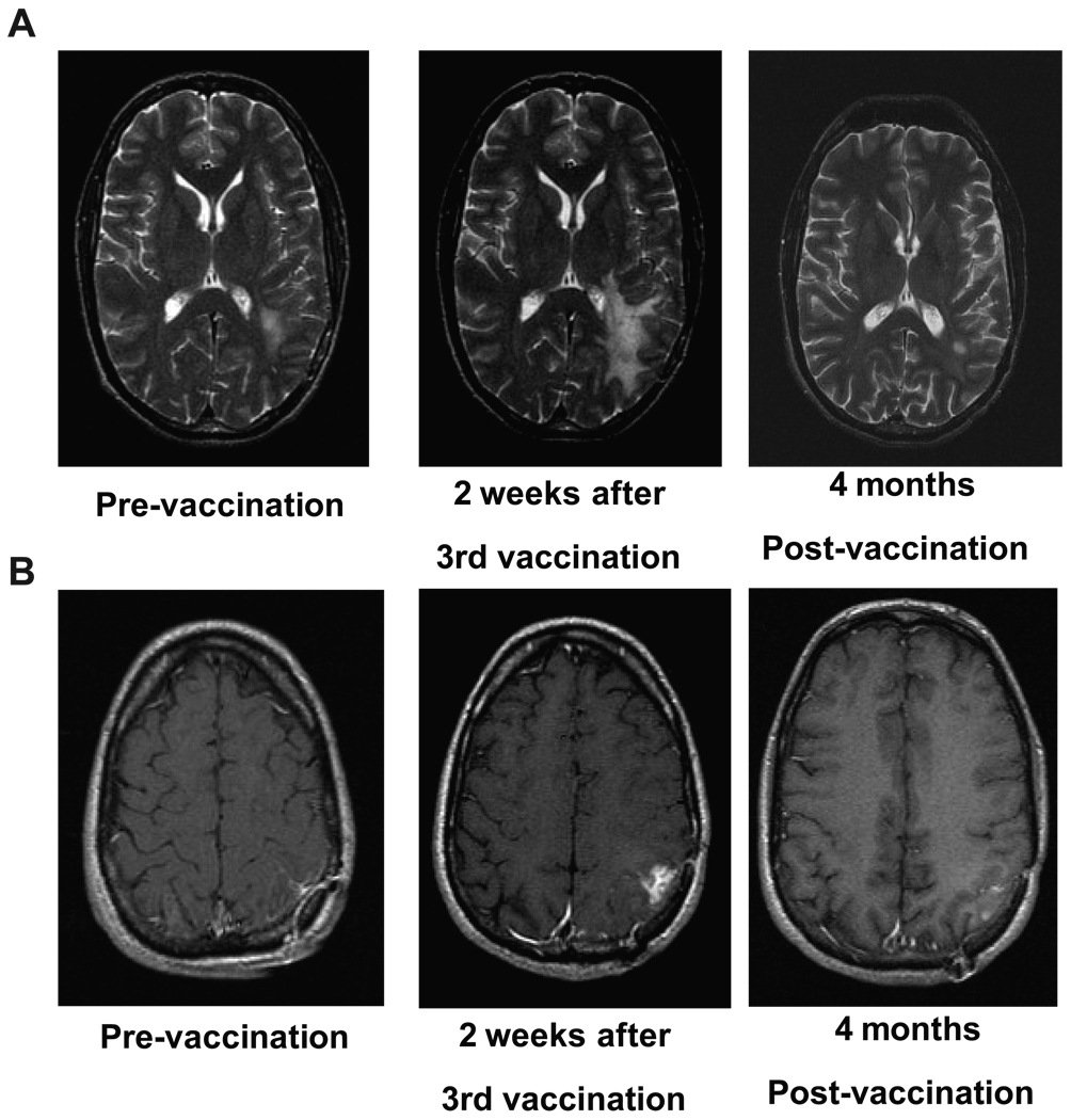
DC vaccinations were well-tolerated, with no major adverse events (NCI Common Toxicity Criteria grade 3 or 4) observed in any subject during the vaccine cycles (Table 1). There were no clinical or radiological signs of EAE or other autoimmune reactions in any patient. There were anecdotal cases of transient increased T2/FLAIR and enhancing lesions on MRI after DC vaccination, which may have suggested inflammatory responses post DC vaccination, particularly in the mesenchymal gene-clustered cohort of patients (Fig. 1). However, these MRI changes resolved in due course and did not require surgical intervention. The appearance and disappearance of such MRI findings, presumed to be related to vaccination and neuroinflammation, was noted in three of our patients (GBM 1–2, 1–3, and 5–4). These three patients were in the mesenchymal subgroup and are still alive over five years from the initial diagnosis of glioblastoma. Nausea/vomiting, headache and fatigue, diarrhea, low-grade fever and and pain/itching at the injection site were the most common symptoms associated with the treatment (Table 1). Local lymphadenopathy was observed in one patient, temporally coinciding with the expansion of HCMV-specific T cell expansion (23). In patients who concomitantly received 5% imiquimod cream or poly-ICLC with DC vaccination in the booster phase, no new additional toxicities were reported. Two patients consistently reported transient fevers (≥103° F) with each DC + poly-ICLC injection. Cumulatively, these data suggest a low toxicity profile for autologous tumor lysate-pulsed DC plus TLR agonists at all DC dose levels tested.
Figure 1.
MRI changes after DC vaccination. Transient increase in MRI T2/FLAIR lesions (A) and contrast enhancement (B) observed in a primary, newly diagnosed glioblastoma patient following DC vaccination (patient GBM5-4). Axial T2/FLAIR (A) and T1/contrast (B) MRI scans taken at 2 weeks pre-vaccination, 2 weeks post-vaccination, and 4 months later.
Systemic cytokine responses and regulatory T-cell populations following DC vaccination with TLR agonists
Others have assessed systemic immune responsiveness from autologous tumor lysate-pulsed DC vaccination by either delayed type hypersensitivity skin testing (DTH) (6, 12) or by restimulating PBMC with lysate-pulsed DC in vitro, followed by assessment of interferon-gamma (IFN-γ) (10, 12). However, the correlations with clinical outcome have not been consistent. In this trial, we elected to assess for more global systemic cytokine responses and changes in regulatory T cell (Treg) frequency that may be induced by our vaccination strategy.
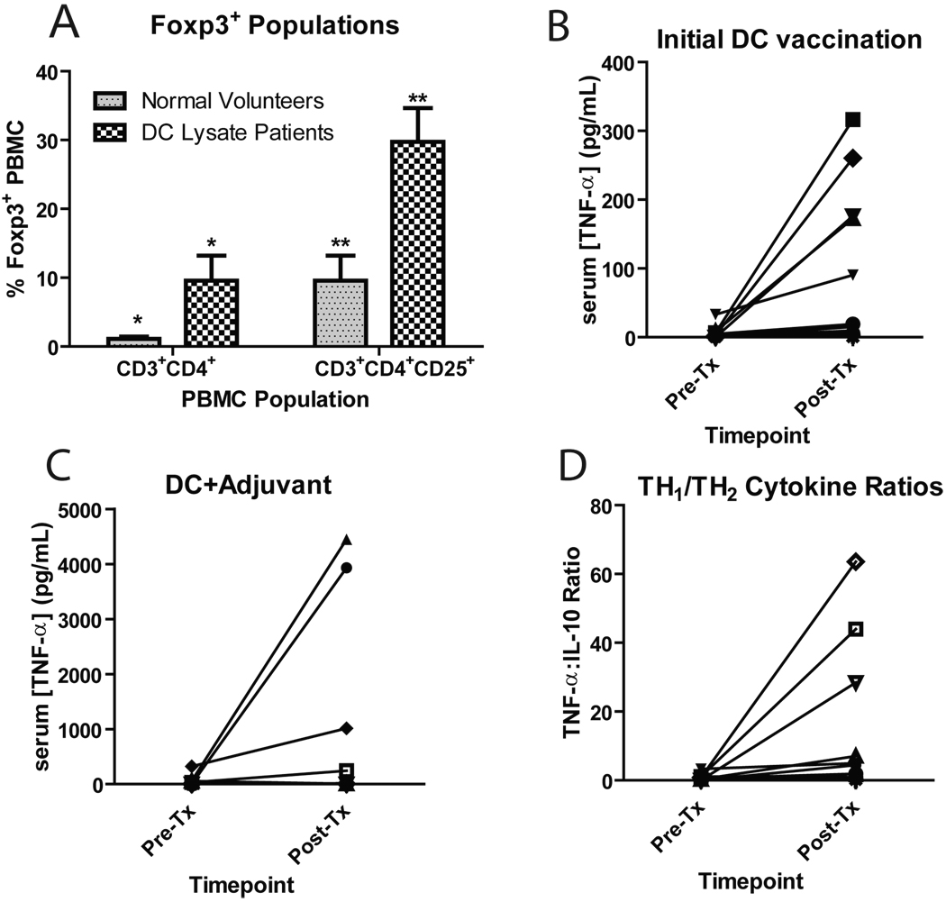
Peripheral blood changes in the frequency of CD3+CD4+Foxp3+ T cells were compared prior to and after DC vaccination for patients with available pre and post-treatment PBMC. We observed that glioblastoma patients on this clinical trial possessed increased frequencies of peripheral blood CD3+CD4+Foxp3+ or CD3+CD4+CD25+Foxp3+ lymphocytes compared with normal volunteers (Fig. 2A). However, at the time points measured, there were no relevant changes in the frequency of this lymphocyte population after immunotherapy that statistically correlated with clinical outcome (data not shown).
Figure 2.
Peripheral blood immune monitoring data. (A) PBMC’s from normal volunteers and DC trial patient pre-vaccination timepoints were thawed and stained for the expression of CD3, CD4 and CD25, followed by the intracellular labeling of Foxp3. Stained cells were acquired on a BD FacsCalibur flow cytometer and analyzed using FloJo software. The frequencies of CD3+CD4+Foxp3+ and CD3+CD4+CD25+Foxp3+ PBMC’s between normal volunteers and glioblastoma patients enrolled in this trial are compared. (*p=0.04; **p=0.01) (B,C) Serum cytokine responses, measured pre- and day 14 post-vaccination, after the initial course of DC vaccination (B) or after booster DC vaccinations with either 5% imiquimod or poly ICLC (C). Serum from patients enrolled on this clinical trial was thawed, labeled with cytometric bead array (CBA) antibody-coated beads, washed and subjected to analysis on a BD FacsCalibur flow cytometer together with cytokine standards. Quantitative assessment of cytokine levels was accomplished with a Microsoft Excel-based CBA software program. (D) Th1/Th2 cytokine ratios. Raw cytokine data for serum TNF-α and IL-10 at each timepoint were divided to generate a Th1:Th2 ratio.
To assess the cytokine microenvironment after DC vaccination with and without the addition of TLR agonists, we performed cytometric bead arrays from patient serum during the time course of the clinical trial to evaluate Th1 and Th2-type cytokine levels. Detectable increases in serum TNF-α and IL-6 were observed after DC vaccination (Fig. 2B, Suppl. Fig. 1A). However, the serum cytokine levels were variable between patients and the magnitude of changes did not seem to correlate with clinical outcome. Log-fold elevations in serum TNF-α and IL-6 were observed after booster DC vaccinations with either 5% imiquimod cream or 20 µg/kg poly ICLC (Fig. 2C, Suppl. Fig. 1B). To assess whether the Th1/Th2 cytokine balance might be relevant, we calculated ratios of each Th1-type cytokine with Th2-type cytokines to generate an effector/regulatory cytokine ratio (Fig. 2D). However, such information was also not significantly associated with the clinical outcome (data not shown), although our sample numbers may have been too small to detect statistical significance.
Dose escalation
A typical dose escalation scheme was performed with autologous tumor lysate-pulsed DC vaccination, using 1, 5 and 10 million DC administered intradermally. A fixed amount of lysate (100 µg) was added to the DC and incubated overnight prior to injection. The patient characteristics and survival data for each dose cohort are outlined in (Supplementary Table 2). In this dose escalation trial, there was no relationship between increasing DC dose and toxicity or specific adverse events of any kind. There were also no DC dose-dependent differences in immunological responses tested. As seen in Supplementary Table 2, the median overall survival was actually longer in the 1 million DC dose cohort compared with the higher dose cohorts. However, these differences in OS were not statistically significant, given the small sample size in each dose cohort and age differences between groups.
Survival analysis
Although this Phase I clinical trial was not powered to detect clinical efficacy, tumor response was monitored by clinical and MRI assessments at baseline (within one month prior to therapy), and every eight weeks thereafter as surrogate markers for clinical response and tumor status. Objective clinical data are summarized below and are listed in Table 1. When considering all 23 glioblastoma patients enrolled in this clinical trial (newly diagnosed and recurrent patients), the median time to tumor progression (TTP) was 15.9 months. The median overall survival time (OS), taken from the date of initial surgical diagnosis of glioblastoma, was 31.4 months. Overall survival from the time of initial diagnosis at one, two and three years was 91%, 55% and 47%, respectively. If we include only those who received the DC vaccine in the newly diagnosed setting (n=15), the median overall survival is 35.9 months, with a mean follow-up time of over four years, and one, two and three-year survival rates of 93%, 77% and 58%, respectively. For recurrent patients that enrolled in our vaccine trial (n=8), the median overall survival was 17.9 months from the time of initial glioblastoma diagnosis. OS was significantly longer for those who received DC vaccination at initial diagnosis compared to those who enrolled in this trial at the time of recurrence (p=0.03; Supplementary. Fig. 2).
Microarray gene expression profiling
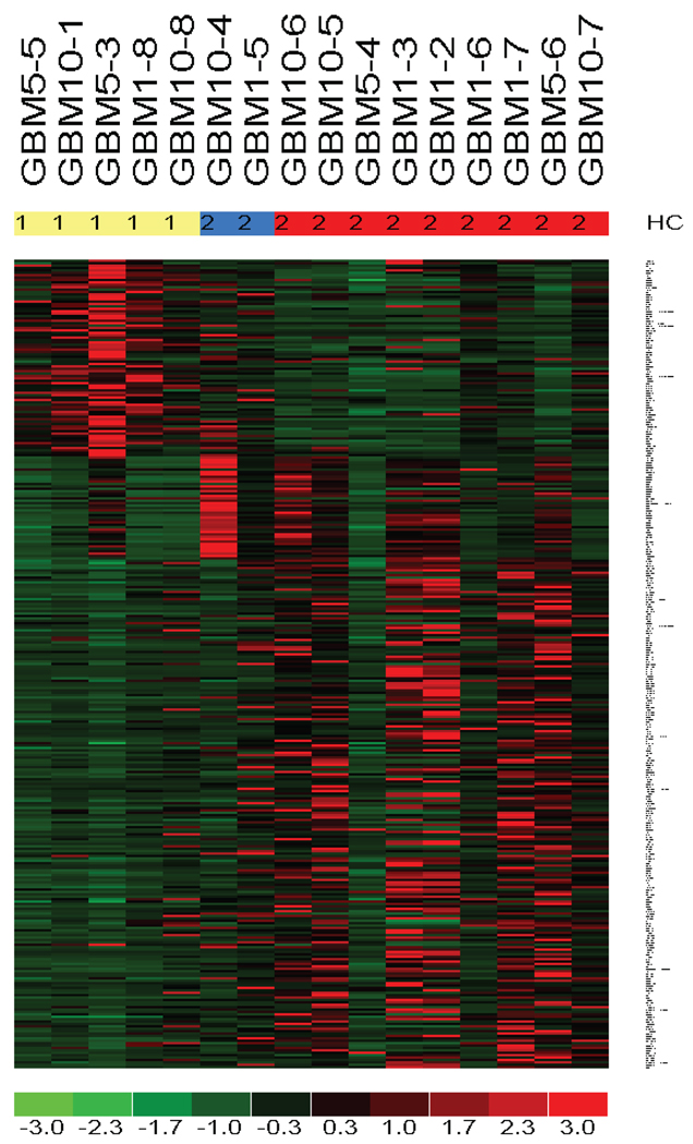
Since gene expression patterns have been shown to be highly correlated with survival in various cancers, we investigated whether the genetic signature of glioblastomas (20) was associated with clinical outcome in this DC immunotherapy trial. In patients where available pre-treatment tumor samples were available, we performed microarray-based gene expression classification as previously published (20, 21). As shown in Figure 3, gene expression profiling of our pre-treatment tumor samples produced the typical proneural (PN), proliferative (Pro) and mesenchymal (Mes) hierarchical clusters, using probesets previously described by our group (20, 21). Furthermore, we validated these hierarchical clusters using the UCSF-Genentech and TCGA probesets (24, 25), which yielded similar gene expression signatures for our DC lysate patients (data not shown).
Figure 3.
Microarray-based, expression profiling of pre-treatment glioblastoma samples from DC vaccine patients. Total RNA was isolated from frozen, surgically-resected tumors and subjected to global gene expression classification using Affymetrix human U133 Plus 2.0 microarray chips. Sufficient fresh-frozen tissue was available for extraction of high-quality RNA (without amplification) in 17 of the cases. Proneural (HC1, yellow legend), Proliferative (HC2A, blue legend), and Mesenchymal (HC2B, red legend) gene expression signatures were identified using probesets previously published (21). Heat maps were created using the dChip microarray software program.
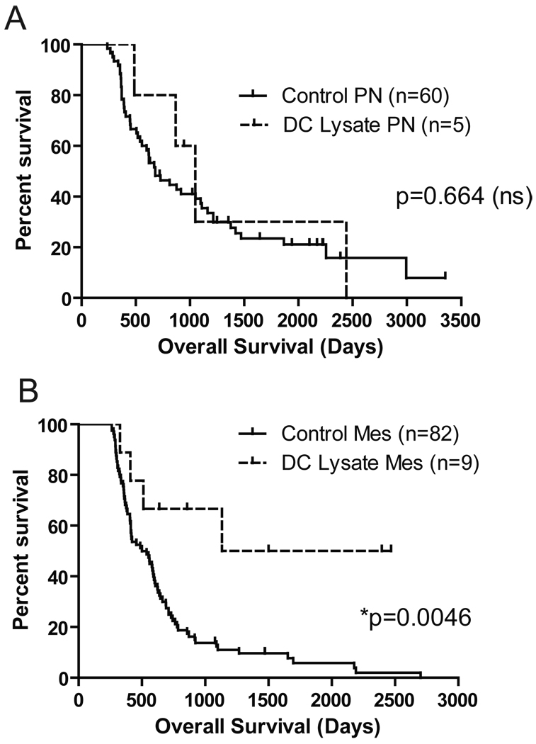
The mesenchymal gene expression signature is defined by overexpression of many inflammatory-associated genes. Thus, we hypothesized that there might be a difference in the clinical outcome of patients on our trial that could be linked to the local microenvironment of the original tumor. In order to control for any selection bias that might have been introduced by the requisite eligibility criteria for patients receiving the DC vaccine (i.e., subjects needing to be alive and off steroids long enough for vaccine preparation and administration), we eliminated any control patients that died within ~250 days of initial diagnosis for the purposes of our comparative analysis. We also stratified for patients who received radiation alone vs. radiation plus concurrent temozolomide chemotherapy after initial surgical resection, and found no statistical difference in these two groups when the early progressors (OS<250 days) were eliminated. As shown in Figure 4, patients enrolled on our trial with the proneural gene expression signature had an overall survival that was indistinguishable from a set of 60 contemporary proneural tumors analyzed from UCLA and three other institutions (21) (p=0.664; Fig. 4A). In contrast to this, patients in our DC vaccine trial with mesenchymal gene expression signatures had a significantly extended survival compared with 82 concurrently collected tumors that were found to have these same gene expression signatures (p=0.0046; Fig. 4B). While these data are not intended to represent efficacy, such information is noteworthy because glioblastoma patients with mesenchymal gene expression patterns typically have the worst prognosis and are the most refractory to current therapies (21, 24, 25).
Figure 4.
Extended survival in DC vaccinated patients with mesenchymal gene expression signatures, but not in patients with a proneural signature. The overall survival time of DC vaccine patients expressing a (A) Proneural (PN) gene signature (n=5) or (B) Mesenchymal (Mes) gene signature (n=9) was compared with the survival generated from a control, multi-institutional dataset of PN (n=60) or Mes glioblastomas (n=82; solid lines) previously published by our group (21). To accurately account for the potential bias associated with the time delay needed to generate the DC vaccine, we omitted control patients that experienced early progression (<250 days). PN comparison: p=0.664 (not statistically different, ns); Mes comparison (p=0.0046) by the Log-rank (Mantel-Cox) Test calculated in GraphPad software.
Gene expression signature and tumor-infiltating lymphocytes
The density and location of T lymphocyte accumulation within certain solid tumors have been associated with extended survival (26, 27), and recent evidence suggests that such a correlation may exist in malignant glioma (28). However, an association with the subtype of tumor or treatment modality has not been addressed.
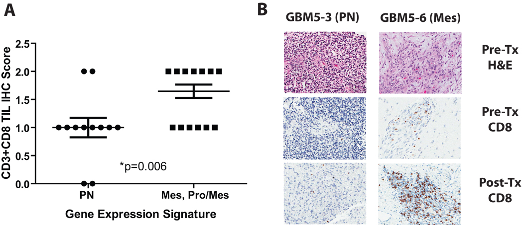
Since the mesenchymal expression signature includes numerous genes associated with inflammation, and tumor-specific T cells are known to be attracted to pro-inflammatory signals, we evaluated whether patients on our DC trial with mesenchymal gene expression signatures also had increased tumor infiltrating lymphocytes (TILs). As shown in Figure 5, tumors with a mesenchymal gene expression signature had significantly increased CD3+ and CD8+ TILs compared with PN tumors (p=0.006). Although our sample size is small, we also found qualitatively increased CD3+ and CD8+ TIL density after DC vaccination in tumors resected at recurrence (Fig. 5B). In post-DC vaccinated tumors resected/biopsied at the time of recurrence, increases in CD3+ and CD8+ TILs were associated with the mesenchymal gene expression profile, but not necessarily with the dose of DC given. Such findings point to a potential mechanism by which distinct glioblastoma tumor subtypes might be differentially responsive to immune-based therapies.
Figure 5.
Increased density of CD3+ and CD8+ lymphocytes in Mes gene expression groups compared with PN tumor sections. (A) 3 µm paraffin-embedded adjacent tissue sections from DC vaccinated patients were stained separately with CD3 and CD8 antibodies and scored in a blinded fashion by a neuropathologist (WHY). The IHC scores were compared between samples known to be PN (n=5) vs. Mes tumor samples (n=9). *p=0.006 by two-tailed t test calculated in GraphPad software. (B) Representative hematoxylin & eosin staining and CD8 IHC staining (pre- and post-DC vaccination) of a PN and Mes glioblastoma showing increased CD8+ TILs in the Mes glioblastoma. Original magnification: × 400.
DISCUSSION
In this Phase I study, we report the safety, feasibility, and bioactivity of a vaccine comprised of autologous DC pulsed with autologous tumor lysate as an adjuvant following surgical resection with standard chemo-radiotherapy. Unlike our previous reported DC vaccination strategy (8) and those reported by other groups (6, 9–13, 29), we included “booster” vaccinations with the innate immune response modifiers, 5% imiquimod (Aldara™) or poly-ICLC (Hiltonol™) based on our pre-clinical studies suggesting that pro-inflammatory innate immune signals could enhance DC activation, trafficking to lymph nodes, and the priming of anti-tumor antigen-specific T lymphocytes (15). There were no dose-limiting toxicities and no detectable differences in safety or efficacy among the three DC dose levels tested. Of note, there was a significant difference in the average age of patients in the 10 million DC cohort compared to the other dose cohorts, which could influence the difference in overall survival. However, another possible hypothesis is that this trend in the data was a reflection of a dilutional decrease in antigens available for presentation by DC at the highest DC dose cohort (10×106 cells), given that the quantity of lysate was fixed (at 100 µg per dose) despite the increased DC cell dose.
The concomitant administration of 5% imiquimod or poly ICLC with DC vaccination was also found to be safe and did not result in any additional toxicity or adverse events. To our knowledge, this is the first report of the use of TLR agonists in conjunction with DC vaccination strategies in brain tumor patients. Because TLR agonists were used only in patients in the booster phase, it is unclear whether or to what extent the addition of the TLR agonists contributed to the potential efficacy and overall survival of these patients. Furthermore, imiquimod and poly-ICLC are two different biological agents, targeting different TLRs. Imiquimod activates TLR-7, while poly-ICLC activates TLR-3, but both induce pro-inflammatory cytokine secretion. These complexities make it somewhat difficult to determine how these innate immune modifiers actually contributed to our study endpoints. Nevertheless, this current study establishes the safety of these TLR agonists in conjunction with glioma lysate-loaded DC, and further Phase II studies directly comparing these TLR agonists at the time of initial vaccination (not only in the booster phase) are currently underway.
While the number of glioblastoma patients entered in this Phase I clinical trial was not powered to measure efficacy, the clinical results of this trial are still noteworthy. The median OS from the time of initial surgical diagnosis was 31.4 months for all glioblastoma patients (n=23) treated in this study, including both those enrolled as newly diagnosed and recurrent tumor patients. For those treated in the newly diagnosed setting, the OS was 35.9 months; and the OS was 17.9 months for those who received vaccination at recurrence. In addition, we have had three patients survive over six years to date. Such statistics are compelling in the face of the expected median survival for this disease, which is currently still reported as about 14 months for newly diagnosed patients that receive standard surgery, radiation and temozolomide chemotherapy (4, 30, 31). This compares favorably even when compared with published data for the best clinically defined prognostic group of glioblastoma patients (Recursive Partitioning Analysis, RPA class III: age < 50 years and KPS ≥ 90), whose 2-year survivals were 40% and 29% for RPA III and IV patients, respectively, following treatment with standard radiation and temozolomide (31). Such data is also favorable compared to other recent brain tumor DC-based vaccine trials without booster injections and TLR adjuvants, where the OS was reported as 21.4 months (mean, 11 newly diagnosed and 23 recurrent glioblastoma patients) (10) and 9.6 months (median) in a recurrent glioblastoma population (6).
Glioblastomas are primarily identified by histologic features assigned to cytologically malignant, mitotically active, necrosis-prone tumors established by the World Health Organization (WHO) (32). Such histologic features are generally associated with patient survival, together with performance status, extent of surgical resection, and age. Yet, histologically identical tumors can behave in different ways; a situation that may underlie the biology of this heterogeneous disease. More recently, extensive genetic profiling of these tumors has been able to identify molecularly classifiable subgroups of glioblastoma (i.e., proneural, proliferative, and mesenchymal subtypes),(20, 21, 24, 33–38) which can better predict survival than conventional histopathologic analysis. Such new classification techniques are of interest so that patients can be more appropriately stratified for new treatment strategies (20).
The mesenchymal subgroup of glioblastomas typically have a poorer prognosis than the more common proneural subgroup (21, 24). However, in our study, patients with the mesenchymal gene expression signatures had significantly extended survival compared with a large, multi-institutional cohort (n=82) of glioblastoma samples of the same molecular subgroup treated with various other therapies. No such survival difference was observed in patients from this clinical trial with proneural signatures, compared to other control glioblastoma subjects of the proneural subgroup (n=60). Admittedly, such comparisons with concurrent and historical controls are not meant to imply efficacy, since this Phase I trial did not have a prospectively matched, placebo-controlled arm. Although some prognostic factors, such as age and Karnofsky performance status, were relatively matched in our comparison groups, the extent of surgical resection was not directly compared between the patients in this trial and our concurrent/historical controls. Since we need adequate amounts of tumor (>2 grams) to generate the autologous vaccines, tumor resectability was taken into account in the eligibility criteria. Therefore, it is possible that the extent of surgical resection may have been greater in our DC vaccinated patients compared to concurrent/historical controls, which could have influenced our survival results. Nevertheless, the median OS (31.4 mo.) of our DC-vaccinated patients is still noteworthy, when compared to large series of glioblastoma patients who underwent gross total tumor resections and were treated with concomitant chemo-radiotherapy, where the median survival was reported to be 18.6 months (31).
It is unclear whether the extended survival of our patients with mesenchymal gene expression signatures is a direct result of the vaccine effects, or good responses to follow-up therapies after failing the vaccine. Since mesenchymal signatures represent glioblastoma subgroups that are more resistant to conventional therapy, it can be speculated that DC vaccination somehow makes these tumors more susceptible to subsequent treatments (39). Because adjuvant temozolomide treatment was coordinated into the schedule of the DC booster vaccinations, this is a difficult distinction to make from our study design. Nevertheless, our results do suggest that mesenchymal gene expression signatures express elevated inflammatory gene transcripts and possess an increased density of tumor-infiltrating CD3+ and CD8+ lymphocytes compared with glioblastomas expressing other genetic signatures. As such, we hypothesize that the expression of inflammatory genes (e.g., IL-1R, TNF-α signaling factors, and chemokines) may facilitate the priming and trafficking of tumor-specific T cells into the tumor parenchyma, which might be enhanced by DC vaccination and innate immune response modifiers. Hence, the mesenchymal gene expression signature may have a direct impact on the bioactivity of the vaccine itself, irrespective of post-vaccine therapy. Prospectively designed, randomized, multi-center Phase III clinical trials will be required to validate such hypotheses and proof of clinical benefit remains to be established.
Overall, the results reported here may provide novel insights for prospective patient selection in future immunotherapy studies and lend additional credence for the ability of genetic expression signatures to impart relevant data for personalized cancer treatment. Based on the results of this Phase I trial, we will continue developing more advanced clinical trials with this particular approach. We currently are planning a randomized, multi-center Phase II/III clinical trial of DC vaccination for newly diagnosed glioblastoma (DCVax-Brain™), which will hopefully help to further define which subgroups of patients may respond to tumor vaccination strategies. This in turn may lead to further optimization and refinements of related trials of DC-based vaccines for patients with glioblastoma, with the ultimate goal of developing novel immunotherapeutic strategies for brain cancer patients.
Statement of Translational Relevance
The selective identification of patients who will respond to a particular therapy is of paramount importance, especially for patients diagnosed with malignant glioma. Patients diagnosed with glioblastoma (WHO grade IV) have an expected 5 year survival of less 3.3%. In this study, we report the results of a phase I clinical trial in which glioblastoma patients were treated with a personalized immunotherapy approach, comprised of autologous tumor lysate-pulsed dendritic cell vaccination. In addition, we utilized gene expression profiling to identify a group of patients with a particular gene expression signature (mesenchymal glioblastoma) that had longer survival following DC vaccination, compared to contemporary/historical control patients of the same gene expression subgroup, who did not receive vaccination. This signature was associated with inflammatory transcripts and enhanced tumor infiltrating T lymphocytes. Thus, these results suggest that gene expression signatures may be able to identify an immunogenic subgroup of glioblastoma that could be more responsive to immune-based therapies.
Supplementary Material
Acknowledgments
We thank Timothy Cloughesy, M.D., and Albert Lai, M.D., Ph.D. for helpful discussions and comments.
Research Support. This work was supported in part by NIH/NCI grants K01-CA111402 and RO1-CA123396 (to RMP), R01 CA 112358 (to LML), the Philip R. and Kenneth A. Jonsson Foundations, the Neidorf Family Foundation, the Ben & Catherine Ivy Foundation and Northwest Biotherapeutics, Inc. RMP is the recipient of the Howard Temin NCI Career Development award and STOP Cancer Career Development award. Flow cytometry was performed at the UCLA Jonsson Comprehensive Cancer Center (JCCC) Core Facility and gene expression was performed in the JCCC Gene Expression Shared Resource, which are supported by the NIH award CA16042. Microarray analysis was supported by the Carson Foundation. Tissue acquisition and IHC was supported by the UCLA Brain Tumor Translational Resource (BTTR), while the General Clinical Research Center (GCRC) was supported by M01-RR00865.
Footnotes
Disclaimers. The authors do not have any conflict of interests in this work.
REFERENCES
- 1.Deorah S, Lynch CF, Sibenaller ZA, Ryken TC. Trends in brain cancer incidence and survival in the United States: Surveillance, Epidemiology, and End Results Program, 1973 to 2001. Neurosurg Focus. 2006;20:E1. doi: 10.3171/foc.2006.20.4.E1. [DOI] [PubMed] [Google Scholar]
- 2.Cohen MH, Li Shen Y, Keegan P, Pazdur R. FDA Drug Approval Summary: Bevacizumab (Avastin(R)) as Treatment of Recurrent Glioblastoma Multiforme. Oncologist. 2009 doi: 10.1634/theoncologist.2009-0121. [DOI] [PubMed] [Google Scholar]
- 3.Lai A, Filka E, McGibbon B, et al. Phase II Pilot Study of Bevacizumab in Combination With Temozolomide and Regional Radiation Therapy for Up-Front Treatment of Patients With Newly Diagnosed Glioblastoma Multiforme: Interim Analysis of Safety and Tolerability. Int J Radiat Oncol Biol Phys. 2008 doi: 10.1016/j.ijrobp.2007.11.068. [DOI] [PubMed] [Google Scholar]
- 4.Stupp R, Mason WP, van den Bent MJ, et al. Radiotherapy plus concomitant and adjuvant temozolomide for glioblastoma. N Engl J Med. 2005;352:987–996. doi: 10.1056/NEJMoa043330. [DOI] [PubMed] [Google Scholar]
- 5.Yang MY, Zetler PM, Prins RM, Khan-Farooqi H, Liau LM. Immunotherapy for patients with malignant glioma: from theoretical principles to clinical applications. Expert Rev Neurother. 2006;6:1481–1494. doi: 10.1586/14737175.6.10.1481. [DOI] [PubMed] [Google Scholar]
- 6.De Vleeschouwer S, Fieuws S, Rutkowski S, et al. Postoperative adjuvant dendritic cell-based immunotherapy in patients with relapsed glioblastoma multiforme. Clin Cancer Res. 2008;14:3098–3104. doi: 10.1158/1078-0432.CCR-07-4875. [DOI] [PubMed] [Google Scholar]
- 7.Kikuchi T, Akasaki Y, Abe T, et al. Vaccination of glioma patients with fusions of dendritic and glioma cells and recombinant human interleukin 12. J Immunother. 2004;27:452–459. doi: 10.1097/00002371-200411000-00005. [DOI] [PubMed] [Google Scholar]
- 8.Liau LM, Prins RM, Kiertscher SM, et al. Dendritic cell vaccination in glioblastoma patients induces systemic and intracranial T-cell responses modulated by the local central nervous system tumor microenvironment. Clin Cancer Res. 2005;11:5515–5525. doi: 10.1158/1078-0432.CCR-05-0464. [DOI] [PubMed] [Google Scholar]
- 9.Walker DG, Laherty R, Tomlinson FH, Chuah T, Schmidt C. Results of a phase I dendritic cell vaccine trial for malignant astrocytoma: potential interaction with adjuvant chemotherapy. J Clin Neurosci. 2008;15:114–121. doi: 10.1016/j.jocn.2007.08.007. [DOI] [PubMed] [Google Scholar]
- 10.Wheeler CJ, Black KL, Liu G, et al. Vaccination elicits correlated immune and clinical responses in glioblastoma multiforme patients. Cancer Res. 2008;68:5955–5964. doi: 10.1158/0008-5472.CAN-07-5973. [DOI] [PubMed] [Google Scholar]
- 11.Yamanaka R, Abe T, Yajima N, et al. Vaccination of recurrent glioma patients with tumour lysate-pulsed dendritic cells elicits immune responses: results of a clinical phase I/II trial. BrJCancer. 2003;89:1172–1179. doi: 10.1038/sj.bjc.6601268. [DOI] [PMC free article] [PubMed] [Google Scholar]
- 12.Yamanaka R, Homma J, Yajima N, et al. Clinical Evaluation of Dendritic Cell Vaccination for Patients with Recurrent Glioma: Results of a Clinical Phase I/II Trial. Clin Cancer Res. 2005;11:4160–4167. doi: 10.1158/1078-0432.CCR-05-0120. [DOI] [PubMed] [Google Scholar]
- 13.Yu JS, Liu G, Ying H, Yong WH, Black KL, Wheeler CJ. Vaccination with tumor lysate-pulsed dendritic cells elicits antigen-specific, cytotoxic T-cells in patients with malignant glioma. Cancer Res. 2004;64:4973–4979. doi: 10.1158/0008-5472.CAN-03-3505. [DOI] [PubMed] [Google Scholar]
- 14.Yu JS, Wheeler CJ, Zeltzer PM, et al. Vaccination of malignant glioma patients with peptide-pulsed dendritic cells elicits systemic cytotoxicity and intracranial T-cell infiltration. Cancer Res. 2001;61:842–847. [PubMed] [Google Scholar]
- 15.Prins RM, Craft N, Bruhn KW, et al. The TLR-7 agonist, imiquimod, enhances dendritic cell survival and promotes tumor antigen-specific T cell priming: relation to central nervous system antitumor immunity. J Immunol. 2006;176:157–164. doi: 10.4049/jimmunol.176.1.157. [DOI] [PubMed] [Google Scholar]
- 16.Zhu X, Nishimura F, Sasaki K, et al. Toll like receptor-3 ligand poly-ICLC promotes the efficacy of peripheral vaccinations with tumor antigen-derived peptide epitopes in murine CNS tumor models. J Transl Med. 2007;5:10. doi: 10.1186/1479-5876-5-10. [DOI] [PMC free article] [PubMed] [Google Scholar]
- 17.Yang I, Kremen TJ, Giovannone AJ, et al. Modulation of major histocompatibility complex Class I molecules and major histocompatibility complex-bound immunogenic peptides induced by interferon-alpha and interferon-gamma treatment of human glioblastoma multiforme. J Neurosurg. 2004;100:310–319. doi: 10.3171/jns.2004.100.2.0310. [DOI] [PubMed] [Google Scholar]
- 18.Bigner DD, Pitts OM, Wikstrand CJ. Induction of lethal experimental allergic encephalomyelitis in nonhuman primates and guinea pigs with human glioblastoma multiforme tissue. J Neurosurg. 1981;55:32–42. doi: 10.3171/jns.1981.55.1.0032. [DOI] [PubMed] [Google Scholar]
- 19.Day A, Carlson MR, Dong J, O'Connor BD, Nelson SF. Celsius: a community resource for Affymetrix microarray data. Genome Biol. 2007;8:R112. doi: 10.1186/gb-2007-8-6-r112. [DOI] [PMC free article] [PubMed] [Google Scholar]
- 20.Freije WA, Castro-Vargas FE, Fang Z, et al. Gene expression profiling of gliomas strongly predicts survival. Cancer Res. 2004;64:6503–6510. doi: 10.1158/0008-5472.CAN-04-0452. [DOI] [PubMed] [Google Scholar]
- 21.Lee Y, Scheck AC, Cloughesy TF, et al. Gene expression analysis of glioblastomas identifies the major molecular basis for the prognostic benefit of younger age. BMC Med Genomics. 2008;1:52. doi: 10.1186/1755-8794-1-52. [DOI] [PMC free article] [PubMed] [Google Scholar]
- 22.Thumann P, Moc I, Humrich J, et al. Antigen loading of dendritic cells with whole tumor cell preparations. Journal of immunological methods. 2003;277:1–16. doi: 10.1016/s0022-1759(03)00102-9. [DOI] [PubMed] [Google Scholar]
- 23.Prins RM, Cloughesy TF, Liau LM. Cytomegalovirus immunity after vaccination with autologous glioblastoma lysate. N Engl J Med. 2008;359:539–541. doi: 10.1056/NEJMc0804818. [DOI] [PMC free article] [PubMed] [Google Scholar]
- 24.Phillips HS, Kharbanda S, Chen R, et al. Molecular subclasses of high-grade glioma predict prognosis, delineate a pattern of disease progression, and resemble stages in neurogenesis. Cancer Cell. 2006;9:157–173. doi: 10.1016/j.ccr.2006.02.019. [DOI] [PubMed] [Google Scholar]
- 25.Verhaak RG, Hoadley KA, Purdom E, et al. Integrated genomic analysis identifies clinically relevant subtypes of glioblastoma characterized by abnormalities in PDGFRA, IDH1, EGFR, and NF1. Cancer Cell. 2010;17:98–110. doi: 10.1016/j.ccr.2009.12.020. [DOI] [PMC free article] [PubMed] [Google Scholar]
- 26.Camus M, Tosolini M, Mlecnik B, et al. Coordination of intratumoral immune reaction and human colorectal cancer recurrence. Cancer Res. 2009;69:2685–2693. doi: 10.1158/0008-5472.CAN-08-2654. [DOI] [PubMed] [Google Scholar]
- 27.Galon J, Costes A, Sanchez-Cabo F, et al. Type, density, and location of immune cells within human colorectal tumors predict clinical outcome. Science. 2006;313:1960–1964. doi: 10.1126/science.1129139. [DOI] [PubMed] [Google Scholar]
- 28.Yang I, Tihan T, Han SJ, et al. CD8+ T-cell infiltrate in newly diagnosed glioblastoma is associated with long-term survival. J Clin Neurosci. 2010 doi: 10.1016/j.jocn.2010.03.031. [DOI] [PMC free article] [PubMed] [Google Scholar]
- 29.Heimberger AB, Sun W, Hussain SF, et al. Immunological responses in a patient with glioblastoma multiforme treated with sequential courses of temozolomide and immunotherapy: case study. Neuro Oncol. 2008;10:98–103. doi: 10.1215/15228517-2007-046. [DOI] [PMC free article] [PubMed] [Google Scholar]
- 30.Mirimanoff RO, Gorlia T, Mason W, et al. Radiotherapy and temozolomide for newly diagnosed glioblastoma: recursive partitioning analysis of the EORTC 26981/22981-NCIC CE3 phase III randomized trial. J Clin Oncol. 2006;24:2563–2569. doi: 10.1200/JCO.2005.04.5963. [DOI] [PubMed] [Google Scholar]
- 31.Stupp R, Hegi ME, Mason WP, et al. Effects of radiotherapy with concomitant and adjuvant temozolomide versus radiotherapy alone on survival in glioblastoma in a randomised phase III study: 5-year analysis of the EORTC-NCIC trial. Lancet Oncol. 2009;10:459–466. doi: 10.1016/S1470-2045(09)70025-7. [DOI] [PubMed] [Google Scholar]
- 32.Louis DN, Ohgaki H, Wiestler OD, et al. The 2007 WHO classification of tumours of the central nervous system. Acta Neuropathol. 2007;114:97–109. doi: 10.1007/s00401-007-0243-4. [DOI] [PMC free article] [PubMed] [Google Scholar]
- 33.Beetz C, Bergner S, Brodoehl S, et al. Outcome-based profiling of astrocytic tumours identifies prognostic gene expression signatures which link molecular and morphology-based pathology. Int J Oncol. 2006;29:1183–1191. [PubMed] [Google Scholar]
- 34.Li A, Walling J, Ahn S, et al. Unsupervised analysis of transcriptomic profiles reveals six glioma subtypes. Cancer Res. 2009;69:2091–2099. doi: 10.1158/0008-5472.CAN-08-2100. [DOI] [PMC free article] [PubMed] [Google Scholar]
- 35.Mischel PS, Shai R, Shi T, et al. Identification of molecular subtypes of glioblastoma by gene expression profiling. Oncogene. 2003;22:2361–2373. doi: 10.1038/sj.onc.1206344. [DOI] [PubMed] [Google Scholar]
- 36.Nutt CL, Mani DR, Betensky RA, et al. Gene expression-based classification of malignant gliomas correlates better with survival than histological classification. Cancer Res. 2003;63:1602–1607. [PubMed] [Google Scholar]
- 37.Shai R, Shi T, Kremen TJ, et al. Gene expression profiling identifies molecular subtypes of gliomas. Oncogene. 2003;22:4918–4923. doi: 10.1038/sj.onc.1206753. [DOI] [PubMed] [Google Scholar]
- 38.Shirahata M, Iwao-Koizumi K, Saito S, et al. Gene expression-based molecular diagnostic system for malignant gliomas is superior to histological diagnosis. Clin Cancer Res. 2007;13:7341–7356. doi: 10.1158/1078-0432.CCR-06-2789. [DOI] [PubMed] [Google Scholar]
- 39.Wheeler CJ, Das A, Liu G, Yu JS, Black KL. Clinical responsiveness of glioblastoma multiforme to chemotherapy after vaccination. Clin Cancer Res. 2004;10:5316–5326. doi: 10.1158/1078-0432.CCR-04-0497. [DOI] [PubMed] [Google Scholar]
Associated Data
This section collects any data citations, data availability statements, or supplementary materials included in this article.


