Abstract
Clinically approved inhibitors of HIV-1 protease function via a competitive mechanism. A particular vulnerability of competitive inhibitors is their sensitivity to increases in substrate concentration, as may occur during virion assembly, budding and processing into a mature, infectious viral particle. Advances in chemical synthesis have led to the development of new chemical libraries with high diversity using rapid in-solution syntheses. These libraries have been previously shown to be effective at disrupting protein-protein and protein-nucleic acid interfaces. We have screened 44,000 compounds from such a library to identify inhibitors of HIV-1 protease. One compound was identified that inhibits wild type protease, as well as a drug-resistant protease with 6 mutations. Moreover, analysis of this compound suggests an allosteric, non-competitive mechanism of inhibition and may represent a starting point for an additional strategy for anti-retroviral therapy.
Keywords: enzyme, non-competitive inhibitors, kinetics, high-throughput screening
INTRODUCTION
HIV infection continues to be a worldwide health crisis, with over thirty-three million infected people worldwide [1]. Despite improvements in antiretroviral therapeutic development, drug resistance remains a major obstacle to effective control of infection in HIV-infected patients. Numerous advances and improvements have been made in drugs targeting the viral protease, required for maturation of virions into infectious particles [2]. However, common mutations associated with protease inhibitor drug resistance appear in drug-experienced patients, and in certain subtypes of the virus in drug-naÔve patients as well [3], often leading to virologic failure and the onset of disease progression. Drug resistance complicates the use of therapeutics in the treatment of HIV infection, necessitating an ongoing search for novel therapeutics targeting the viral protease.
HIV-1 protease is a 22-kDa homodimeric aspartic protease consisting of two 99-residue polypeptide chains that self-assemble to form the enzymatically active dimer. Currently, all FDA-approved protease inhibitors (PIs) are in the same mechanistic class, i.e. competitive inhibitors that bind the active site of the protease, preventing the association of the protease with substrates and resulting in disruption of virion maturation [2]. One drawback of competitive inhibitors is that similar active site mutations can deleteriously affect small molecule binding in the active site, leading to increased risk of cross-resistance to other competitive inhibitors. An additional potential pitfall is that competitive inhibitors are sensitive to substrate concentrations [4]. An alternative to competitive inhibitors has been the identification of inhibitors that target non-active site regions of the protease, such as the dimer interface [5, 6], flaps [7, 8], or other non-substrate active site regions [9]. Moreover, inhibitors that utilize non-competitive, uncompetitive, or mix-mode mechanisms have also been identified [5–8, 10, 11]. A potential advantage of a non-competitive mechanism will be the insensitivity to substrate concentrations, which may better maintain a therapeutic threshold in the substrate-rich virion. The improved therapeutic threshold and potential insensitivity to current resistance mutations may be more efficacious at inhibiting viral replication and the emergence of drug-resistance.
To identify inhibitors that may target other features of the protease structure to inhibit its function, we screened a library of compounds previously shown to provide inhibitors of protein-protein and protein-nucleic acid interactions [12, 13]. One compound, compound 1, was found to inhibit wild type protease, from the NL4-3 strain of HIV-1, in the low micromolar range. Moreover, compound 1 also inhibited a multidrug resistant (MDR) protease containing 6 mutations associated with PI resistance [14]. The kinetics findings for wild type protease demonstrated a mechanism of inhibition consistent with non-competitive inhibition, and cross-competitive inhibition studies with compound 1 and Pepstatin A, a competitive inhibitor, implicated a non-active site binding affect. Taken together, these findings suggest that compound 1 functions as a non-competitive, allosteric protease inhibitor.
MATERIALS AND METHODS
Enzyme Activity Assays
HIV-1 protease enzymatic activity was assayed as described previously [15], using the fluorescently labeled anthranilyl protease substrate Abz-Thr-Ile-Nle-p-nitro-Phe-Gln-Arg-NH2 (H-2992, Bachem, CA) [16]. In brief, bacterially purified HIV-1 protease was mixed with inhibitor compounds in a reaction buffer containing 25 mM MES, pH 5.6, 200 mM NaCl, 5% DMSO, 5% glycerol, 0.0002% Triton X-100, and 1 mM dithiothreitol in a prewarmed 96-well plate. All clones used for protease bacterial expression were generated from NL4-3 wild type or a multidrug resistant (MDR) protease containing 6 mutations (L24I/M46I/F53L/L63P/V77I/V82A), termed 6X, associated with resistance to saquinavir, nelfinavir, ritonavir, and TL3, as described [14]. The enzyme-substrate reaction was started by the addition of fluorescently labeled substrate and the reaction progress was measured by fluorescence intensity using an FLx-800 fluorescence plate reader (BioTek, VT). For IC50 determinations, final reaction concentrations were 25 nM protease, 30 µM substrate (the approximate Km), and 0.001–600 µM of inhibitor.
Chemical Library
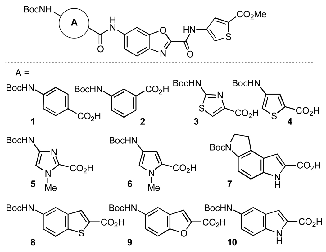
The Boger laboratory has established and reported previously on a collection of chemical libraries [17] consisting of approximately 66,000 compounds prepared by using solution phase technology with liquid–liquid acid–base extraction purification, evaluated for composition and purity, and stored for further assessment [18, 19]. Figure 1 shows a representative diagram of chemical scaffolds and substitutions used to generate the library. From the original library, 44,000 compounds were evaluated in our study. The lead compounds 1 and 2 (Figure 2) identified from the screening were synthesized, then evaluated for composition and purity, below, before use [18, 19].
Figure 1. Representative group of chemical substituents that are used in the reaction in the context of the compound scaffold.
Each substituent is found at circle site labeled A, in this case generating 10 different compounds. For additional information see Materials and Methods and [18, 19].
Figure 2. Compounds used in this study, (A) compound 1 and (B) compound 2.
Compound 1 was identified through protease – substrate screens of the original chemical library, see text, whereas compound 2 is a derivative of compound 1 with the Boc and methyl groups removed from the ends of the compound.
Compound 1. 1H NMR (400 MHz, DMSO-d6, 25 °C) ḍı̃ı84̣s, 1H), 10.74 (s, 1H), 9.86 (s, 1H), 8.39 (d, J = 1.9, 1H), 8.14 (dd, J = 8.3, 1.6 Hz, 2H), 8.08 (s, 1H), 7.92 (d, J = 8.8, 1H), 7.83 (dd, J = 8.9, 1.9, 1H), 7.39 (s, 1H), 3.84 (s, 3H), 1.49 (s, 9H); MS-ESI (m/z) calcd for [C24H22N4O7S2+Cl]− 577.1; found: 577.1.
Compound 2. 1H NMR (400 MHz, DMSO-d6, 25 °C) ḍı̃ı8̣ıs, 1H), 10.85 (s, 1H), 8.41 (d, J = 1.8 Hz, 1H), 8.08 (dd, J = 15.1, 1.4 Hz, 2H), 8.03 (s, 1H), 7.94 (d, J = 8.6 Hz, 1H), 7.84 (dd, J = 8.7, 1.6 Hz, 1H), 7.69 (m, 2H), 7.55 (m, 1H); MS-ESI (m/z) calcd for [C18H12N4O5S2+H]+ 429.0; found: 429.0.
Michaelis-Menten Kinetics Measurements
For Michaelis-Menten kinetics measurements, protease substrate was titrated from 1 to 200 µM. To assay for promiscuous inhibition, 0.001–0.01% Triton X-100 was added to the reaction. To assess inhibitor specificity horseradish peroxidase (Sigma) activity was assayed in 14 mM potassium phosphate, pH 6.0, 0.5% hydrogen peroxide with an enzyme concentration of 5 nM. The reaction was initiated with the addition of the substrate o-phenylenediamine at concentrations from 20 µM to 100 µM in 100 mM sodium phosphate and 50 mM sodium citrate, pH 5.0. Kinetics constants were determined by nonlinear regression of initial reaction velocities as a function of inhibitor concentration using Prism v5.0c (GraphPad Software, San Diego, CA). IC50 values were fit with the following equation:
Michaelis-Menten kinetics constants were fit to the following equation:
Ki constants for compounds 1 and 2 were fit to the following equation:
Cross-competitive Inhibitor Measurements
A variation of Yonetani and Theorell analysis was used to evaluate the binding mode of compound 1 [20, 21]. The use of the variation of Yonetani and Theorell analysis, as discussed by Martinez-Irujo et al [21], takes into account the binding interactions on an enzyme of competitive and non-competitive inhibitors. The cross-competitive inhibitor assessment was accomplished by varying the concentration of Pepstatin A (Roche), a competitive inhibitor, at a fixed concentration of compound 1, a non-competitive inhibitor, while keeping the substrate and protease concentrations constant. The experimental conditions for assessing protease function were identical to those used to determine IC50. Pepstatin A was used at concentrations from 0.6 to 3.0 µM, while compound 1 was held constant at 45, 30, 20, or 0 µM. The determination of the interaction term, γ, that defines the degree to which binding of one inhibitor influences the binding of the second inhibitor, was determined utilizing Prism v5.0c (Graphpad Software, San Diego, CA) using the following equation [21]:
Docking Studies of Compound 1
The docked conformation of compound 1 with HIV protease was generated using AutoDock Vina 1.02 [22]. The high resolution HIV-1 protease structure 2HS1 was chosen as the receptor. Two overlapping search spaces were used, each measuring 25 × 32 × 40 Å, which together spanned chain A of this structure. The darunavir molecule bound in the active site was preserved. In each docking run, 9 conformations were reported, only the most favorable is detailed below. Three-dimensional coordinates for the ligand were determined using Corina [23]. Other docking parameters were kept to their default values.
RESULTS AND DISCUSSION
We have screened a library of compounds previously shown to inhibit protein-protein and protein-nucleic acid interactions [12, 13, 17, 24]. A chemically diverse library of 44,000 compounds was synthesized using a solution phase combinatorial synthesis as previously described [13, 18, 19, 25]. To facilitate synthesis and screening, some compounds were synthesized as part of a screened mixture, with some mixtures containing up to 10 related but distinct compounds. A representative group of compounds from which the lead compounds emerged is shown in Figure 1.
Compounds were screened initially for the ability to inhibit the wild type HIV-1 protease, obtained from the NL4-3 virus, in a real-time kinetics assay using a fluorogenic substrate at a concentration equal to the Km. Compounds that had significant affects on the baseline fluorescent signal, which was designated as 10% above baseline independent of the substrate peptide, were excluded from further screening. Assay conditions were chosen to reduce the possibilities of false positives resulting from promiscuous inhibitors, including minimizing compound aggregate formation by the inclusion of detergent and reducing compound incubation time [26, 27]. Compound groups that showed greater than 50% inhibition of wild type protease at a compound concentration of 20 µM were then tested against 6X protease, a multidrug resistant (MDR) protease containing 6 mutations (L24I/M46I/F53L/L63P/V77I/V82A) associated with resistance to saquinavir, nelfinavir, ritonavir, and TL3, identified here as 6X [14].
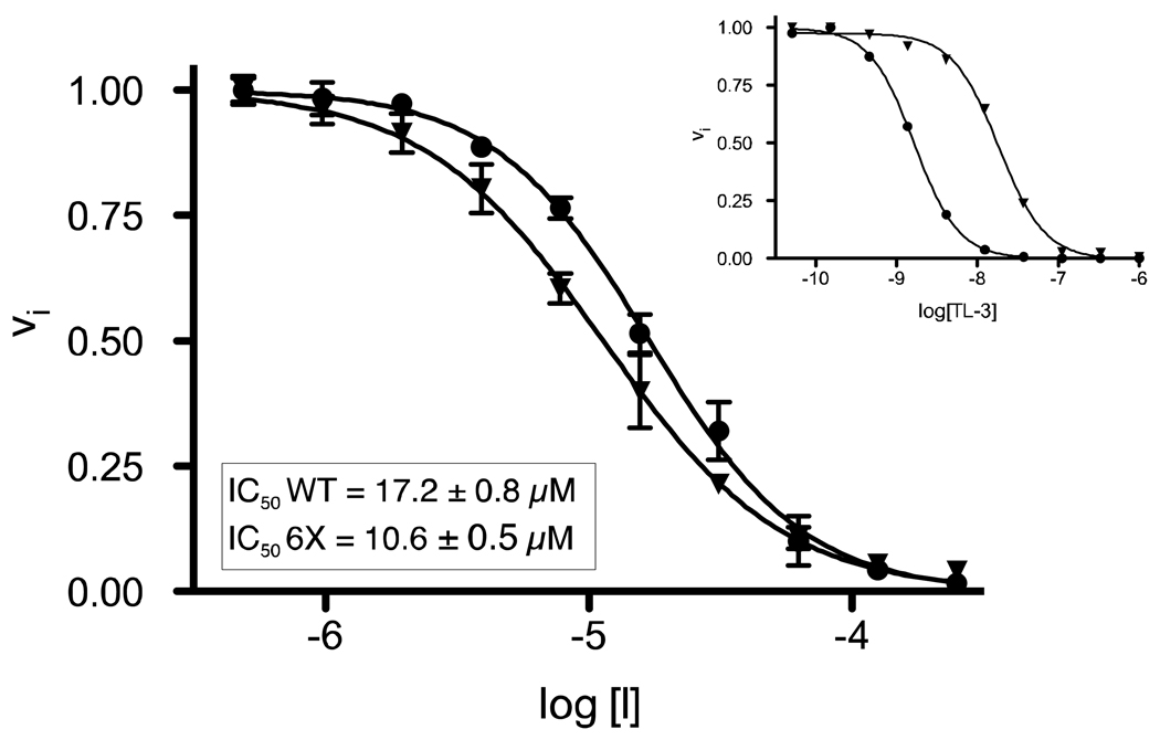
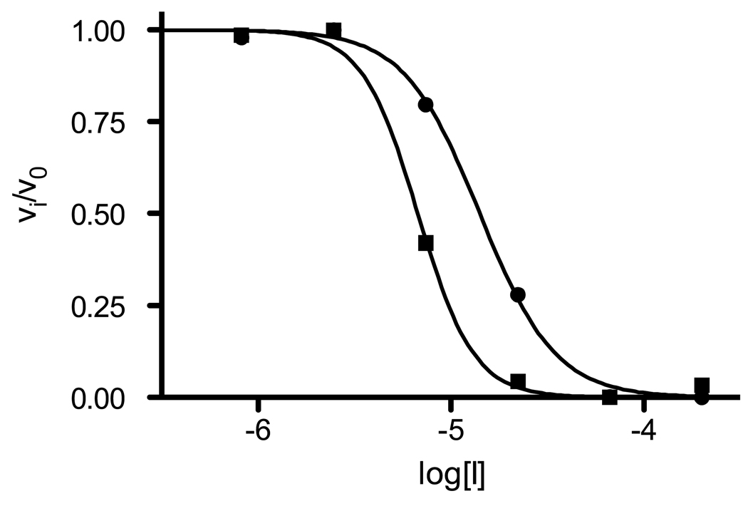
Compound families showing greater than 50% inhibition against both wild type and 6X proteases were then deconvoluted and synthesized as individual compounds. These compounds were then tested individually against wild type and MDR 6X proteases. Individual compounds again showing greater than 50% inhibition at a concentration of 20 µM were selected for more detailed kinetics analyses. One compound, compound 1 (Figure 2), was found to inhibit wild type protease in the low micromolar range (Figure 3). Furthermore, compound 1 also showed low micromolar inhibition of the 6X protease. The half maximal inhibitory concentrations, IC50, were determined against wild type and 6X proteases and found to be 17 µM against wild type protease and 11 µM against the 6X protease (Figure 3). Thus, compound 1 is effective in inhibiting both wild type and a MDR 6X protease at a similar IC50.
Figure 3. IC50 titration of compound 1 against wild type and the 6X multi-drug resistant proteases.
Foreground, evaluation of compound 1, log [I], against wild type (●) and 6X multi-drug resistant (▼) proteases. Inset: For comparison, titration of TL-3, log[TL-3], a protease inhibitor which is effective against the wild type (●) protease, but not the multi-drug resistant 6X protease mutant [14] (▼), is shown. Results from nonlinear regression indicate that the IC50s are within a factor of 2 of each other for wild type and multi-drug resistant 6X protease mutant. IC50 curve fitting was performed as described in the Materials and Methods. ± values indicate the standard error.
To address whether compound 1 was a general enzymatic inhibitor we evaluated whether the compound altered horseradish peroxidase function at various concentrations. No measurable effect on the Michaelis-Menten kinetics of the reaction was observed at any of the compound concentrations evaluated, suggesting that compound 1 is not a general enzymatic inhibitor (data not shown). Furthermore, the inhibitory activity of compound 1 on HIV-1 protease was not abrogated by the addition of non-ionic detergents further strengthening the case that compound 1 is not a promiscuous inhibitor (data not shown) [26, 27].
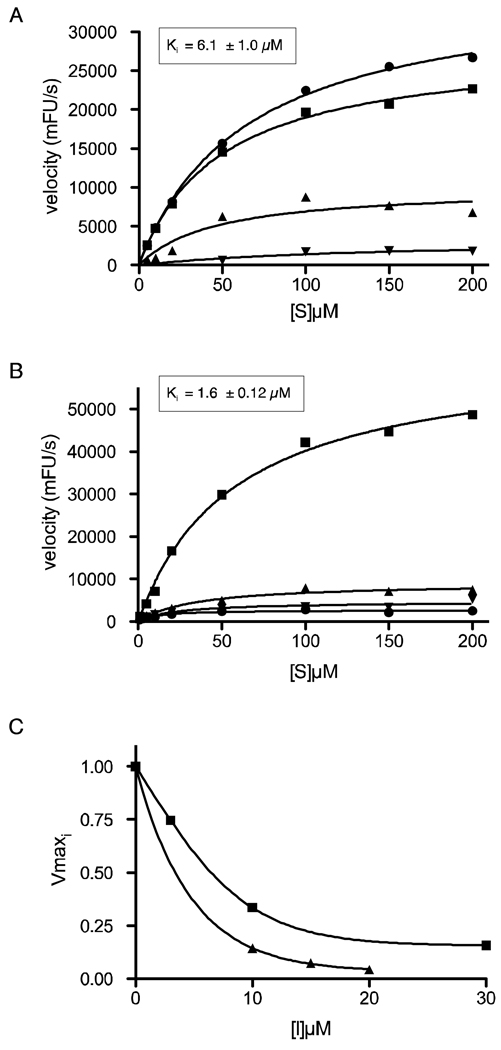
In order to identify the minimal chemical moieties necessary for PI activity, we synthesized a derivative library based on compound 1 and screened each fragment against wild type protease independently. While the majority of the derivatives showed significantly reduced inhibition of protease activity with IC50s ranging from 20 to greater than 1000 µM, one derivative, compound 2 (Figure 2), showed more potent inhibitory activity. Compound 2 is similar to compound 1, but with the Boc and methyl groups removed. When the inhibitory activity of compound 2 was compared to compound 1, it showed a slight decrease in both the IC50 and Ki values as determined with the fluorogenic protease substrate assay (Figures 3 and 4). Therefore, both compounds were active on wild type protease and compound 1 demonstrated activity against the MDR 6X protease mutant.
Figure 4. Michaelis-Menten kinetics of compound 1 and 2 against wild type protease.
(A) Compound 1 was used at 0 (●), 3 (■), 10 (▲), and 30 (▼) µM and (B) compound 2 was used as 0 (■), 10 (▲), 15 (▼), and 20 (●) µM over a range of µM substrate concentrations [S] for determination of the Michaelis-Menten kinetics. This is a representative result from 1 of 3 experiments. (C) Nonlinear regression of Vmax as a function of compound 1 (■) or 2 (▲) concentration. Shown is a representative experiment and standard errors for individual points varied less then 10% of the mean and curve fitting is described in the Materials and Methods. ± values indicate the standard error.
We next determined the effect of compounds 1 and 2 on the Km and Vmax of wild type protease with reactions performed within a range of substrate concentrations from 1 to 200 µM, centered around the Km of 30 µM, at several inhibitor concentrations, from 0 to 30 µM. The values for the initial velocities were then fit to a Michaelis-Menten model using nonlinear regression to determine the dose-dependent effects of the compounds on the Km and Vmax for HIV protease, as shown in Figure 4, panel A, compound 1, and panel B, compound 2. When we measured protease activity as a function of both substrate concentration and inhibitor concentration, and used nonlinear regression to fit the resulting initial velocities to a Michaelis-Menten model, we observed a curvilinear response in Vmax as a function of increasing concentration of both compounds (Figure 4, panel C). The results are consistent with a non-competitive mechanism of inhibition.
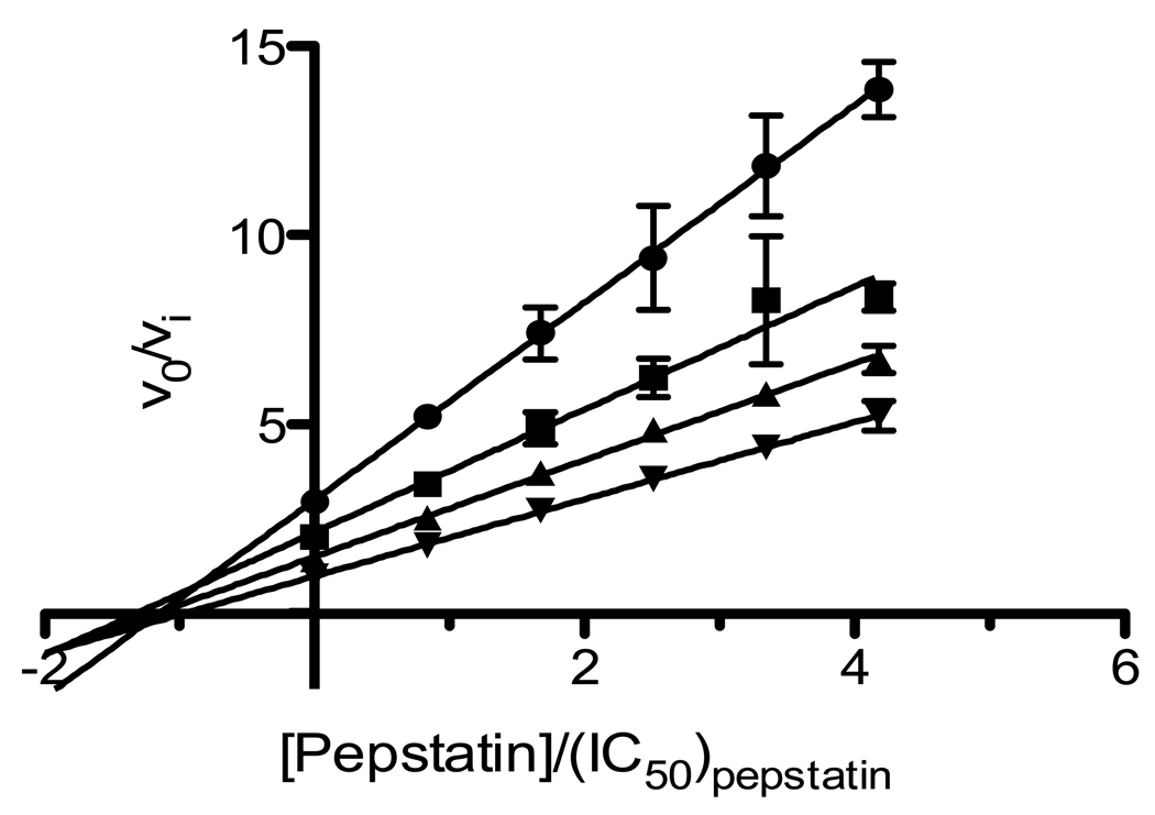
To glean further insights into the underlying molecular process of protease inhibition by compound 1, we utilized a variation of Yonetani and Theorell analysis [20, 21] to evaluate the binding mode. Since the inhibition by compound 1 is consistent with a non-competitive mechanism, which might predict that substrate may still bind to the protease active site when compound 1 is bound, Pepstatin A was used as a cross-competitive inhibitor for analysis. This method of inhibitor cross-competitive analysis allows determination of the degree to which the binding of compound 1 to protease influences the binding of the second inhibitor to the active site [4, 20, 28]. The choice of Pepstatin A was based on the ability to inhibit protease [29], the well established biochemical and structural reports of its binding location in the active site [30], that it has a competitive inhibition mechanism [31, 32], and the reported use of Acetyl-pepstatin A for inhibitor cross-competitive studies for non-active site inhibitors [5]. The graphical findings from a representative cross-competitive study utilizing compound 1 and Pepstatin A is shown Figure 5. In each case, non-parallel lines were obtained which converged at the x-axis, which is consistent with the interpretation that compound 1 and Pepstatin A may bind independent sites [4, 20, 21]. The interaction term, γ, that defines the degree to which the binding of one inhibitor to the enzyme influences the binding of the second inhibitor can be determined through interpolation of the x-intercept or calculated [4, 20, 21]. A small γ value (<1) signifies a synergistic interaction between the inhibitors, whereas a large γ value (1>) indicates mutual antagonism, and in the case that γ = 1, the inhibitors bind to the enzyme in an independent manner. Calculation of γ yielded approximately 1, consistent with compound 1 binding to a protease site independent of Pepstatin binding in the active site. These finding are consistent with compound 1 binding and providing inhibition through a site independent of the active site.
Figure 5. Yonetani and Theorell plot of v/vi versus concentration of Pepstatin A and compound 1.
0 µM of compound 1 (▼); 20 µM of compound 1 (▲); 30 µM of compound 1 (■); 45 µM of compound 1 (●) with varying concentrations of Pepstatin A. Each point was in triplicate and this is a representative result from 1 of 4 experiments. Assay conditions and curve fitting described in Materials and Methods.
Given our findings from the inhibitor cross-competitive study indicating that compound 1 was not binding in the active site, we investigated whether compound 1 functions as a dimerization inhibitor. A number of compounds have been reported to promote inhibition through disruption of protease dimerization [5, 6]. To address whether compound 1 disrupts dimerization we utilized a tethered homodimeric protease, formed by a direct repeat of protease monomers linked by a 5 amino acid sequence [33]. The IC50 of compound 1 was found to be similar for both the non-covalent wild type protease dimer and the covalently tethered dimer protease, as shown in Figure 6. Since compound 1 was active against the protease-tethered dimer, this implies that dimerization disruption is not required for inhibitory activity.
Figure 6. IC50 titrations of compound 1 against wild type and tethered dimer proteases.
Compound 1 [I] demonstrates similar inhibitory efficacy against wild type (●) and the tethered protease dimer (■). Standard errors for individual points varied less then 10% of the mean and shown is a representative experiment. Assay conditions described in the Materials and Methods
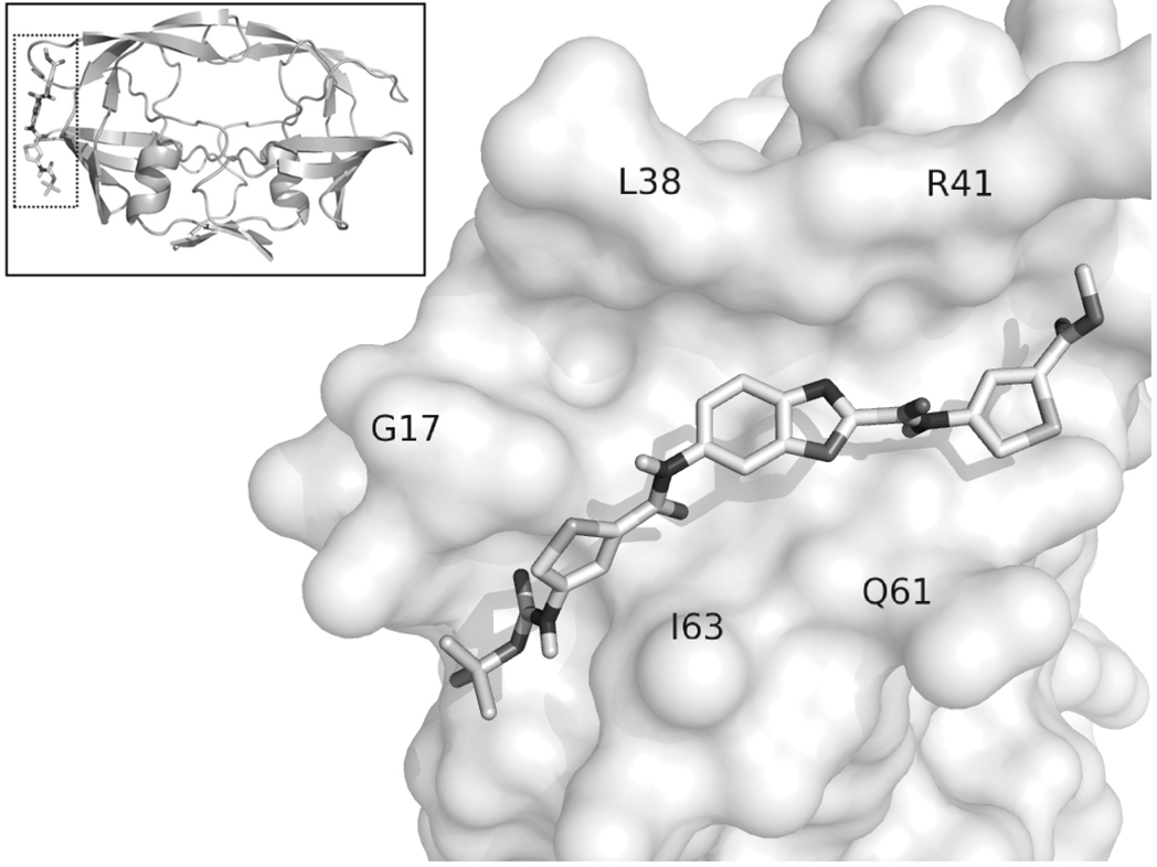
As compound 1 was shown to have a distinct binding location from Pepstatin A and not promote dimer interface disruption, possible binding modes were explored using molecular docking. Focusing the search on the outside surface of the protein, a low-energy conformation was discovered which placed compound 1 in a long, solvent exposed cleft, termed the exo site [34], as shown in Figure 7. The exo site is composed of distinct regions that include the elbow, cantilever, and fulcrum components of the protease. Molecular dynamic simulations of protease flap movement relative to the exo site has indicated that the exo site is compressed when the flaps are open and is extended when the flaps are closed [34]. Moreover, the exo site has been shown, via a fragment-based screen, to accommodate small molecules [35]. The predicted binding energy from the compound 1 docking simulation (−7.2 kcal/mol) corresponds to a Ki of 5.2 µM, very close to the experimentally observed Ki value of 6.1 µM. Together with the biochemical findings presented herein, the docked compound 1 conformation supports a plausible allosteric binding mechanism which is consistent with structural data [35]. It is tempting to speculate that binding of compound 1 to the exo site influences flap dynamics, perhaps by locking the flaps closed and rendering the protease unable to bind substrate. A number of recent reports have implicated novel compounds that disrupt flap movement, thereby altering enzymatic function [36, 37].
Figure 7. Docked conformation showing compound 1 bound outside of the HIV-1 protease (2HS1) active site.
A space-filling rendering of the exo site showing the location of the solvent exposed cleft and binding of compound 1. The exo site is a feature of the protease altered by movement of the flaps. Insert of protease shows the area that is magnified. The predicted binding energy of this conformation was −7.2 kcal/mol, equivalent to a Ki of 5.2 µM.
Currently, all approved protease inhibitors are competitive inhibitors, which target the active site. Given the rise in protease inhibitor resistant HIVs, new inhibitors with novel inhibitory mechanisms are needed. Non-active site, allosteric inhibitors may avoid mediating selective pressure associated with active site inhibitors, which result in drug resistance mutations. The identification of compound 1 from a novel library of diverse compounds was found to inhibit both wild type and a multidrug resistant protease, through a non-competitive, allosteric mechanism. This compound provides a rationale starting point from which to chemically investigate novel inhibitory mechanisms that may provide another avenue of viral suppression.
Acknowledgments
FUNDING
Funding from the NIH to M.J.G. [5T32AI007354], M.W.C [5T32NSO412119], B.E.T. and J.H.E. [GM083658, GM48870 and AI40882] and D.L.B. [CA78045], and CFAR support [3 P30 AI036214-13S1] is gratefully acknowledged. This is publication MEM 20132 from The Scripps Research Institute.
Abbreviations used
- FDA
Food and Drug Administration
- MDR
multidrug resistant
- MS-ESI
mass spectrometry–electrospray ionization
- PI
protease inhibitor
- 6X
HIV protease containing L24I/M46I/F53L/L63P/V77I/V82A mutations
Footnotes
AUTHOR CONTRIBUTION
Rolf Muller, Jeremiah Savage, and Ying C. Lin screened the combinatorial chemical library for HIV-1 protease inhibitory activity. Ying C. Lin and Jeremiah Savage produced the protease for the biochemical assays and protease screening. Sukwon Hong, Wei Jin, and Landon R. Whitby synthesized, determined the composition and purity of the library as well as deconvoluted the library. Michael J. Giffin and Max W. Chang designed and performed all biochemical analyses on the selected protease inhibitors. Max W. Chang performed all docking studies. John H. Elder, Dale L. Boger, and Bruce E. Torbett were involved in the design and interpretation of the results. Michael J. Giffin, Max W. Chang, and Bruce E. Torbett were primarily involved in writing the manuscript. Bruce E. Torbett and Max W. Chang edited the final manuscript.
REFERENCES
- 1.UNAIDS. Joint United Nations Programme on HIV/AIDS. Geneva: 2008. 2008 Report on the global AIDS epidemic. [Google Scholar]
- 2.Anderson J, Schiffer C, Lee SK, Swanstrom R. Viral protease inhibitors. Handb. Exp. Pharmacol. 2009:85–110. doi: 10.1007/978-3-540-79086-0_4. [DOI] [PMC free article] [PubMed] [Google Scholar]
- 3.Bennett DE, Camacho RJ, Otelea D, Kuritzkes DR, Fleury H, Kiuchi M, Heneine W, Kantor R, Jordan MR, Schapiro JM, Vandamme AM, Sandstrom P, Boucher CA, van de Vijver D, Rhee SY, Liu TF, Pillay D, Shafer RW. Drug resistance mutations for surveillance of transmitted HIV-1 drug-resistance: 2009 update. PLoS ONE. 2009;4:e4724. doi: 10.1371/journal.pone.0004724. [DOI] [PMC free article] [PubMed] [Google Scholar]
- 4.Copeland RA. Evaluation of enzyme inhibitors in drug discovery : a guide for medicinal chemists and pharmacologists. Hoboken, N.J.: Wiley-Interscience; 2005. [PubMed] [Google Scholar]
- 5.Bowman MJ, Byrne S, Chmielewski J. Switching between allosteric and dimerization inhibition of HIV-1 protease. Chem. Biol. 2005;12:439–444. doi: 10.1016/j.chembiol.2005.02.004. [DOI] [PubMed] [Google Scholar]
- 6.Hwang YS, Chmielewski J. Development of low molecular weight HIV-1 protease dimerization inhibitors. J. Med. Chem. 2005;48:2239–2242. doi: 10.1021/jm049581j. [DOI] [PubMed] [Google Scholar]
- 7.Judd DA, Nettles JH, Nevins N, Snyder JP, Liotta DC, Tang J, Ermolieff J, Schinazi RF, Hill CL. Polyoxometalate HIV-1 protease inhibitors. A new mode of protease inhibition. J. Am. Chem. Soc. 2001;123:886–897. doi: 10.1021/ja001809e. [DOI] [PubMed] [Google Scholar]
- 8.Kovalevsky AY, Ghosh AK, Weber IT. Solution kinetics measurements suggest HIV-1 protease has two binding sites for darunavir and amprenavir. J. Med. Chem. 2008;51:6599–6603. doi: 10.1021/jm800283k. [DOI] [PMC free article] [PubMed] [Google Scholar]
- 9.Broglia R, Levy Y, Tiana G. HIV-1 protease folding and the design of drugs which do not create resistance. Curr. Opin. Struct. Biol. 2007;18:60–66. doi: 10.1016/j.sbi.2007.10.004. [DOI] [PubMed] [Google Scholar]
- 10.Asante-Appiah E, Chan WW. Synergistic binding of inhibitors to the protease from HIV type 1. Biochem. J. 1996;315(Pt 1):113–117. doi: 10.1042/bj3150113. [DOI] [PMC free article] [PubMed] [Google Scholar]
- 11.Sperka T, Pitlik J, Bagossi P, Tozser J. Beta-lactam compounds as apparently uncompetitive inhibitors of HIV-1 protease. Bioorg. Med. Chem. Lett. 2005;15:3086–3090. doi: 10.1016/j.bmcl.2005.04.020. [DOI] [PubMed] [Google Scholar]
- 12.Eubanks LM, Hixon MS, Jin W, Hong S, Clancy CM, Tepp WH, Baldwin MR, Malizio CJ, Goodnough MC, Barbieri JT, Johnson EA, Boger DL, Dickerson TJ, Janda KD. An in vitro and in vivo disconnect uncovered through high-throughput identification of botulinum neurotoxin A antagonists. Proc. Natl. Acad. Sci. USA. 2007;104:2602–2607. doi: 10.1073/pnas.0611213104. [DOI] [PMC free article] [PubMed] [Google Scholar]
- 13.Stover JS, Shi J, Jin W, Vogt PK, Boger DL. Discovery of Inhibitors of Aberrant Gene Transcription from Libraries of DNA Binding Molecules: Inhibition of LEF-1-Mediated Gene Transcription and Oncogenic Transformation. J. Am. Chem. Soc. 2009;131:3342–3348. doi: 10.1021/ja809083d. [DOI] [PMC free article] [PubMed] [Google Scholar]
- 14.Buhler B, Lin YC, Morris G, Olson AJ, Wong CH, Richman DD, Elder JH, Torbett BE. Viral evolution in response to the broad-based retroviral protease inhibitor TL-3. J. Virol. 2001;75:9502–9508. doi: 10.1128/JVI.75.19.9502-9508.2001. [DOI] [PMC free article] [PubMed] [Google Scholar]
- 15.Giffin MJ, Heaslet H, Brik A, Lin YC, Cauvi G, Wong CH, McRee DE, Elder JH, Stout CD, Torbett BE. A copper(I)-catalyzed 1,2,3-triazole azide-alkyne click compound is a potent inhibitor of a multidrug-resistant HIV-1 protease variant. J. Med. Chem. 2008;51:6263–6270. doi: 10.1021/jm800149m. [DOI] [PMC free article] [PubMed] [Google Scholar]
- 16.Toth MV, Marshall GR. A simple, continuous fluorometric assay for HIV protease. Int. J. Pept. Protein. Res. 1990;36:544–550. doi: 10.1111/j.1399-3011.1990.tb00994.x. [DOI] [PubMed] [Google Scholar]
- 17.Boger DL, Desharnais J, Capps K. Solution-phase combinatorial libraries: modulating cellular signaling by targeting protein-protein or protein-DNA interactions. Angew Chem. Int. Ed. Engl. 2003;42:4138–4176. doi: 10.1002/anie.200300574. [DOI] [PubMed] [Google Scholar]
- 18.Cheng S, Comer DD, Williams JP, Myers PL, Boger DL. Novel Solution Phase Strategy for the Synthesis of Chemical Libraries Containing Small Organic Molecules. J. Am. Chem. Soc. 1996;118:2567–2573. doi: 10.1016/0968-0896(96)00069-7. [DOI] [PubMed] [Google Scholar]
- 19.Cheng S, Tarby CM, Comer DD, Williams JP, Caporale LH, Myers PL, Boger DL. A solution-phase strategy for the synthesis of chemical libraries containing small organic molecules: a universal and dipeptide mimetic template. Bioorg. Med. Chem. 1996;4:727–737. doi: 10.1016/0968-0896(96)00069-7. [DOI] [PubMed] [Google Scholar]
- 20.Yonetani T, Theorell H. Studies on Liver Alcohol Hydrogenase Complexes. 3. Multiple Inhibition Kinetics in the Presence of Two Competitive Inhibitors. Arch. Biochem. Biophys. 1964;106:243–251. doi: 10.1016/0003-9861(64)90184-5. [DOI] [PubMed] [Google Scholar]
- 21.Martinez-Irujo JJ, Villahermosa ML, Mercapide J, Cabodevilla JF, Santiago E. Analysis of the combined effect of two linear inhibitors on a single enzyme. Biochem. J. 1998;329(Pt 3):689–698. doi: 10.1042/bj3290689. [DOI] [PMC free article] [PubMed] [Google Scholar]
- 22.Trott O, Olson AJ. AutoDock Vina: improving the speed and accuracy of docking with a new scoring function, efficient optimization, and multithreading. J. Comput. Chem. 2010;31:455–461. doi: 10.1002/jcc.21334. [DOI] [PMC free article] [PubMed] [Google Scholar]
- 23.Gasteiger J, Rudolph C, Sadowski J. Automatic generation of 3D-atomic coordinates for organic molecules. Tetrahedron Computer Methodology. 1990;3:537–547. [Google Scholar]
- 24.Boger DL, Fink BE, Hedrick MP. Total Synthesis of Distamycin A and 2640 Analogues: A Solution-Phase Combinatorial Approach to the Discovery of New, Bioactive DNA Binding Agents and Development of a Rapid, High-Throughput Screen for Determining Relative DNA Binding Affinity or DNA Binding Sequence Selectivity. J. Am. Chem. Soc. 2000;122:6382–6394. [Google Scholar]
- 25.Lee AM, Rojek JM, Spiropoulou CF, Gundersen AT, Jin W, Shaginian A, York J, Nunberg JH, Boger DL, Oldstone MB, Kunz S. Unique small molecule entry inhibitors of hemorrhagic fever arenaviruses. J. Biol. Chem. 2008;283:18734–18742. doi: 10.1074/jbc.M802089200. [DOI] [PMC free article] [PubMed] [Google Scholar]
- 26.McGovern SL, Caselli E, Grigorieff N, Shoichet BK. A common mechanism underlying promiscuous inhibitors from virtual and high-throughput screening. J. Med. Chem. 2002;45:1712–1722. doi: 10.1021/jm010533y. [DOI] [PubMed] [Google Scholar]
- 27.Feng BY, Shelat A, Doman TN, Guy RK, Shoichet BK. High-throughput assays for promiscuous inhibitors. Nat. Chem. Biol. 2005;1:146–148. doi: 10.1038/nchembio718. [DOI] [PubMed] [Google Scholar]
- 28.Asante-Appiah E, Chan WW. Analysis of the interactions between an enzyme and multiple inhibitors using combination plots. Biochem. J. 1996;320(Pt 1):17–26. doi: 10.1042/bj3200017. [DOI] [PMC free article] [PubMed] [Google Scholar]
- 29.Seelmeier S, Schmidt H, Turk V, von der Helm K. Human immunodeficiency virus has an aspartic-type protease that can be inhibited by pepstatin A. Proc. Natl. Acad. Sci. USA. 1988;85:6612–6616. doi: 10.1073/pnas.85.18.6612. [DOI] [PMC free article] [PubMed] [Google Scholar]
- 30.Fitzgerald PM, McKeever BM, VanMiddlesworth JF, Springer JP, Heimbach JC, Leu CT, Herber WK, Dixon RA, Darke PL. Crystallographic analysis of a complex between human immunodeficiency virus type 1 protease and acetyl-pepstatin at 2.0-A resolution. J. Biol. Chem. 1990;265:14209–14219. [PubMed] [Google Scholar]
- 31.Dunn BM, Gustchina A, Wlodawer A, Kay J. Subsite preferences of retroviral proteinases. Methods Enzymol. 1994;241:254–278. doi: 10.1016/0076-6879(94)41068-2. [DOI] [PubMed] [Google Scholar]
- 32.Dunn BM, Goodenow MM, Gustchina A, Wlodawer A. Retroviral proteases. Genome Biol. 2002;3 doi: 10.1186/gb-2002-3-4-reviews3006. Reviews 3006.1–3006.7. [DOI] [PMC free article] [PubMed] [Google Scholar]
- 33.Cheng YS, Yin FH, Foundling S, Blomstrom D, Kettner CA. Stability and activity of human immunodeficiency virus protease: comparison of the natural dimer with a homologous, single-chain tethered dimer. Proc. Natl. Acad. Sci. USA. 1990;87:9660–9664. doi: 10.1073/pnas.87.24.9660. [DOI] [PMC free article] [PubMed] [Google Scholar]
- 34.Perryman AL, Lin JH, McCammon JA. HIV-1 protease molecular dynamics of a wild-type and of the V82F/I84V mutant: possible contributions to drug resistance and a potential new target site for drugs. Protein Sci. 2004;13:1108–1123. doi: 10.1110/ps.03468904. [DOI] [PMC free article] [PubMed] [Google Scholar]
- 35.Perryman AL, Zhang Q, Soutter HH, Rosenfeld R, McRee DE, Olson AJ, Elder JH. Fragment-Based Screen against HIV Protease. Chem. Biol. Drug Design. 2010;75:257–268. doi: 10.1111/j.1747-0285.2009.00943.x. [DOI] [PMC free article] [PubMed] [Google Scholar]
- 36.Bottcher J, Blum A, Dorr S, Heine A, Diederich WE, Klebe G. Targeting the open-flap conformation of HIV-1 protease with pyrrolidine-based inhibitors. Chem. Med. Chem. 2008;3:1337–1344. doi: 10.1002/cmdc.200800113. [DOI] [PubMed] [Google Scholar]
- 37.Damm KL, Ung PM, Quintero JJ, Gestwicki JE, Carlson HA. A poke in the eye: inhibiting HIV-1 protease through its flap-recognition pocket. Biopolymers. 2008;89:643–652. doi: 10.1002/bip.20993. [DOI] [PMC free article] [PubMed] [Google Scholar]


