Abstract
The evolution of ideas about the way we see color was closely linked to physical theories of light. Proponents of both corpuscular and wave theories viewed light as a continuous spectrum. This was not easily reconciled with the fact that, for the human eye, all colors can be matched by mixture of three primaries. Physicists such as Mayer who described trichromatic color matching often assumed that there were just three types of rays in the spectrum. This argument was finally resolved by Thomas Young, who noted that trichromatic color matching was consistent with a continuous spectrum if there were just three receptors in the eye. This kind of conceptual mistake, in this case the confusion of the properties of the visual system with physical properties of light, has been common in the history of color science. As another example, the idea of trichromacy was disputed by those who viewed color sensations as opponent processes, red-green, blue-yellow and black-white. The discovery of color-opponent neurons in the visual pathway has partly resolved this dilemma. Much of the physiological substrate of the way we detect and distinguish colors is now established, but the link between the signals leaving the retina and the way we name and order colors is still poorly defined.
Keywords: visual system, trichromatic color matching
Introduction
Color is an integral part of our experience. It is used to detect objects, to define their contours, and judge their properties. A prime example of its use is the detection of fruit, such as cherries or raspberries, in foliage, and judgment of their degree of ripeness. Full trichromatic vision, based on long-wavelength (L), middle-wavelength (M) and short-wavelength (S) sensitive visual pigments, is unique to primates among mammals. These pigments are separated into individual cone photoreceptors. Other mammals usually have an S pigment in a small proportion of cones plus another pigment in the middle-wavelength region of the spectrum. This may provide them with an ability to distinguish colors along a blue-yellow dimension, but the details are uncertain. In any event, primates appear to have developed a highly sophisticated visual system capable of detecting both the borders between objects and their surface characteristics, such as color and brightness.
This review is concerned with the development of ideas about color vision in humans, especially from a biological viewpoint; an excellent overview from a more psychophysical perspective can be found in Mollon [1]. Views of human color vision are closely bound up with theories about the nature of light, especially at the beginnings of color science in the 17th to 19th centuries. At that time there was no clear distinction between the properties of light itself, the properties of the eye and retina (i.e., the presence of the three pigments in three cone types with associated postreceptoral mechanisms) and color percepts which arise at a higher level in the brain, such as long-range effects like color contrast, in which the color of one field is affected by the color of the surrounding area. The resolution of these issues has provided a substantial challenge and many major figures in science and letters have attempted contributions, not always successfully. Physicists were responsible for most major advances in color science until into 20th century, and this review begins with an overview of theories of the physical nature of light after Newton, who set color science in motion by publication of the Opticks [2].
Theories of light and color in the 18th century
Prior to Newton theories about vision tended to view colors as intermediate stages between black and white. For example, Franciscus Aguilonius, a Belgian Jesuit, wrote a volume on mathematics, optics and vision in 1613 in which black and white were considered the extreme colors and red, yellow and blue as intermediates. Aguilonius was interested in color mixture and it is possible to interpret his scheme in a three-dimensional context [3]. However, Newton was the first to describe colors in a way familiar to a present day student, and there is a very modern feel to the methods sections in the Opticks. It is well known that he bought the prisms used in these classic experiments in a market nearby Cambridge. These prisms were adult toys through which one could view colored fringes at the edges of objects, an observation which Goethe took very seriously, as discussed in a later section.
A pencil of light originating from a hole in his window blind was dispersed by Newton's prism to provide a spectrum. A most elegant series of control experiments excluded the possibility of artifact due to inhomogeneities in the glass or other factors. The spectrum was described a sequence of colors – red, orange, yellow, green, blue, indigo and violet - and Newton showed by inserting a second prism, with appropriate masks, that each band of color could not be further divided into component parts. Different colors were of different refrangibility. Recombination of the spectrum with a second prism near the first resulted in a reconstitution of white. A remarkable further series of observations follow, which can be recommended to any graduate student as a model for construction of a scientific thesis. The demonstration that white was a composite color was novel and controversial.
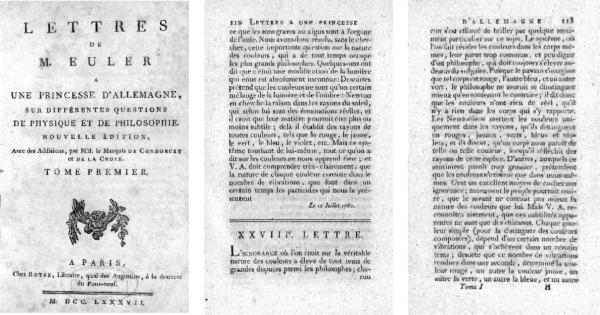
Newton's experiments are well known, but two issues are worthy of emphasis. Newton viewed the spectrum as a continuous distribution `Rays fell, appear tinged with this series of colors, violet, indigo, blue, green, yellow orange, red, together with all their intermediate degrees in a continual succession perpetually varying. So that there appeared as many degrees of colours as there were sorts of rays differing in refrangibility' (Book One, Part II, PROP. II Theor. II). Secondly, Newton followed a corpuscular theory of light. `Are not the rays of light very small bodies emitted from shining substances' (Book 3, Pt. I, Qu. 29). It is certainly possible to find quotations in support of a wave theory in the Opticks, as Young did in his Bakerian lecture [4]. However, the major contemporary of Newton to support the wave theory was Huygens, followed in the next century by Euler. In 1769 Euler published a physics textbook `Lettres a une Princesse d'Allemagne' set in the form of a series of letters to Princesse d'Anhalt-Dessau, niece of the king of Prussia, to whom Euler had acted as tutor in natural science [5]. The title page is shown in Fig. 1, together with the pages from which the following quotation is taken.
Fig. 1.
Euler was a proponent of the wave theory of light. This was an alternative to the corpuscular theory of light favored by Newton. Both theories implicitly assumed a continuous spectrum, in the one case a function of wavelength, in the other a function of weight or bigness or particles. Contrary to this view was the fact that different colors can be matched by mixing three primaries, which was interpreted as reflecting the existence of three different rays. Euler wrote a physics textbook under the guise of a series of letters to a German princess whom he had tutored. The title page shown is from a 1787 edition of these letters, although the text appears to have been written in 1760. The negative comments in Lettre XXVIII about the subjective approach to colors were likely provoked by Tobias Mayer's proposal in 1758 that all colors could be matched by mixing three primaries.
“The Newtonians assign the difference between colors in rays, which the distinguish into red, yellow, green blue and violet; and they say that a body appears of this or that color, if it reflects rays of that particular type. Others, for whom this theory is too straightforward, suggest that colors only exist within our percepts. This is an excellent way of hiding one's ignorance; otherwise the common man might believe that the savant does not know any more about the nature of colors than he himself. But V.A. {the princess} will easily recognize that these apparent subtleties are no more than quibbles. Each simple color (to distinguish them from compound colors) depends on a certain number of oscillations achieved in a certain time; so that a particular number of oscillations per second determines the color red, another the color yellow, another green, another blue; and another violet, since these are the simple of colors of the rainbow.”
The experimental evidence for the wave theory was not strong, and some of its support derived from an analogy with sound, which was well established as a wave phenomenon. An analogy between the seven colors of the Newtonian spectrum and the seven notes in a musical octave found its way into the Opticks. In any event, both the corpuscular and wave theories assumed a continuous spectrum. Euler's outburst about unnamed `others', who believed in subjective color perceptions, is likely to have been provoked by an alternative view of color that was formulated in the mid 18th century.
The University of Göttingen, Tobias Mayer, Georg-Christof Lichtenberg and Thomas Young
The University of Göttingen was founded in 1733 at the instigation of George II of England, who was also the Elector of the state of Lower Saxony. His father, George I, had acceded to the English throne in 1715 and George II felt that an independent University for Lower Saxony was appropriate. This rescued Göttingen from a long period of economic depression following the devastation of Europe during the 30 years war. The University, named the Georgia-Augusta, was given a liberal constitution and developed free of the theological strictures which hampered natural science at longer-established German universities, and the Georgia Augusta rapidly became one of the foremost scientific universities in Europe. It remained so until the pogroms that afflicted the German universities in the 1930s.
Tobias Mayer was born in Marbach on the Neckar in 1723. He grew up in difficult circumstances, but trained himself in mathematics, and from 1746 worked at the Hohmannische Kartographische Buro in Nuremberg, making maps. Within the framework provided by the Newtonian revolution, astronomical tools for accurate cartography were in rapid development for military and industrial purposes, and Mayer soon became a leading figure in European astronomy. In 1750, he was offered the Professorship of applied mathematics at the Georgia–Augusta and moved there in 1751, where he soon was appointed director of the newly built observatory. He is mostly remembered for his role in the development of an astronomical technique for determining longitude at sea [6]. After several naval catastrophes, the major being the destruction of a large portion of the British fleet on the Scillly Isles in 1707, the British government offered a major prize (£10,000) for discovery of an accurate method of longitude estimation. Many unusual suggestions were proposed, and one has found a place in modern literature [7]. For a method of longitude estimation to win the prize, it was necessary to ascertain local time relative to Greenwich mean time with an accuracy of a minute or less. Mayer's method was calibrated on measurements of lunar occultations of stars (the time of which could be measured very precisely) and involved star sights compared to lunar position and accurate knowledge of lunar motion. This led to a sophisticated analysis of optical, gravitational and other physical factors influencing lunar orbit. His only serious competitor was John Harrison, the English inventor of the marine chronometer. Mayer died in 1762 but a part the prize, shared with Harrison, was awarded to his widow in 1765. Since chronometers were expensive (at least initially) and seamen could never guarantee their reliability, Mayer's astronomical tables and chronometers remained in parallel use up to the end of the 19th century [6].
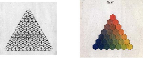
Mayer was interested in vision; he published in 1755 an article on the relation between visual acuity and luminance level, which is a remarkable early example of mathematical psychophysics [8]. The empirical aspects of generating colors by mixing pigments were well known among practicing artists; one could match any color with three pigments. Le Blon [9] began production of color prints based on three different color plates (plus black to help produce dark colors) around 1720. Mayer due to his experience in printing maps must have been aware of these practicalities, and in 1758 gave a public lecture in which he put forward for the first time a formal mathematical exposition of the color triangle now familiar to most schoolchildren. The example constructed later by Lichtenberg is shown in Fig.2B. The lecture on the topic in 1758 was reported at length in a scientific periodical, the “Göttingische Anzeige von gelehrten Sachen,” [10] but a full manuscript remained unpublished at Mayer's death in 1762. It was left to Georg–Christoph Lichtenberg, at the request of George III, to collate and publish in 1775 many of Mayer's papers. Most manuscripts have to do with astronomy, but a full description of Mayer's triangle is included. He used red, yellow, and blue as primaries, and postulated 12 discriminable steps along each edge (Fig. 2A), to give a triangle with 91 elements. Mayer then describes an additive set of equations for producing any desired colour. The triangle was then expanded into a solid, in which white and black points above and below the plane of the triangle could be reached by 12 successive mixture steps.
Fig. 2.
A. The color triangle of Tobias Mayer was the first formal treatment of the principle, known to artists and dyers, that all colors can be produced by mixing three pigments. Each primary could be mixed with the others in proportions ranging from 0–12. The space was extended into three dimensions toward black and white by adding such pigments. B. Lichtenberg published Mayer's theory, and attempted to provide a color plate in the printed version. He experimented with Mayer's mixtures using dry, powdered pigments, as had Mayer. This provides an approximation to additive color mixture, although brightness is reduced and whites appear a dirty grey. Printing involves pigments in solution, which results in a subtractive color mixture. Lichtenberg had much difficulty providing adequate print reproductions of the triangle. The difference between mixing pigment powders and in solution was attributed to chemical changes; the difference between additive and subtractive mixture was not appreciated.
Mayer's main interest was practical; he wished to construct a color order system in which any desired color could be derived by mixing others. However, his triangle was obviously relevant to contemporary physical theories of light, and the assumption that all colors could be produced by mixing three primaries was apparently incompatible with the idea of a continuous spectrum. For example, the Newtonian experiment, in which a part of the spectrum (such as orange) was shown to be indivisible by insertion of a second prism, is difficult to reconcile with Mayer's formulation. The status of Mayer's ownviews on the subject are uncertain; in the lecture report he is quoted as saying there may be only three types of ray in the spectrum. And Euler, as a proponent of wave theory, reacted vigorously.
Lichtenberg provided an elegant [11] expansion of Mayer's approach in a commentary included in the Opera Inedita. He first acknowledges Lambert's contribution and then modifies Mayer's formulation so that each primary is normalized to the sum of all three, as in current practice in the Commision International d'Eclairage (CIE) standard color space. The second section of his commentary has largely to do with the difficulties encountered in practical color mixture, especially in the preparation of a hand-colored plate included in each volume.
Georg-Christof Lichtenberg was a remarkable figure. Although physically handicapped and a hunchback, probably due to rickets in childhood, he was a professor in Göttingen from 1771 until his death in 1799, becoming a professor of physics in 1775. He was a distinguished essayist and critic, although much of his work remained unpublished in his lifetime. Today his books of aphorisms [12] are part of German literary culture. Scientifically, his main achievement was the description of electrical field lines, which are still sometimes called “Lichtenbergische Figuren”. He is said to have demonstrated to students the lightness of hydrogen by filling a pig's bladder with it and letting the bladder rise in the lecture hall, and then to show its explosive properties he ignited it with an electric spark. It is not surprising his lectures were famous throughout Europe.
His scientific interests were wide-ranging, including vision. He was well acquainted with Sömmerring, professor of medicine in Mainz, who provided the first description of the fovea as a hole in the middle of human retinae post-mortem, reported in the Göttingsche Anzeige in 1794. Sömmerring originally suggested that the hole was the blind spot, but Lichtenberg wrote that the foveal pit was too central. With Sommerring and others [13] he brought out a tract on optometry, “Pflichten gegen die Augen”. Lichtenberg was certainly aware of ideas about trichromacy which were circulating at that time.
George Walls [14] initiated modern-day interest in the work of an English glassmaker by the name of George Palmer and Mollon [15] has traced more details of this elusive figure. Palmer published a pamphlet in 1775 in which he proposed that there are three different types of receptor in the retina, for red, green and blue. However, Palmer failed to make the conceptual leap to the conclusion that there was indeed a continuous spectrum and that it is the three pigments which make the color triangle possible. Palmer, like Mayer, assumed there were three types of ray.
This pamphlet, now rare, is the first explicit suggestion of three receptors, or cone types, in the retina. Although Palmer was a businessman rather than scientist, his pamphlet attracted interest, and his theory was reviewed in 1781 in a German scientific periodical, “Magazin für das neueste aus der Physik und Naturgeschichte”, which was edited by Georg-Christof Lichtenberg's brother, L.C. Lichtenberg in Gotha, not far from Göttingen. Co-editor of the Magazin was Voigt, who appears to have written the Palmer article [16], which was entitled “Des Herrn Giros von Gentilly Muthmassungen über die Gesichtsfehler bey Untersuchngen der Farben” (The speculations of Herr Giros von Gentilly about visual deficiencies when examining colors). Giros von Gentilly was a pseudonym of Palmer. The first descriptions of color blindness had appeared a few years earlier, as described in a later section, and the review describes both Palmer's theory and his suggestion that absence of one of the receptors was the cause of color blindness. This does not appear in the original pamphlet, so how Voigt was informed of Palmer's speculation on this issue is unknown. In a later pamphlet [17], Palmer did specifically suggest that lack of a receptor was the reason for color blindness. In any event, G.C. Lichtenberg would have known of this review; the concept of trichromacy was in the air.
Thomas Young was a third polymath after Mayer and Lichtenberg. He was elected to the Royal Society in 1794 when only 21, for his work on the mechanisms of visual accommodation; he showed accommodation took place in the lens rather than by a change in the eye's shape [18]. He spent 18 months in Göttingen in 1795–6 to write a doctoral thesis. He and Lichtenberg shared an interest in physiological optics, and Young writes that Lichtenberg was one of the few professors with whom it was possible to become personally acquainted [19]. Young cites the article in (L.C) Lichtenberg's magazine discussing Palmer, and it seems likely that he and Lichtenberg discussed color. Unfortunately, there is no recorded correspondence between them; Lichtenberg only noted Young's visits (“Heute Thomas Young bey mir”) in his diary, and not the topics of their discussions.
In 1801 Young delivered the Bakerian lecture to the Royal Society [4] which for the first time put the wave theory of light on a firm experimental basis through an explanation of interference fringes. Almost as an aside, Young makes the conceptual leap to trichromacy, with a continuous spectrum but just three receptors in the eye, in the famous passage:
“Now it is almost impossible to conceive each sensitive point of the retina to contain an infinite number of particles, each capable of vibrating in perfect unison with every possible undulation, it becomes necessary to suppose the number limited, for instance, to the three principal colours, red, yellow, and blue, of which the undulations are related in magnitude nearly as the numbers 8,7 and 6…. Allowing this statement, it appears that any attempt to produce a musical effect from colours will be unsuccessful.”
After Young
Young's Bakerian lecture stimulated much dispute, especially in England. The wave theory of light was not generally accepted for several decades, and then largely due to the experimental efforts of European natural philosophers, such as Fresnel and Foucault. The trichromatic theory of vision likewise found little favor. A flavor for the disputatious nature of the discussions around light and color can be acquired from the interchange between Lichtenberg and Goethe.
Goethe believed he had disproved the Newtonian approach to color in a famous passage. “Aber wie verwundert war ich, als die durch's Prisma angeschaute weiße Wand nach wie vor weiß blieb, daß nur da, wo ein Dunkles dran steiß, sich eine mehr oder weniger entschiedene Farbe zeigte, daß zuletzt die Fensterstäbe am allerlebhaftesten farbig erschienen, indessen am lichtgrauen Himmel draußen keine Spur von Färbung zu sehen war. Es bedurfte keiner langen Überlegung, so erkannte ich, daß ein Gränze notwendig sei, um Farben hervorzubringen, und ich sprach wie durch einen Instinkt sogleich vor mich laut aus, daß die Newtonische Lehre falsch sei.” (But how amazed I was, as the white wall looked at through the prism stayed white as before, that only where there was a dark boundary, did a more or less distinct color appear, so that in the end the window bars appeared of the most lively color, while on the light grey sky outside no trace of color could be seen. It needed little thought before I realized, that an edge is necessary to evoke colors, and I spoke to myself instinctively, out loud, that the Newtonian doctrine was false). It is easy to see the error in Goethe's approach, but his Farbenlehre [20] contains a wide variety of interesting descriptions of color phenomena. Many are concerned with what one would now call color induction, the induction or modification of color by the surrounding area [21]. These effects are now recognized as complex and largely of cortical (rather than retinal) origin, and are likely to be associated with color constancy, the visual system's attempt to correctly identify colors despite changes in the illuminant, from, say, daylight to candlelight. His correspondence with Lichtenberg was aimed at recruiting Lichtenberg's support for his color theories [22, 23]. Goethe had published his `Beiträge zur Optick' in the 1790s, and in the third part is a section on induced colors, Färbige Schatten (colored shadows). Goethe sent a draft to Lichtenberg who answered in a most polite manner, while leaving no doubt that he disagreed with Goethe. Lichtenberg was a confirmed Newtonian and anglophile, having visited England on at least two occasions, and was on good terms with English natural philosophers. His reply to Goethe is a remarkably modern assessment of the nature of color appearance and color constancy with a highly sophisticated approach [23]. However, Goethe was a nobleman, with a ministerial position in Weimar, and Lichtenberg was a mere professor from a bourgeois background, and so Lichtenberg had to be circumspect. But it is instructive that at the time `colored shadows' were discussed in the same context as the nature of light or trichromatic theory. Today, the psychophysical analysis of induced colors is largely a separate topic from what one might call `front-end' trichromacy (cones and retinal mechanisms). Induction effects are much studied psychophysically [21] and of major practical importance, although the physiological underpinnings are almost certainly in the cortex rather than the retina, and their details are virtually unknown.
Vigorous opposition to both Newton's and Young's views was also extensive in the established scientific community. A misleading factor was the discovery of absorption lines in the sun's spectrum [24], which appeared to separate four different regions, or colors, within it. These were at first interpreted as divisions of differently colored rays, rather than absorption by elements in the sun's atmosphere. Another point of view was espoused by Brewster [25], who was a major figure in optics, having established his reputation by descriptions of the polarization of light. He became interested in spectral absorption by glass filters. Viewing spectra through such filters after prismatic refraction, he fell into the same fallacy as Goethe; he viewed the eye as an instrument of physical measurement rather than an organ whose properties were determined by three different opsins. He was led to a conclusion diametrically opposite to Newton, that light of a given color, e.g., yellow, could be refracted to different extents by a given glass, and was strongly opposed to a wave theory of light. Brewster was an influential figure, and his theory received wide discussion. He vigorously opposed the new ideas put forward by Helmholtz, Maxwell and others in the 1850s.
One major obstacle to further advances was the failure to distinguish between additive and subtractive color mixture. The difference is illustrated in Fig. 4. Two spectra are indicated, with absorption in the short and long wavelength regions of the spectrum. If two lights with these spectra are combined, the resultant is a straightforward additive mixture, as indicated. If two liquid pigments are mixed, the result is termed a subtractive mixture, and the resultant has quite a different spectrum. Only those rays not absorbed by either pigment are reflected. The illustration shown is only schematic, but is pertinent to the controversy at that time about whether the mixture of yellow and blue gives rise to white (an additive mixture) or green (as can happen with a subtractive mixture).
Fig. 4.
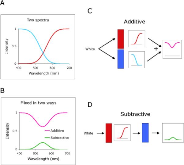
Additive and subtractive color mixture. A. Two example spectra, containing either short- or long-wave-length light. B. The resultant spectra following additive or subtractive mixture are quite different. C,D. How this occurs can be visualized by considering appropriate filters. If these are placed in parallel, and the light from each filter combined, then the resultant is an additive mixture. If they are placed in series, the light from the first filter is filtered again by the second, and the resultant is a subtractive mixture.
In practice, the different results obtained by mixing light and pigments was well recognized. For example, Gamaus [26], a Göttingen student, quotes Lichtenberg in a transcription of Lichtenberg's lectures. “Farbe (color) heisst das reine Licht durch das Prisma; pigmentum hingegen die gefärbten Körper, oder die färbenden Stoffe. Colores kann man also nicht kaufen”. (Color is the pure light from the prism; pigments on the other hand are the colored bodies or the coloring dye. Colors are thus not for sale). For the experiments on color mixture that Lichtenberg tested when exploring Mayer's color triangle, he mixed dry pigments, which approximate an additive color mixture when viewed from a distance (assuming the grain size is larger than the wavelength of light), although yielding colors of lower lightness level. A mix of yellow and blue dry pigments gives a dirty grey rather than a white. The different results obtained in painting or printing when mixing colored solutions of the same pigments, i.e., subtractive mixture, were attributed to chemical changes; for example, pH dependent effects on the color of solutions were well known. Another common method of testing colored mixtures was by spinning a disc with different colored sectors, a technique later refined by Maxwell. This gives an approximately additive mixture, but again it is difficult to generate bright colors. In any event, the failure to provide a satisfactory explanation for the differences found when mixing light compared to pigments hindered further progress.
The establishment of trichromacy
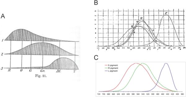
Following Lichtenberg's death in 1799 Göttingen scientists became less interested in vision. However, it is perhaps appropriate that the next great German vision scientist, Hermann von Helmholtz, was largely responsible for establishing the trichromatic theory. Helmholtz was a further universal figure, having invented the ophthalmoscope and measured the velocity of nerve conduction before turning to color. His first contribution was to explain the difference between additive and subtractive color mixture by considering absorption at an object's surface in terms of layers of thin filters [27]. He then undertook a sophisticated series of color mixture experiments using prism spectra and special masks (V and Z shaped). One issue was creation of white by mixing different spectral colors, i.e., specifying what we would now call complementary colors. He at first could only create white by mixing blue and yellow. His failure to find other pairs led him to doubt the Young hypothesis. However, shortly afterward Grassmann [28] introduced a three-dimensional vector formulation which stressed the continuous nature of color transitions (his three vectors correspond to hue, brightness and saturation in modern terminology). Helmholtz, prompted by Grassmann's approach and with more sophisticated equipment eventually showed that most wavelengths had complementary colors, except for a region in the green part of the spectrum for which a purple is the complementary hue. The color diagram of Helmholtz is the first with a modern appearance and is included in Fig. 5. At the same time that Helmholtz was pursuing his experiments, James Clerk Maxwell, later to develop electromagnetic theory, also began color mixture measurements. In the first experiments, he used a rotating colored disc, mounted on a spinning top, as had others before him. However, instead of painting differently colored sectors on its surface, three colored paper discs with radial slits were so arranged that from trial to trial the degree of overlap and thus their relative proportions could be varied. There were two such areas, a central circular one, surrounded by an annular region. The observer had to change the proportions of the papers in, say, the central region to match the surrounding annulus. These were the first color matching experiments as the technique would now be understood. Soon after this, Maxwell devised an apparatus for mixing any three spectral lights to match white, or daylight [29]. Both sets of apparatus were carefully calibrated and color mixtures could be described in a mathematical space. Linear transformation permitted these data to be used to derive the mixtures of three primaries required to match any spectral light.
Fig. 5.
Spectra of the visual pigments. A. Helmholtz drew speculative pigment spectra with peaks in the red-orange, green and violet. B. König and Dieterici proposed more reliable estimates of the spectra. The curves R, G and V represent their estimates. Note that the peaks of the RG curves are separated by only ~25 nm in mid-spectrum. The k and d curves are the authors estimates of their own pigments; they differ in the short wavelength part of the spectrum which they attributed to differences in macular pigment.
Maxwell's unequivocal and formal demonstration of trichromatic color mixing provided the best evidence for Young's trichromatic theory so far produced. His experiments are the direct ancestor of the color mixture experiments of Wright [30] and others that formed the experimental foundation of the CIE color system which now forms the basic standard for specification of color in scientific and industrial applications. Descriptions of the development of this system can be found elsewhere [30]. But from a physiological viewpoint, definition of the spectral sensitivities of the visual receptors, or pigments, became a prime objective. This could be achieved through color matching by color deficient observers. Maxwell had already recognized that the pigments would have broad spectra, so that a given wavelength excites more than one pigment, a fact not adequately recognized by Helmholtz. Maxwell found among his experimental subjects four such observers. He recognized that they lacked a visual pigment, and that determining the ratio of intensities of two spectral lights confused by such observers provides a potential means of specifying the missing pigment absorption spectra. But the history of the discovery of color blindness is topic for itself.
Color blindness
It is now known that about 8% of the male Caucasian population suffer from some degree of red-green color blindness. The deficit is sex-linked, since the M and L opsin genes are found on the X chromosome. Female observers with red-green deficiency are thus rare (~0.64%). It is remarkable that red-green color blindness remained unremarked for so long, considering that primates as a whole seem to have invested considerable effort in developing a red-green chromatic system. There is anecdotal evidence that a few renaissance artists were `very bad at colors', and restricted their production to black-white drawings and engravings [31]. However, the first serious descriptions of color deficiency appeared beginning in the mid 17th century. These were anecdotal accounts of individuals, often in color-related trades, who seemed to have serious problems in distinguishing colors along the red-green axis. Their identification and the nature of their defects is romantically ambiguous (Harris the shoemaker and his brother, the sea captain [32], Colardeau, a French poet, described by the Abbé Rozier in 1977, an apothecary from Strasborg [16]). It is very likely that such individuals suffered from red-green color blindness, since, for example, Harris complained about his inability to identify cherries among green leaves. George Palmer was the first to suggest that such individuals lacked one of the three visual pigments in the eye. Color deficiency was of interest, and Lichtenberg suggested that one advertise for such individuals for financial reward. Young also suggested that color blindness could result from lacking a visual pigment [33].
The best early description of color blindness is that of Dalton, the famous chemist and early proponent of the atomic theory [34]. He noted that he (and his brother) misnamed certain colors, especially under certain lighting conditions. In particular, deep red colors were difficult to identify. Together with earlier evidence, this description from a respected figure established color deficiency as a real phenomenon. Young read this description, and used it to support the hypothesis of color blindness being due to the lack of a color receptor in the eye. However, Dalton thought that his problems of color discrimination originated from tinting of his optic media, the vitreous and aqueos humors. Dalton willed his eyes to science. After his death, much later in 1844, his eyes were investigated, but no colors were visible in the media. A perspicacious doctor cut a hole in the back of one of Dalton's eyes and viewed the world through it. No color distortions were apparent. This supported the idea of a pigment deficiency rather than a problem in the optic media, although a number of other hypotheses had also been proposed [35]. From Dalton's description, his form of color blindness was thought to be protanopia, a lack of the (L) red pigment, but fragments of Dalton's eye remained preserved in Manchester, England. With modern molecular techniques, it proved possible to genetically specify Dalton's form of color blindness, and it seems he lacked the green (M) rather than the L pigment [36].
Daltons's autobiographical description helped establish red-green color blindness as a scientific and practical problem. However, until the trichromatic theory became generally accepted, further advances were slow. One problem was reliance on anecdotal evidence and personal description rather than objective testing. Color- defective observers try and name colors in the same way as color normals, and so their personal descriptions need not be that helpful. Seebeck [37] was the first to use objective testing methods. He presented his subjects with a large selection of colored papers to be sorted into a series and then measured observers spectral sensitivity with prismatic colors. He distinguished two classes of defective; both made red-green confusions, but the second group was worse at red-blue distinctions, and `they have, which was not true for the first class, only a weak perception of the least refracted rays', i.e., long wavelengths. These classes correspond to deuteranopes (lacking the M opsin) and protanopes (lacking the L opsin). Seebeck also was the first to convincingly describe a color-blind female, whose father also (necessarily) had the same type of deficiency; the inherited nature of color blindness had been noted before but again Seebeck's description provided more reliable evidence.
Although further progress was slow, color blindness was now a well recognized syndrome and color vision of industrial workers, such as railway engineers, began to be tested; confusing red and green signal flags was found to have potentially catastrophic consequences. It was again Maxwell who set the field in motion once more. He only knew of one type of color defective, probably a protanope. Using the spinning top he showed that such defectives only need two colors (plus black) to make a match to daylight. Such a demonstration was direct evidence in favor of the Young hypothesis. Further, Maxwell realized that by finding the relative intensities of two spectral lights confused by color defective observers, it would be possible to deduce the spectral sensitivity of the missing visual pigment. The assumption is that in color defectives one pigment is missing rather than modified, which is only true for a subset of color defectives, as discussed in the final section of this review. Also, at that time tritanopes, who lack the S-cone pigment, had not been described. However, König and Dieterici [38] were able to describe pigment spectra which are not too different from those accepted today. Fig. 5 shows Helmholtz's suggestions for the pigment spectra from his Physiologische Optik [39] compared to those of König and Dieterici and modern measurements. Helmholtz's spectra are not very accurate (he had no direct evidence) but the König and Dieterici curves are quite good. Despite these advances, the trichromatic theory remained controversial, and one reason for this was grounded in conceptions of color space.
Color circles and spaces
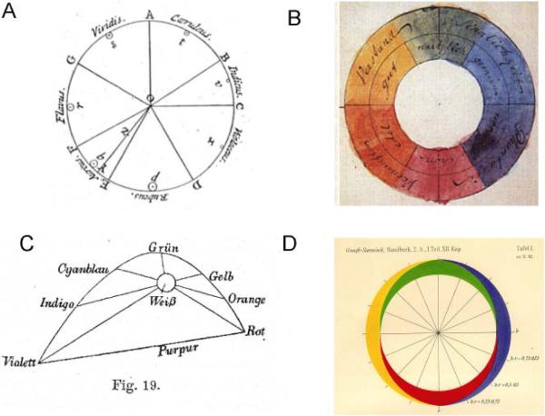
Parallel to discussions about the nature of light and the presence or absence of different receptor types, there was a continual attempt to portray colors in a two-dimensional space, with the aim of a graphic representation of colors. Fig. 6 shows some examples. With this procedure, at first sight a straightforward attempt to graphically portray physical results, other psychophysical properties of color vision became important, and the resolution of these issues is still incomplete.
Fig. 6.
Versions of color circle and color spaces. A. Newton's color circle was divided according to an analogy with the octave scale, as described in the text. Violet (Violaceus) directly abuts onto red (Ruber). B. Goethe's color circle was inscribed with personal qualities (yellow is Gut (good) and Verstand (thought or intelligence)). However, purple is now a part of the circle. C. Helmholtz constructed a color space based on his investigations of complementary spectral colors. The spectral locus forms an arc with the ends connected by a straight line along which the non-spectral colors, purples, are mapped. D. Hering sketched a color circle in which red-green and blue yellow axes define the intermediate colors.
The first color circle shown is that of Newton based on the prismatic spectrum. There is a continuum from red to violet with no purple. The extents of the different areas were mapped according to an analogy with sound `With the center O and radius OD describe a Circle ADF, and distinguish its Circumference into seven Parts DE, EF, FG, GA, AB, BC, CD, proportional to the seven Musical Tones or intervals of the eight Sounds, Sol, la, fa, sol, la, mi, fa, sol. contained in an eight.…'. The analogy to sound was dismissed out of hand by Young (see above). However, Newton's color circle was a novelty, with the assumption that the center was white. Mayer's color triangle was more of empirical attempt at color mixture, but color circles of different designs were constructed by others. That of Goethe (Fig. 6B) included purple between red and violet. He associated different colors with different physical feelings and emotional states, which found little acceptance. The third example is that of Helmholtz, which was derived from his color matching experiments designed to determine complementary colors. Helmholtz's color space was derived from physical measurement. Those of Newton and others were inherently influenced by other considerations. One manifestation of this was the vigorous discussion about whether red, green and blue or red, yellow and blue should be considered primary colors for color mixture. Mayer and Young suggested red, yellow and blue, but Young later changed his position to support red, green and blue. Brewster (whose theory of light was aberrant but influential) viewed yellow as a primary color. In fact, Maxwell realized that in color mixture experiments it was irrelevant which primaries were used. In practice, it is desirable that they be well separated in the spectrum, but the principles of color mixture remain the same.
Arguments about the primaries probably reflected the tendency to mix up the physical primaries of color matching with the psychophysical concepts of unique hues. Hering [40] was the first to bring this issue to prominence. He postulated that four colors, red, yellow green and blue (plus black and white making six) had a unique status. It is impossible to conceive of a bluish yellow or reddish green but a reddish yellow (orange) is a familiar concept. The color theory he proposed stated that there are red-green, blue-yellow and black-white opponent processes in the human retina. However, the evidence for this was sparse; colored after images were one piece of evidence, but not compelling. The arguments about whether yellow and green should be primary colors may have reflected the tendency, when viewing a color circle, to be influenced not only by the principles of color mixture, but by the inherent tendency to classify colors in an opponent scheme. It now appears there are opponent processes in the retina after the receptors, as described in the next section, but they unfortunately do not precisely correspond to the Hering opponent axes. This remains one of the main unresolved issues in color vision.
It is thus important to distinguish color spaces based on color matching (which are defined by the spectra of the visual pigments, and are physically specifiable) and those defined by opponent processes and more complex psychophysical interactions. As an example of the latter, color naming principles seem to be common in humans no matter whether they live in cities, jungles or arctic snows [41], despite wide differences in the light spectra reflected by these local environments. But this issue is outside the scope of the current review.
Modern developments: cones and their opsins
Despite the difficulties posed by the obvious perceptual presence of opponent colors, the trichromatic theory became generally accepted. However, biological as opposed to psychophysical evidence proved elusive. There were attempts to measure the spectra of light reflected back out of the human eye in normal and color blind observers before and after bleaching a cone pigment and calculating the spectra by looking at the difference [42, 43], but this only achieved measurements for the M and L opsins; the S cones are too sparse. Biological confirmation first became possible with measurement techniques that permitted estimation of light absorption [44] or electrical responses [45] of single cones. The Young-Helmholtz-Maxwell view received final confirmation.
Psychophysical estimation of the spectra of the visual pigments became more sophisticated and accurate, but the principles remained the same. Confusions of color-blind observers permit inference of the pigment spectra, and this analysis was aided by the description of a rare form of color blindness, tritanopia, caused by absence of the S cones [46]. This deficiency is not sex linked, and is an autosomal defect on chromosome 7; the interested reader may find further details elsewhere [47]. The spectra calculated by Smith and Pokorny [48] were for many years in general use by the vision community for designing experiments, and the latest revisions [49] only differ in very minor ways.
Testing methods for color deficiency became of practical importance, and three main types are in use. A typical example of plate tests are the Isihara plates [50]. These consist of a disc of colored dots, for example greens and blues. One or two numbers are constructed by dots of different colors, e.g., brown. Color normal observers pick up the numbers without difficulty, but color deficient observers see the wrong numbers or none at all. Different sets of color dots can distinguish between different types of color deficiency. Another type of test is the ordering test, on the principle used by Seebeck. An example is the Farnsworth-Munsell test [51] in which different colored caps around the color circle must be ordered into progression. Confusion between caps indicates color deficiency, and the actual caps that are confused indicate the type of deficiency. The most informative test is the Rayleigh match, proposed by Lord Rayleigh, the famous physicist. The commercial implementation of the test is the Nagel anomoloscope, described in 1907 [52]. The principle is that two monochromatic red and green primaries are mixed and matched to a yellow in intensity and color. The use of longer wavelengths means that the S cone plays no role in the match. For color normal observers, the proportion of red and green primaries is strongly delimited and very reproducible, both between observers and for repeat measurements. For protanopes and deuteranopes, lacking the L and M cone, the yellow primary can be matched just by one of the red/green primaries. However, it is found that only a fraction of the 8% color defectives males are true dichromats (1% protanopes, 2% deuternaopes). Others only show partial loss. König and Dieterici were aware of such individuals, but the Nagel anomoloscope provided a simple and reliable clinical test. Their match is different from color normals, and much more variable. This indicates a loss of red-green discriminative ability. Peak absorptions of the M and L pigments in normals are ~535 and 561 nm respectively. It was hypothesized that in the partial color blind the pigment spectra are closer together than in normal observers.
Modern molecular approaches have elucidated the genetics of color blindness and confirmed this suggestion [47]. The L and M cone opsin genes form a tandem array close together on the X chromosome, which probably occurred through a gene duplication early in primate evolution. The gene that is expressed in a given cone is determined by molecular mechanisms upstream from the array. There is a high degree in homology between the opsin genes, and if unequal crossing over occurs during meiosis, one of the resulting chromosomes can contain either just a single L or M opsin gene (while the other contains three genes). Male individuals with just one opsin gene on their X chromosome are protanopes or deuteranopes (although other genetic patterns can also lead to dichromacy). But unequal crossing over can also produce hybrid M/L genes. Only a few amino acids in the opsin gene (perhaps 6–7) determine spectral tuning. The hybrid gene produces a resultant opsin with a spectral tuning lying between that of the standard M and L types, the exact location (and the severity of the deficiency) depending on the actual amino acids substituted in the hybrid gene. If it is the M opsin gene which is affected the resulting defect is termed deuteranomaly (3%), if the L opsin gene it is termed protanomaly (2%). The closer the pigments in peak wavelength, the poorer the chromatic discrimination and the greater the variation in the Rayleigh match.
A contentious issue for many years was whether, in dichromats, the lacking cone types (M or L) were simply not present, resulting in a lower cone density, or whether they were filled with the `wrong' pigment (see Pokorny, Verriest, Smith and Pinckers [53] for review). The latter became accepted as the case, but recent evidence shows that a small proportion of dichromats do show a cone loss, due to a defect in one of the opsin genes associated with pigment expression, rather than aberrant spectral tuning [54]. These individuals have abnormally low cone densities. Remarkably, the loss of a significant proportion of cones causes little psychophysical deficit.
Vision in red-green color blind humans is limited to two cones, the S cone and another in the middle- to long-wavelength range. Such humans are able to judge surface properties of objects quite well, at least if the objects are large. One might ask if other mammals which just have an S and one other cone have equivalent visual capabilities. But it seems likely that most lower mammals possess a more restricted visual experience than color-blind humans, with emphasis on detection of boundaries between objects, although this is speculation.
Modern developments: postreceptoral pathways
There is a prescient description of the mechanism of visual transduction by Elliot. He suggested [55] that different wavelengths of light caused resonant vibrations in the appropriate receptors. “If the red-making vibrations fall on the eye, they excite the red-making vibrations in that part of the retina whereon they impinge, but do not excite the others because they are not in unison with them… From hence it may be understood that the rays of light do not cause colors in the eye any otherwise than by the mediations of the vibrations or colors liable to be excited in the retina… We are therefore perhaps to consider each of these vibrations or colors in the retina, as connected with a fibril in the optic nerve. That the vibration being excited, the pulses thereof are communicated to the nervous fibril, and by that communicated to the sensory, or mind, where it occasions, by its action, the respective color to be perceived…”. Elliot however did not suggest that there were only three receptor types in the retina. The elicitation of some form of vibrations in the retina by light was a common theme in literature of the time. However, Elliot's suggestion was not widely appreciated. The idea of a tuned transducer [1] took some time to become established, as did the idea that only by comparison of the activity of such transducers could the color of a light be deduced. This principle is now dignified by the title of the Principle of Univariance; a single receptor will confound wavelength and intensity, it is only by comparison of receptors that colors can be identified.
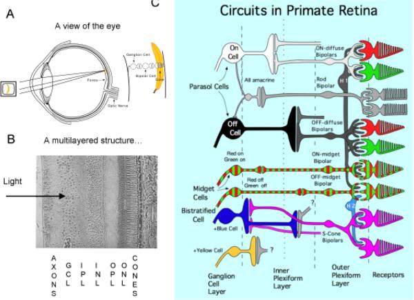
The status of the color opponent theory has radically changed in the last 50 years. A firmer psychophysical foundation was provided by Hurvich and Jameson [56]. It will be remembered that the unique hues, red, yellow, green and blue were assigned special status. Hurvich and Jameson asked observers to add, say, a wavelength identified as unique green, to a test wavelength, for example, in the orange region of the spectrum, until the resulting mixture appeared neither reddish not greenish. Similar procedures can be carried out for the other unique hues. The resulting color valence functions provided more convincing psychophysical evidence for the presence of opponent chromatic channels in perception. The discovery of cells in the primate lateral geniculate nucleus [57], and in the retina [58], of cells which were excited by one part of the spectrum and inhibited by another seemed at first to provide a spectacular confirmation of the opponent theory. Fig. 7 shows a sketch of the current view of primate retina (see Lee [59] for further information). The eye is shown in A, and objects are focused on the receptor layer that lies at the back of the eye. The receptors contact bipolar cells, which contact ganglion cells, which send their axons to the lateral geniculate nucleus and brain. In B is shown a histological section of the retina, which is a strongly laminated structure. The initials refer to the different layers, as described in the legend. Light passes through the neural layers to reach the receptors, except in the fovea where the neurons are pushed to one side. Fig. 7C shows a wiring diagram. This looks complex, but the principle for all ganglion cells is the same. There are parasol cells, which receive input from just the M and L cones, and send their axons to the magnocellular layers of the geniculate. They may be excited by light (on cells) or dark (off cells) stimuli, and form the substrate of a luminance or achromatic channel in psychophysics. A red-green system, the midget cells, are shown with red green zebra stripes; they send their axons to the parvocellular layers of the geniculate. These are excited by red light and inhibited by green light or vice versa, and form the basis of a psychophysical channel responsible for detection of red-green chromatic changes. They achieve this color opponency by receiving excitatory input from one cone (M or L) and inhibitory input from the other. Lastly, two further cell types are excited by blue light and inhibited by yellow light or vice versa. These cell types are responsible for detection of colors along the blue-yellow axis. Again excitation or inhibition from the S cones opposed by the other cell types forms the basis of this behavior. Despite this evidence for separate red-green and blue-yellow physiological channels that correspond well with psychophysics for detection of chromatic changes, the results of Hurvich and Jameson experiments (which really have to do with the subjective impression of, say, redness or greenness) do not correspond all that well with physiological findings, so that, for example, the location of unique blue is not that expected from physiological measurement. The reason for this discrepancy is one of the most intriguing unsolved problems in color vision [60].
Fig. 7.
Current view of the retina. A. A cross-section of the eye; images are focused on the receptor layer and signals pass through bipolar cells to the ganglion cells, which send axons to the brain via the optic nerve. B The retina is highly laminated structure. The cone receptors are at back, and the Outer Nuclear Layer (ONL) contains the cell bodies of cones and horizontal cells. Outer Plexiform Layer (OPL) contains a synaptic network between cones, bipolar cells and horizontal cells. The Inner Nuclear Layer (INL) contains cell bodies of bipolar cells and amacrine cells, and the Inner Plexiform Layer (OPL) contains a synaptic network between bipolar cells, amacrine cellls and ganglion cells. The Ganglion Cell Layer (GCL) contains ganglion cell bodies which send their axons to the brain. C. A wiring diagram of the primate retina. There are three main cell systems which send axons to the cortex via the lateral geniculate nucleus. Each receive input from the cones, and signals pass through specialized bipolar cells to the ganglion cells. Amacrine and horizontal cells are interneurones which aid local processing of signals. There is an achromatic, luminance pathway beginning in the parasol ganglion cells, a red-green pathway beginning in the midget ganglion cells and a blue-yellow pathway made up of small bistratified cells and another type. These color opponent systems receive opponent input from the different cones, but the color channels do not map perfectly onto the psychophysical opponent systems.
The development of molecular biological techniques to identify the opsin genes confirmed that among mammals only primates have three cone pigments and trichromatic vision. There are significant differences between Old- and New-World primates [61], and it seems likely that trichromatic vision evolved close to the time these two groups divided. Anatomical and physiological observations are consistent with the idea that the midget, red-green system took over a ganglion cell class that was previously involved in spatial vision [62]. Knowledge of the evolution of color vision in primates may provide a framework to interpret some of the unusual features of this system. In any event, it is clear that primates underwent a major remodeling of the visual pathway to accommodate the red-green channel of vision. It is thus remarkable that such a large proportion of human males suffer from color deficiency without many ill effects. On the other hand, incidence of color blindness is significantly lower in primitive, tribal populations than in Caucasian males [47]. Also, a genetic search for color deficiency in macaque monkeys yielded only two examples in a sample of over 3000 individuals [63], both from one troop, which probably indicates a recent mutation. Such mutations must normally be highly disadvantageous if they are strongly selected against in the wild. It would thus appear that red-green color vision remains important for native primate populations, but the selection factors responsible are still in dispute.
Conclusion
Much has been neglected in this review. For example, Schrödinger [64] produced a line element theory of color discrimination of great originality. Some of the disputes about which colors are primaries are recounted in an entertaining account by Sherman [65]. Disputes about color vision have tended to be contentious, and generated more heat than light. It now seems that many of these disputes arose through category errors. The first of these was to confound the physical properties of light with those of the human visual system, as in the dispute between Mayer and Euler. Another was to mix up the laws governing trichromatic color matching (which are essentially physical laws applied to spectral absorption by the pigments) with the predisposition to sort and name colors to fit perceptual color spaces, as in the identification of the unique hues. Much of the dispute about primary colors are likely related to this confusion. Lastly, perceived color not only depends on spectral composition, but on the color of surrounding areas. Chevreul [66] was one of the first to make a systemic study of this, in order to improve the rendering of colors within tapestries. Some colors, such as brown, are only seen as such when set in a lighter surround. Such colors are the subject of much of Goethe's Farbenlehre, with it's anti-Newtonian bias. Acute experimenters realized this and tried to control for such effects. Helmholtz, for example, with his color mixing experiments with prismatic light, was very careful to exclude all extraneous regions of the spectrum from his field of view so as to avoid such induction effects. Viewing colors in anything other than what is termed aperture mode, i.e., with a dark surround, is liable to influence the percept.
In view of all the confounding factors, it is a tribute to the scientific acumen of early color scientists, such as Newton, Young, Helmholtz and Maxwell, to have come so far with the limited technical means available to them.
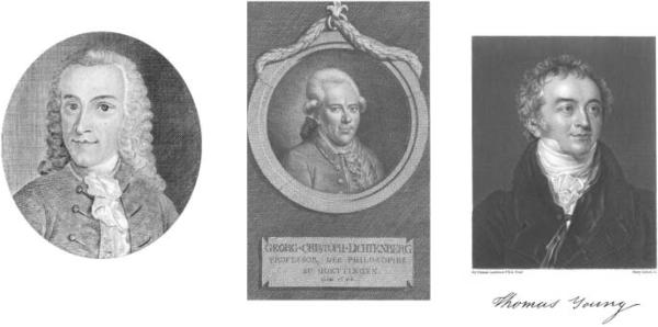
Fig. 3.
Tobias Mayer, Georg-Christof Lichtenberg and Thomas Young are notable for their scientific contributions in multiple fields. Tobias Mayer was primarily an astronomer, Lichtenberg and Young physicists, but all were polymaths contributing to many areas. Tobias Mayer was a Professor in Göttingen from 1749 to his death in 1762, Lichtenberg was a Professor from 1775 to 1799, and Thomas Young was a student in Göttingen from 1794 to 1795.
Acknowledgement
This work was partially supported by NIH EY 13112.
References
- 1.Mollon JD. The origins of modern color science. In: Shevell SK, editor. The science of color. Elsevier; Oxford, U.K.: 2003. pp. 1–40. [Google Scholar]
- 2.Newton I. Opticks. Innus.; London: 1704. [Google Scholar]
- 3.von Campenhausen C, Pfaff C, Schramme J. Three dimensional interpretation of the color system of Aguilonius/Rubens 1613. Color Res App. 2000;26:S17–9. [Google Scholar]
- 4.Young T. On the theory of light and colours. Phil Trans Royal Soc (London) 1802;92:12–48. [Google Scholar]
- 5.Euler M. Lettres a une Princesse d'Allemagne sur differentes questions de physique et de philosophie. Vol. 1. Royez; Paris: 1787. [Google Scholar]
- 6.Forbes EG. The birth of navigational science. National Maritime Museum; London: 1974. [Google Scholar]
- 7.Eco U. The island of the day before. Harcourt Brace; New York, USA: 1995. [Google Scholar]
- 8.Mayer T. Erfahrungen über die Schärfe des Gesichts. Göttingsche Anzeige von gelehrten Sachen. 1754:401–2. [Google Scholar]
- 9.Blon JCL. Il Colorito: Or the harmony of colouring in painting. London: 1721. [Google Scholar]
- 10.Mayer T. Ein Entwurf einer Messung der Farben. Göttingsche Anzeigen von gelehrten Sachen. 1758;147:1385–9. [Google Scholar]
- 11.Mayer T. In: Opera inedita. Lichtenberg GC, editor. Göttingen: 1775. [Google Scholar]
- 12.Lichtenberg GC. Südelbuche. Marizverlag; Wiesbaden: 2006. [Google Scholar]
- 13.Sömmerring ST. Adams, Busch und Lichtenberg über einige Pflichten gegen die Augen. Frankfurt am Main. 1794 [Google Scholar]
- 14.Walls GL. The G. Palmer story. Journal of the History of Medicine and Allied Sciences. 1956;11:66–96. doi: 10.1093/jhmas/xi.1.66. [DOI] [PubMed] [Google Scholar]
- 15.Mollon JD. `….aus dreyerly Arten von Membranen oder Molekülen': George Palmer's legacy. In: Cavonius CR, editor. Color Vision Deficiencies XIII. Kluwer; Dordrecht, Holland: 1997. [Google Scholar]
- 16.Voigt JH. Des Herrn Giros von Gentilly Muthmassungen uber die Gesichtsfehler bei Untersuchung der Farben. Magazin fur das Neuste aus der Physik und Naturgeschichte. 1781;1:57–61. [Google Scholar]
- 17.Palmer G. Lettre sur les moyens de produire, la nuit, une lumière pareille à celle du jour. L'auteur; Paris: 1785. [Google Scholar]
- 18.Young T, Brocklesby R. Observations on vision. Phil Trans Royal Soc (London) 1793;83:168–78. [Google Scholar]
- 19.Peacock G. The life of Thomas Young. London: 1855. [Google Scholar]
- 20.von Goethe JW. Zur Farbenlehre. 1810 [Google Scholar]
- 21.Shevell SK. Color appearance. In: Shevell SK, editor. Color vision. Elsevier; Oxford UK: 2003. pp. 149–90. [Google Scholar]
- 22.Joost U. “Da ich…. Den bunten Schatten nachlaufe, wie ehmals als Knabe den Schmetterlingen” - Lichtenberg, Goethe, and the coloured shadows. Col Res Appl. 2000 [Google Scholar]
- 23.Lichtenberg GC. Lichtenberg's letter to Goethe on “Färbige Schatten”. Col Res Appl. 2002;27:300–303. [Google Scholar]
- 24.Wollaston WH. A method of examining refractive and dispersive powers by prismatic reflection. Phil Trans Royal Soc (London) 1802;42:365–380. [Google Scholar]
- 25.Brewster D. Transactions of the Royal Society. Vol. 9. Edinburgh: 1822. Description of a monochromatic lamp for microscopic purposes, with remarks on the absorption of the prismatic rays by coloured media; p. 433. [Google Scholar]
- 26.Lichtenberg GC. In: Lichtenberg über Luft und Licht. Gamaus G, editor. Geistinger`s Buchhandlung; Vienna and Trieste: 1811. [Google Scholar]
- 27.Helmholtz H. Über der Theorie der zusammengestzten Farben. Annalen der Physik. 1852;94:1–28. [Google Scholar]
- 28.Grassmann H. Zur Theorie der Farbenmischung. Ann Phys Lpz. 1853;89:60–84. English trans Philos Mag 1854;7:254-264. [Google Scholar]
- 29.Maxwell JC. On the theory of the compound colours and the relations of the colours in the spectrum. Phil Trans Royal Soc (London) 1860;150:57–84. [Google Scholar]
- 30.Wright WD. Golden Jubilee of the CIE. Society of Dyers and Colourists; Bradford: 1981. The historical and experimental background to the 1931 CIE system of colorimetry; pp. 3–18. [Google Scholar]
- 31.Lanthony P. Daltonism in painting. Col Res Appl. 2001;26:S12–S16. [Google Scholar]
- 32.Huddart J. An account of persons who could not distinguish colours. Phil Trans Royal Soc (London) 1777;67:260–5. [Google Scholar]
- 33.Young T. A course of lectures on natural philosophy and the mechanical arts. J. Johnson; London: 1807. [Google Scholar]
- 34.Dalton J. Extraordinary facts relating to the vision of colours. Memoirs of the Literary and Philosophical Society of Manchester. 1798;5:28–43. [Google Scholar]
- 35.Sherman PD. Colour vision in the Nineteenth Century. Adam Hilger; Bristol: 1981. [Google Scholar]
- 36.Hunt DM, Dulai KS, Bowmaker JK, Mollon JD. The chemistry of John Dalton's colour blindness. Science. 1995;267:984–8. doi: 10.1126/science.7863342. [DOI] [PubMed] [Google Scholar]
- 37.Seebeck A. Annalen der Physik. Leipzig: 1837. Über den bei mancher Personen vorkommenden Mangel an Farbensinn; p. 42. [Google Scholar]
- 38.König A, Dieterici C. Sitzungsberichte der Preussische Akademie der Wissenschaften. Berlin: 1886. Die Grundempfindungen und ihre Intensitats-Vertheilung im Spectrum; pp. 805–29. [Google Scholar]
- 39.von Helmholtz H. Handbuch der Physiologischen Optik. 2nd ed Voss; Hamburg: 1896. [Google Scholar]
- 40.Hering E. Zur Lehre vom Lichtsinne. Carl Gerold`s Sohn; Wien: 1878. [Google Scholar]
- 41.Berlin B, Kay P. Basic color terms. 1969 [Google Scholar]
- 42.Rushton WAH. A cone pigment in the protanope. J Physiol. 1963;168:345–59. doi: 10.1113/jphysiol.1963.sp007196. [DOI] [PMC free article] [PubMed] [Google Scholar]
- 43.Rushton WAH. A foveal pigment in the deuteranope. J Physiol. 1965;176:24–37. doi: 10.1113/jphysiol.1965.sp007532. [DOI] [PMC free article] [PubMed] [Google Scholar]
- 44.Bowmaker JK, Dartnall HJA. Visual pigments of rods and cones in a human retina. J Physiol. 1980;298:501–11. doi: 10.1113/jphysiol.1980.sp013097. [DOI] [PMC free article] [PubMed] [Google Scholar]
- 45.Baylor DA, Nunn BJ, Schnapf JL. Spectral sensitivity of cones of the monkey Macaca Fascicularis. J Physiol. 1987;390:145–60. doi: 10.1113/jphysiol.1987.sp016691. [DOI] [PMC free article] [PubMed] [Google Scholar]
- 46.Wright WD. The characteristics of tritanopia. J Opt Soc Am. 1952;42:509–20. doi: 10.1364/josa.42.000509. [DOI] [PubMed] [Google Scholar]
- 47.Sharpe LT, Stockman A, Jägle H, Nathans J. Opsin genes, cone photopigments, color vision and color blindness. In: Gegenfurtner KR, Sharpe LT, editors. Color vision: from genes to perception. Cambridge University Press; Cambridge: 1999. pp. 3–51. [Google Scholar]
- 48.Smith VC, Pokorny J. Spectral sensitivity of the foveal cone photopigments between 400 and 500 nm. Vision Res. 1975;15:161–71. doi: 10.1016/0042-6989(75)90203-5. [DOI] [PubMed] [Google Scholar]
- 49.Stockman A, MacLeod DI, Johnson NE. Spectral sensitivities of the human cones. J Opt Soc Am A. 1993;10:2491–521. doi: 10.1364/josaa.10.002491. [DOI] [PubMed] [Google Scholar]
- 50.Ishihara S. Tests for colour-blindness 16 plates edition. Kanehara Shuppan; Tokyo: 1962. [Google Scholar]
- 51.Farnsworth D. The Farnsworth-Munsell 100 hue and dichotomous tests for color vision. J Opt Soc Am. 1943;33:568–78. [Google Scholar]
- 52.Nagel WA. Zwei apparate fur die augenartzliche Funktionsprufung: Adaptometer und kleines spektralphotometer (Anomaloskop) Ztschr Augenheilk. 1907;17:201. [Google Scholar]
- 53.Pokorny J, Smith VC, Verriest G, Pinckers AJLG, editors. Congenital and acquired color vision defects. Grune and Stratton; New York: 1979. [Google Scholar]
- 54.Carroll J, Neitz M, Hofer H, Neitz J, Williams DR. Functional photoreceptor loss revealed with adaptive optics: an alternate cause of color blindness. Proc Natl Acad Sci. 2004;101:8461–6. doi: 10.1073/pnas.0401440101. [DOI] [PMC free article] [PubMed] [Google Scholar]
- 55.Elliot J. Elements of the branches of natural philosophy connected with medicine. J. Johnson; London: 1786. [Google Scholar]
- 56.Hurvich LM, Jameson D. An opponent-process theory of color vision. Psych Rev. 1957;6:384–404. doi: 10.1037/h0041403. [DOI] [PubMed] [Google Scholar]
- 57.DeValois RL, Abramov I, Jacobs GH. Analysis of response patterns of LGN cells. J Opt Soc Am. 1966;56:966–77. doi: 10.1364/josa.56.000966. [DOI] [PubMed] [Google Scholar]
- 58.Gouras P. Identification of cone mechanisms in monkey ganglion cells. J Physiol. 1968;199:533–47. doi: 10.1113/jphysiol.1968.sp008667. [DOI] [PMC free article] [PubMed] [Google Scholar]
- 59.Lee BB. Paths to color in the retina. Australian Journal of Optometry. 2004;87:239–48. doi: 10.1111/j.1444-0938.2004.tb05054.x. [DOI] [PubMed] [Google Scholar]
- 60.Mollon JD, Jordan G. On the nature of unique hues. In: Dickinson C, Murray I, Carden D, editors. John Dalton's colour vision legacy. Taylor & Francis; London: 1997. pp. 381–92. [Google Scholar]
- 61.Silveira LCL, Grünert U, Kremers J, Lee BB, Martin PR. Comparative anatomy and physiology of the primate retina. In: Kremers J, editor. Structure and function of the primate visual system. John Wiley; Chichester, England: 2005. pp. 127–60. [Google Scholar]
- 62.Wässle H, Boycott BB. Functional architecture of the mammalian retina. Physiol Rev. 1991;71:447–80. doi: 10.1152/physrev.1991.71.2.447. [DOI] [PubMed] [Google Scholar]
- 63.Onishi A, Koike S, Ida-Hosanuma M, Imai H, Shichida Y, Takenaka O, Hanazawa A, Komatsu H, Mikami A, Goto S, Surybroto B, Frajallah A, Varavudhi P, Eakavhibata C, Kitahara K, Yamamori T. Variations in long- and middle-wavelength-sensitive opsin gene loci in crab-eating monkeys. Vision Res. 2002;42:281–92. doi: 10.1016/s0042-6989(01)00293-0. [DOI] [PubMed] [Google Scholar]
- 64.Schrödinger E. Ueber das Verhaltnis der Vierfarbenzur Dreifarbentheorie. Sitzber Wien Akad Wiss, Math-naturwiss Kl. 1925:134. Abt. IIa:471-90. [Google Scholar]
- 65.Sherman PD. Colour vision in the 19th century. Adam Hilger; Bristol, England: 1981. [Google Scholar]
- 66.Chevreul ME. The principles of harmony and contrast of colors and their applications to the arts. Reinhold; New York: 1839. 1967. [Google Scholar]


