Abstract
Label-free and real-time single-molecule detection may aid the development of high-throughput biosensing platforms. Molecular fluctuations are a source of noise that often hinders single-molecule identification by obscuring the fine details of molecular identity. In this study, we report molecular identification through direct observation of quantum-fluctuation-induced inelastic noise in single organic molecules. We investigated current fluctuations flowing through a single molecule that is chemically connected to two electrodes. We found increased current oscillations synchronous to electric field excitations of characteristic molecular vibrational modes that contribute to inelastic electron tunnelling. This finding demonstrates a large contribution of charge interaction with nuclear dynamics on noise properties of single-molecule bridges and suggests a potential use of inelastic noise as a valuable molecular signature for single-molecule identification.
 Molecular fluctuations are a source of noise that can impede single-molecule identification. Here, quantum-fluctuation-induced inelastic noise is observed as current fluctuations in individual molecules, suggesting that inelastic noise could be used as a molecular signature.
Recent advances in single-molecule detection techniques have revolutionized our understanding of biological phenomena and opened new opportunities for human gene analysis1,2,3,4,5,6. Unlike ensemble average measurements of macroscopic properties, molecular fluctuations generate a noise that often becomes a serious impediment for identifying important single-molecule signatures. In stark contrast, human sensory apparatus deals with the stochastic nature of biological systems by leveraging the unique ability to use noise for detecting weak electrical signals (via the stochastic resonance mechanism)7,8. Likewise, noise analyses have provided valuable insights into single-molecule science. In particular, shot noise measurements have proven to be a powerful method for evaluating the Fano factors, as well as the number and transparency of channels for charge transmission in individual molecules9,10. In spite of these sophisticated experiments, little is known about noise characteristics in the inelastic regime, in which conducting electrons interact with molecular vibrational degrees of freedom.
Here, we report the observation of inelastic contributions to current noise in single organic molecules connected to two metal electrodes. We analysed conductance fluctuations in a current-carrying molecular bridge under a high field. We found stepwise increases in the current oscillation amplitudes at distinct voltages. Our results suggest that this field-induced current noise originates from inelastic excitations of characteristic vibration modes inherent to target molecules, and thus manifests itself as a valuable molecular signature for single-molecule identification.
Results
Current versus bias voltage in single-molecule junctions
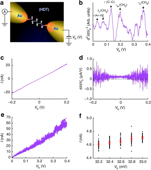
We explored high-field conductance fluctuations in 1,6-hexanedithiol (HDT) single molecules trapped between two Au nanoelectrodes (HDT single-molecule junctions) using a self-breaking technique at 4 K under a low applied voltage of 0.2 V (Fig. 1a,b; Supplementary Figs S1–S3). Figure 1c shows an I–Vb curve measured by a direct current (DC) method. The linear characteristics represent tunnelling electron transport through an HDT molecule, with a large highest occupied molecular orbital-lowest unoccupied molecular orbital (HOMO-LUMO) gap exceeding 6 eV that yields a high carrier injection barrier at the metal–molecule interfaces when bonded to Au electrodes. In addition, we observe that current tends to become unstable with increasing Vb. Correspondingly, the numerical differential conductance exhibits considerable fluctuations at high-Vb regimes (Fig. 1d). We note that the magnitude of the fluctuations is enhanced with increasing Vb.
Figure 1. A measurement scheme for characterizing current fluctuations in single-molecule junctions.
(a) A schematic illustration showing a measurement circuit used to investigate the high-field conductance fluctuations in Au–HDT–Au single-molecule junctions at 4.2 K. (b) IET spectrum of the HDT single-molecule junction formed by a self-breaking technique at 4.2 K. The peaks indicated by arrows can all be assigned to the IETS-active molecular vibrational modes (Supplementary Figs S1–S3 for details). (c) An example of I–Vb characteristics acquired for HDT single-molecule junctions at 1.3 mG0 conductance state. (d) Differential conductance numerically derived from the I–Vb curve in b, revealing considerable fluctuations at high Vb regimes. (e) 50-point current measurements conducted for evaluating the bias voltage-induced conductance fluctuations observed in HDT single-molecule junctions. (f) A magnified view of the plots in e. Black and red dots are the 50-point current data measured at each Vb and the corresponding average current <I>. Error bars denote s.d.
Electric field dependence of current noise in HDT
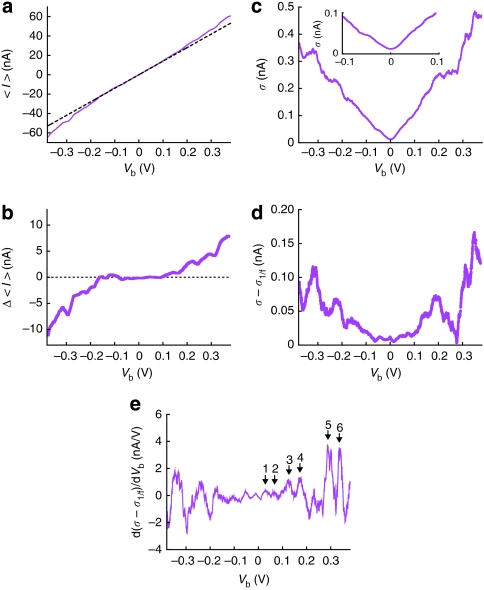
To elucidate the underlying mechanism responsible for the electric-field-induced conductance fluctuations in single HDT molecules, we carried out consecutive 50 current measurements per Vb condition at a sampling rate of 30 Hz during a bias sweep in a range of −0.38 V≤Vb≤0.38 V (Fig. 1e) and examined their average current <I> and current noise σ (Fig. 1f). <I> is plotted as a function of Vb in Figure 2a. We obtained linear <I>–Vb characteristics analogous to the I–Vb curve shown in Figure 1c. Subtracting the elastic tunnelling current Ie from <I> yields the inelastic component Δ<I>=<I>–Ie11,12. We estimated Ie from a linear fit to the <I>–Vb curve in a low bias range of −0.005 V≤Vb≤0.005 V; nonlinearity of elastic tunnelling contributions is anticipated to be negligibly small because of the relatively high tunnelling barrier at the electrode–molecule links expected for the insulating molecular wire of HDTs (Supplementary Fig. S4). By extrapolating linear Ie–Vb to high biases (dotted line in Fig. 2a), we extracted Δ<I> and plotted with respect to Vb (Fig. 2b). The fact that |Δ<I>| rises above 0 A with increasing Vb implies opening of active inelastic channels, rendering an increase in transmission of HDT molecules11,12,13.
Figure 2. Charge transmission and current fluctuations in a single-molecule tunnelling junction.
(a) <I>–Vb characteristics obtained for an HDT single-molecule junction at 4 K. The dotted lines are the elastic current assumed to increase linearly with Vb. (b) Δ<I> plots derived from <I>–Vb curve in a. (c) σ versus Vb plots acquired for an HDT single-molecule junction at 4.2 K. (d) σ plots after subtracting 1/f-like contributions σ1/f. (e) The d(σ–σ1/f)/dVb–Vb curve obtained numerically from d. The characteristic peaks are labelled numerically.
Meanwhile, σ is found to increase with Vb, manifesting more pronounced current fluctuations in the HDT single molecule under high field (Fig. 2c). Moreover, although somewhat obscure at a low-Vb region, σ–Vb plots demonstrate a staircase-like voltage dependence. We first note an overall linear response of σ with respect to Vb. This feature is generally interpreted by Hooge's empirical rule established for bulk materials that states a linear response of the resistance fluctuation-induced 1/f noise amplitude to current14,15. The validity of this rule in atomic- and molecular-scale conductors has been examined recently for various nanomaterials, such as carbon nanotubes, graphenes and atom-sized contacts16,17,18,19. We subtracted the 1/f-like noise σ1/f by linear fitting to σ–Vb plots in the positive and negative Vb regimes. The resulting (σ−σ1/f)–Vb plots reveal clear steps at distinct Vb (Fig. 2d). Measurements on another 40 HDT single-molecule bridges yielded similar results. Numerical differentiation as shown in Figure 2d yields a spectrum with pronounced peaks that emerged at positions corresponding to the σ steps (Fig. 2e). These peaks are located at Vp=30, 66, 127, 172, 300 and 337 mV in the positive side and at Vp=−31, −79, −127, −199, −298 and −369 mV in the negative side (Table 1). The spectrum is therefore almost symmetric with bias polarity.
Table 1. Peaks in the σ-spectrum for an Au–HDT–Au single-molecule junction and an Au–Au single-atom chain.
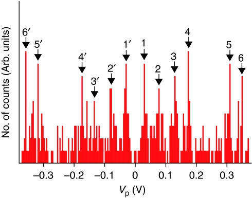
We examined the reproducibility of the characteristic σ steps by statistically analysing σ–Vb characteristics for 41 HDT single-molecule bridges at 1.3 mG0. A histogram constructed with Vp data extracted from the σ spectrum of single HDT molecules contains several peaks at almost symmetric positions with respect to zero bias (Fig. 3). As we see in Table 2, the statistical Vp distribution fairly reproduces the σ spectrum shown in Figure 2e.
Figure 3. A peak voltage histogram constructed with Vp data extracted from the σ spectra of 41 HDT single-molecule junctions formed at 4.2 K.
Peaks are numbered as 1–6 and 1′–6′ in the positive and the negative side, respectively.
Table 2. Peaks in the Vp histogram of Au–HDT–Au single-molecule junctions.
| Peak no. | mV | cm−1 |
|---|---|---|
| Au-HDT-Au | ||
| 1 | 30 | 242 |
| 1′ | −30 | 242 |
| 2 | 78 | 630 |
| 2′ | −80 | 646 |
| 3 | 130 | 1,049 |
| 3′ | −134 | 1,082 |
| 4 | 174 | 1,405 |
| 4′ | −199 | 1,405 |
| 5 | 310 | 2,503 |
| 5′ | −318 | 2,567 |
| 6 | 350 | 2,825 |
| 6′ | −358 | 2,890 |
Noise characteristics of gold single-atom contacts
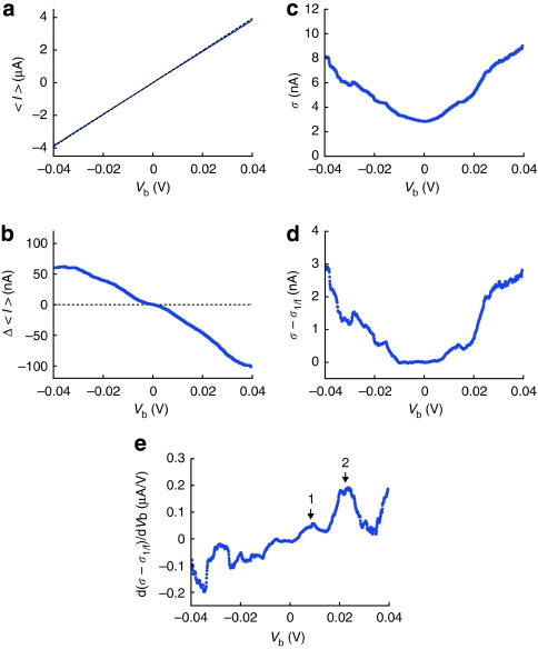
It is interesting to note that the σ peak profile agrees with what appears in the inelastic electron tunnelling (IET) spectrum20 (Fig. 1b). Indeed, we can assign σ peaks to electron energy loss spectroscopy (EELS) and Raman-active vibrational modes21,22, as listed in Table 1. This implies that the high-field current fluctuations in single HDT molecules are of inelastic origin23,24,25. To verify the possible roles of inelastic excitations of vibration modes of single-molecule junctions on current fluctuations, we conducted σ measurements on Au single-atom chains. Forming 1 G0 states using the self-breaking method, with a stretching speed of 6 pm s−1 at 4 K, we exhibited 50-point current measurements in a bias range of −0.04 V≤Vb≤0.04 V. The relatively small bias window was used because Au single-atom contacts tend to become increasingly unstable at bias voltage above 0.05 V presumably because of the local heating and current-induced forces13. <I>–Vb plots in Figure 4a show linear characteristics reflecting the quasi-ballistic nature of electron transport through an Au single-atom contact26. We extract the inelastic component by examining linear fitting to Figure 4a in a bias range from −0.001 V to 0.001 V. In contrast to HDT molecules demonstrating increased transmission via active inelastic channels, Δ<I>–Vb plots in Figure 4b reveal negative contributions of electron–phonon interactions in an Au single-atom chain on the electrical conductance26. This discrepancy can be explained by considering the relative importance of the counteracting effects of elastic and inelastic processes in electron transmission through single-atom and molecule junctions; whereas the inelastic mechanism leads to enhanced transmission, elastic contribution involving virtual phonon excitations decreases transmission27,28,29. Theoretical studies have predicted a shift in the balance towards predominance of inelastic effects as transmission T becomes <0.5 and superior elastic effects for T becomes >0.5, the overall tendency of which agrees with the positive and negative Δ<I> acquired for HDT single-molecule junctions with T~10−3 and Au single-atom chains with T~1, respectively27,28,29. As for the high-field current fluctuations, σ shows a stepwise manner analogous to that found for single HDT molecules (Fig. 4c,d). The corresponding σ spectrum demonstrates peaks at Vp=8 and 22 mV (Fig. 4e). Significantly, we again find good accordance between these peak voltages and the characteristic vibration modes of single-atom wires at energies 10 and 18 meV reported in previous scanning tunnelling microscopy studies30,31 (Table 1). It is therefore corroborated that bias voltage-induced conductance fluctuations can serve as a unique molecular signature for single-molecule identifications.
Figure 4. Inelastic contributions to electrical conductance and current fluctuations in a single-atom quasi-ballistic chain at 4.2 K.
(a) <I>–Vb characteristics of an Au single-atom contact at 4 K. The dotted lines are the elastic component. (b) Δ<I> plots derived from the <I>–Vb curve in a. (c) σ–Vb plots measured for an Au single-atom contact formed at 4.2 K. (d) Plots of (σ–σ1/f) with respect to Vb and (e) the corresponding noise spectrum. Arrows point to the characteristic peaks in the spectrum at the positive Vb range measured.
Discussion
The above results strongly indicate that bias voltage-dependent current oscillations are phonon-assisted phenomena relevant to electron vibration interactions in atomic/molecular bridges. Inelastic tunnelling in molecular systems is known to cause a local heating13,32,33,34. A certain amount of heat is anticipated to pile up in the molecular junctions every time a new inelastic channel sets in. Although this excess heat dissipates via lattice conduction through two electrodes connecting a molecule, local heating effectively raises the junction temperature Teff above the bath temperature13,32,33,34. This may yield a stepwise increase in the thermal noise with increasing Vb, which explains a staircase-like increase of σ observed in single HDT molecules and also in Au single-atom wires. It has been theoretically proposed that the heating rate can be deduced by analysing the bias voltage dependence of Johnson–Nyquist noise in molecular junctions23,25. We therefore sought to extract information regarding electrical heating in HDT junctions from the σ plots in Figure 2d. We evaluated Teff by tentatively considering σT=(σ–σ1/f), where σT is the thermal noise component of σ, and assuming σT √Teff. The relative increase in σT at Vb=0.4 V from the zero-bias value is about 20, from which we obtain Teff~1,600 K at a bath temperature of 4 K. The estimated Teff is obviously too high and contradicts the fact that an HDT molecule can be held stable during a bias sweep without being burned out.
The overestimation of Teff, which is also marked by the fact that the IET spectrum can be observed, suggests significant inelastic noise in the σ–σ1/f of single HDT molecules derived from the stochastic nature of phonon-assisted electron tunnelling and concomitant fluctuations in the phonon-induced energy levels24,35,36. Shot noise measurements have proven to be a powerful method for evaluating Fano factors, as well as the number and transparency of charge transmission channels in atomic/molecular conductors9,10,37. A theoretical model predicts a linear increase in the power spectral density of this shot noise with the bias voltage in a quasi-ballistic Au single-atom chain under a condition Teff>hν/kB, where ν is the frequency of phonon modes38. Indeed, we found a stepwise linear increase in σ–σ1/f after each vibration mode excitation above 0.01 V, as shown in Figure 2e, which in turn suggests a significant local heating in an Au chain that raises its Teff to >hν/kB~230 K at Vb=0.02 V by virtue of the poor contact-cooling efficiency via lattice heat conduction at 4 K. Furthermore, the positive contributions of inelastic channels to noise amplitude found in the present study suggest a weak electron–phonon interaction and symmetric molecule–metal electronic coupling in HDT single-molecule junctions35. (It should be noted that our discussions on inelastic noise is based on a fixed sampling rate. Although the noise amplitude is anticipated to change at different data acquisition speeds, the qualitative arguments on the voltage-specific change in noise amplitudes attributable to the electron–vibration interactions in atomic/molecular conductors still hold.) These results indicate that vibrational mode-specific inelastic noise in single molecules can be more than a useful molecular signature to identify single-molecule junctions: with elaborate theoretical models, it can serve as a unique probe for detailed investigation of the electronic properties of single-molecule bridges.
Methods
Fabrication of mechanically controllable break junctions (MCBJs)
Lithographically defined nano-MCBJs were exploited to form single-molecule bridges. Fabrication procedures of nano-MCBJs are described elsewhere26. In short, we spin-coated a polyimide on a phosphor bronze substrate for electrical insulation. Sub-micrometer-scale Au junctions were then patterned using an electron beam lithography technique and subsequent lift-off processes. The sample was then exposed to reactive ion etching to remove polyimide underneath the junctions. As a result, we obtained Au nanobridges with free-standing length of about 2 μm. In experiments, we first bent a substrate with a three-point bending mechanism and ruptured Au junctions immersed in a dilute toluene solution of test molecules. The sample geometries were designed to provide the damping factor r=Δd/ΔL~3×10−4, where Δd and ΔL denote the displacements of the MCBJ electrodes and the substrate deflection, respectively19. Here, we used HDTs, benchmark molecules the electron transport properties of which have been studied intensively39,40. Subsequently, we evacuated the sample chamber to remove the solvent, leaving HDT molecules adsorbed on a fresh fracture surface of Au junctions. When the vacuum level reached below 10−5 torr, we performed opening and closing of Au junctions repeatedly for hundreds of cycles at room temperatures. At the same time, we examined the evolution of Au–HDT–Au structures by monitoring temporal conductance changes of junctions under a junction-stretching speed νd of 0.6 pm s−1 using a picoammeter (Keithley 6487) under a DC bias voltage of 0.2 V. The measured G-t curves revealed conductance steps at <1G0, suggestive of formations of Au–HDT–Au systems (Supplementary Fig. S1). After this, we slowly cooled the junctions to 4 K.
Formations of Au–HDT–Au single-molecule bridges
We created an HDT single-molecule bridge using a self-breaking method9 under νd=6 pm s−1 and Vb=0.2 V at 4 K. Conductance decreased in a stepwise manner. We stopped junction stretching when a well-defined G-plateau at 1.3×10⁻³ G0 was detected right after a conductance drop from 1 G0, which indicates opening of an atom-sized electrode gap (Supplementary Fig. S2).
Inelastic electron tunnelling spectroscopy measurements
To verify the presence of HDT molecules between two electrodes, we exhibited inelastic electron tunnelling spectroscopy (IETS) analyses by measuring dI/dVb in a bias range of 0.4 V≤Vb≤0.4 V using a lock-in amplifier (SR830, Stanford Research Systems) with a reference AC voltage of 10 mVrms at 1 kHz after forming and holding the single-molecule conductance state (Supplementary Fig. S3). The differential conductance was found to increase in a stepwise manner with Vb, indicating the opening of inelastic channels. d2I/dVb2–Vb characteristics were obtained numerically from the measured dI/dVb curves. Pronounced peaks were observed in the IET spectrum that can be assigned to the IETS-active vibrational modes of Au–alkanedithiol–Au bridges.
Author contributions
M. Tsutsui, M. Taniguchi and T.K. planned and designed the experiments. M. Tsutsui fabricated nano-MCBJs. M. Tsutsui and M. Taniguchi exhibited single-molecule conductance and noise measurements. M. Taniguchi performed data analyses. M. Tsutsui, M. Taniguchi and T.K. wrote the paper.
Additional information
How to cite this article: Tsutsui, M. et al. Single molecule identification via electric current noise. Nat. Commun. 1:138 doi: 10.1038/ncomms1141 (2010).
Supplementary Material
Supplementary Figures S1-S4 and Supplementary References
Acknowledgments
This research is supported partially by the Japan Society for the Promotion of Science (JSPS) through its 'Funding Program for World-Leading Innovative R&D on Science and Technology'.
References
- Nie S. & Emory S. R. Probing single molecules and single nanoparticles by surface-enhanced Raman scattering. Science 275, 1102–1106 (1997). [DOI] [PubMed] [Google Scholar]
- Weiss S. Fluorescence spectroscopy of single biomolecules. Science 283, 1676–1683 (1999). [DOI] [PubMed] [Google Scholar]
- Giepmans B. N. G., Adams S. R., Ellisman M. H. & Tsien R. Y. The fluorescent toolbox for assessing protein location and function. Science 312, 217–224 (2006). [DOI] [PubMed] [Google Scholar]
- Branton D. et al. The potential and challenges of nanopore sequencing. Nat. Biotechnol. 26, 1146–1153 (2008). [DOI] [PMC free article] [PubMed] [Google Scholar]
- Eid J. et al. Real-time DNA sequencing from single polymerase molecules. Science 323, 133–138 (2009). [DOI] [PubMed] [Google Scholar]
- Tsutsui M., Taniguchi M., Yokota K. & Kawai T. Identifying single nucleotides by tunnelling current. Nat. Nanotechnol. 5, 286–290 (2010). [DOI] [PubMed] [Google Scholar]
- Collins J. J., Imhoff T. T. & Grigg P. Noise-enhanced tactile sensation. Nature 383, 770–770 (1996). [DOI] [PubMed] [Google Scholar]
- Bezrukov S. M. & Vodyanoy I. Noise-induced enhancement of signal transduction across voltage-dependent ion channels. Nature 378, 362–364 (2006). [DOI] [PubMed] [Google Scholar]
- Galperin M., Ratner M. A., Nitzan A. & Troisi A. Nuclear coupling and polarization in molecular transport junctions: beyond tunneling to function. Science 319, 1056 (2008). [DOI] [PubMed] [Google Scholar]
- Djukic D. & van Ruitenbeek J. M. Shot noise measurements on a single molecule. Nano. Lett. 6, 789 (2006). [DOI] [PubMed] [Google Scholar]
- Troisi A. & Ratner M. A. Molecular signatures in the transport properties of molecular wire junctions: what makes a junction 'molecular'? Small 2, 172–181 (2006). [DOI] [PubMed] [Google Scholar]
- Stipe B. C., Rezaei M. A. & Ho W. Single-molecule vibrational spectroscopy and microscopy. Science 280, 1732–1735 (1998). [DOI] [PubMed] [Google Scholar]
- Tsutsui M., Taniguchi M. & Kawai T. Local heating in metal-molecule-metal junctions. Nano. Lett. 8, 3293–3297 (2008). [DOI] [PubMed] [Google Scholar]
- Hooge F. N. 1/f noise. Physica B & C 83, 14–23 (1976). [Google Scholar]
- Dutta P. & Horn P. M. Low-frequency fluctuations in solids: 1/f noise. Rev. Mod. Phys. 53, 497–516 (1981). [Google Scholar]
- Collins P. G., Fuhrer M. S. & Zettl A. 1/f noise in carbon nanotubes. Appl. Phys. Lett. 76, 894 (2000). [Google Scholar]
- Lin Y. - M. & Avouris P. Strong suppression of electrical noise in bilayer grapheme nanodevices. Nano. Lett. 8, 2119–2125 (2008). [DOI] [PubMed] [Google Scholar]
- Wu Z. M., Wu S. M., Oberholzer S., Calame M. & Schönenberger C. Scaling of 1/f noise in tunable break junctions. Phys. Rev. B 78, 235421 (2008). [Google Scholar]
- Tal O., Krieger M., Leerink B. & van Ruitenbeek J. M. Electron-vibration interaction in single-molecule junctions: from contact to tunneling regimes. Phys. Rev. Lett. 100, 196804 (2008). [DOI] [PubMed] [Google Scholar]
- Taniguchi M., Tsutsui M., Yokota K. & Kawai T. Mechanically-controllable single molecule switch based on configuration specific electrical conductivity of metal-molecule-metal junctions. Chem. Sci. 1, 247–253 (2010). [Google Scholar]
- Kato H. S., Noh J., Hara M. & Kawai M. An HREELS study of alkanethiol self-assembled monolayers on Au(111). J. Phys. Chem. B 106, 9655–9658 (2002). [DOI] [PubMed] [Google Scholar]
- Bryat M. A. & Pemberton J. E. Surface Raman scattering of self-assembled monolayers formed from 1-alkanethiols: behavior of films at gold and comparison to films at silver. J. Am. Chem. Soc. 113, 8284–8293 (1991). [Google Scholar]
- D'Agosta R., Sai N. & Di Ventra M. Local electron heating in nanoscale conductors. Nano. Lett. 6, 2935–2938 (2006). [DOI] [PubMed] [Google Scholar]
- Haupt F., Novotny T. & Belzig W. Phonon-assisted current noise in molecular junctions. Phys. Rev. Lett. 103, 136601 (2009). [DOI] [PubMed] [Google Scholar]
- Wheeler P. J., Russom J. N., King N. S. & Natelson D. Shot noise suppression at room temperature in atomic-scale Au junctions. Nano. Lett. 10, 1287–1292 (2010). [DOI] [PubMed] [Google Scholar]
- Agraït N., Yeyati A. L. & van Ruitenbeek J. M. Quantum properties of atomic-sized conductors. Phys. Rep. 377, 81–279 (2003). [Google Scholar]
- Viljas J. K., Cuevas J. C., Pauly F. & Hafner M. Electron-vibration interaction in transport through atomic gold wires. Phys. Rev. B 72, 245415 (2005). [Google Scholar]
- Tal O., Krieger M., Leerink B. & van Ruitenbeek J. M. Electron-vibration interaction in single-molecule junctions: from contact to tunneling regimes. Phys. Rev. Lett. 100, 196804 (2008). [DOI] [PubMed] [Google Scholar]
- Shimazaki T. & Asai Y. Theoretical study of the lineshape of inelastic electron tunneling spectroscopy. Phys. Rev. B 77, 115428 (2008). [Google Scholar]
- Gawronski H., Mehlhorn M. & Morgenstern K. Imaging phonon excitation with atomic resolution. Science 319, 930–933 (2008). [DOI] [PubMed] [Google Scholar]
- Agraït N., Untiedt C., Rubio-Bollinger G. & Vieira S. Onset of energy dissipation in ballistic atomic wires. Phys. Rev. Lett. 88, 216803 (2002). [DOI] [PubMed] [Google Scholar]
- Ioffe Z. et al. Detection of heating in current-carrying molecular junctions by Raman scattering. Nat. Nanotechnol. 3, 727–732 (2008). [DOI] [PubMed] [Google Scholar]
- Chen Y.- C., Zwolak M. & Di Ventra M. Local heating in nanoscale conductors. Nano. Lett. 3, 1691–1694 (2003). [Google Scholar]
- Huang Z., Chen F., D'Agosta R., Bennett P. A., Di Ventra M. & Tao N. J. Local ionic and electron heating in single-molecule junctions. Nat. Nanotechnol. 2, 698–703 (2007). [DOI] [PubMed] [Google Scholar]
- Galperin M., Nitzan A. & Ratner M. A. Inelastic tunneling effects on noise properties of molecular junctions. Phys. Rev. B 74, 075326 (2006). [Google Scholar]
- Galperin M., Ratner M. A. & Nitzan A. Molecular transport junctions: vibrational effects. J. Phys. Condens. Matter 19, 103201 (2007). [Google Scholar]
- Birk H., de Jong M. J. M. & Schönenberger C. Shot-noise suppression in the single-electron tunneling regime. Phys. Rev. Lett. 75, 1610 (1995). [DOI] [PubMed] [Google Scholar]
- Chen Y.- C. & Di Ventra M. Effect of electron-phonon scattering on shot noise in nanoscale junctions. Phys. Rev. Lett. 95, 166802 (2005). [DOI] [PubMed] [Google Scholar]
- Xu B. & Tao N. J. Measurement of single-molecule resistance by repeated formation of molecular junctions. Science 301, 1221–1223 (2003). [DOI] [PubMed] [Google Scholar]
- Tao N. J. Electronic transport in molecular junctions. Nat. Nanotechnol. 1, 173–181 (2006). [DOI] [PubMed] [Google Scholar]
Associated Data
This section collects any data citations, data availability statements, or supplementary materials included in this article.
Supplementary Materials
Supplementary Figures S1-S4 and Supplementary References


