Abstract
Botulinum neurotoxins (BoNTs) are among the most potent biological toxins for humans. Of the seven known serotypes (A-G) of BoNT, serotypes A, B and E cause most of the foodborne intoxications in humans. BoNTs in nature are associated with non-toxic accessory proteins known as neurotoxin-associated proteins (NAPs), forming large complexes that have been shown to play important roles in oral toxicity. Using mouse intraperitoneal and oral models of botulism, we determined the dose response to both BoNT/B holotoxin and complex toxins, and compared the toxicities of BoNT/B and BoNT/A complexes. Although serotype A and B complexes have similar NAP composition, BoNT/B formed larger-sized complexes, and was approximately 90 times more lethal in mouse oral intoxications than BoNT/A complexes. When normalized by mean lethal dose, mice orally treated with high doses of BoNT/B complex showed a delayed time-to-death when compared with mice treated with BoNT/A complex. Furthermore, we determined the effect of various food matrices on oral toxicity of BoNT/A and BoNT/B complexes. BoNT/B complexes showed lower oral bioavailability in liquid egg matrices when compared to BoNT/A complexes. In summary, our studies revealed several factors that can either enhance or reduce the toxicity and oral bioavailability of BoNTs. Dissecting the complexities of the different BoNT serotypes and their roles in foodborne botulism will lead to a better understanding of toxin biology and aid future food risk assessments.
Keywords: botulinum neurotoxins, Clostridium botulinum, neurotoxin associated proteins (NAPs), bioavailability, food matrices
1. Introduction
The disease botulism is caused by seven known serotypes of BoNTs that are produced by Clostridium botulinum (alphabetically from BoNT/A-BoNT/G), C. butyricum (BoNT/E), C. baratii (BoNT/F), and C. argentinense (BoNT/G) strains (Hill et al., 2007). Most food-borne intoxications are caused by BoNT serotypes A, B, E and occasionally by serotype F (Arnon et al., 2001; Bigalke and Rummel, 2005; Scarlatos et al., 2005). BoNT is synthesized by the bacterium as a 150 kDa polypeptide (referred to here as holotoxin), which consists of a 50 kDa light chain, and a 100 kDa heavy chain domains linked by a disulfide bond. The heavy chain contains the translocation (N-terminus region of the heavy chain) and cell-binding domain (C-terminus region of the heavy chain) (Arnon et al., 2001; Simpson, 2004). The light chain contains the endopeptidase domain, which cleaves proteins associated with intracellular vesicular transport, such as SNAP25 (synaptosomal-associated protein of 25 kDa) for BoNT/A and VAMP2 (vesicle-associated membrane protein) for BoNT/B, and consequently inhibits acetylcholine release from neurons, leading to muscle paralysis.
BoNTs assemble with non-toxic neurotoxin-associated proteins (NAPs) to form large protein complexes (described here as BoNT complex), known also as “progenitor” toxins. Toxin complexes vary in sizes of 12S, 16S or 19S, also known as M, L and LL toxins of molecular sizes 300 kDa, 500 kDa, and 900 kDa, respectively (Eisele et al., 2011; Hines et al., 2005; Inoue et al., 1996; Ohishi, Sugii, and Sakaguchi, 1977). The 12S toxin is composed of BoNT and a non-toxic, non-hemaglutinin protein (NTNH); the larger complexes of 16S and 19S are formed from BoNT with NTNH and hemaglutinin proteins (HA). NAPs play a substantial role in oral poisoning by protecting the holotoxin from degradation and promoting toxin uptake by epithelial cells (Fujinaga et al., 2009; Simpson, 2004; Sugii, Ohishi, and Sakaguchi, 1977). The molecular size of toxin complexes was shown to be directly proportional to its oral bioavailability; the larger the molecular size, the greater the resistance to degradation by pepsin and/or gastric secretions (Chen, Kuziemko, and Stevens, 1998; Ohishi, Sugii, and Sakaguchi, 1977; Sugii, Ohishi, and Sakaguchi, 1977).
The mechanism by which the BoNT complex traverses the intestinal barrier into the blood stream and then finds its target, the peripheral motor neurons, is not fully understood. BoNT holotoxins, specifically the heavy chain fragments, have been shown to cross the intestinal epithelium via a transcytosis mechanism. This is thought to occur at the apical side of epithelial membranes to reach the basolateral side and then into the blood stream, where the complex presumably breaks apart (Ahsan et al., 2005; Fujinaga, ; Fujinaga et al., 2009; Maksymowych and Simpson, 1998; Maksymowych and Simpson, 2004). Other studies have suggested a role for NAPs such as HA33 to disrupt epithelial cell membranes and thus promoting toxin complex passage (Fujinaga et al., 2009; Jin et al., 2009). Thus, there appears to be at least two mechanisms for BoNT complex translocation, one NAP-independent and the other NAP-dependent.
The organization and arrangement of toxin complex genes is similar for some serotypes of BoNT. For example, the BoNT/B complex contains NAPs NTNH, HA70, HA33 and HA17, and is similar to some BoNT/A subtypes (Hines et al., 2005; Smith et al., 2007). Yet, the BoNT/B complex has been shown to be more toxic than the BoNT/A complex in oral toxicity studies (Ohishi, 1984; Sugii, Ohishi, and Sakaguchi, 1977). The basis for these serotype differences has not been explained. Most studies have been performed with purified holotoxin or complex BoNTs in buffer. The effect of a complex matrix, such as food, on BoNT oral bioavailability is still unclear. Recently, toxin dose effects and oral bioavailability of BoNT/A in a few food matrices has been investigated (Cheng et al., 2008). Few published studies are available on the oral bioavailability of BoNT/B and BoNT/E, both of which are important in foodborne intoxications.
In this study, we determined the in vivo dose-response relationship between purified BoNT/B holotoxin and its toxin complex, and the effect of different food matrices on oral bioavailability. In addition, the oral bioavailability of BoNT/B complex was contrasted with that of BoNT/A complex, revealing important serotype differences in toxicity.
2. Materials and methods
2.1. Materials
Purified BoNT/A and BoNT/B (holotoxins and toxin complexes) and polyclonal rabbit anti-BoNT/A and anti-BoNT/B antibodies were purchased from Metabiologics (Madison, WI). BoNT/A (Hall strain, subtype A1) and BoNT/B (Okra strain, subtype B1) holotoxins were stored at 4 ºC. BoNT/A and BoNT/B complex toxins were aliquoted and frozen at - 20 ºC prior to use. Toxin samples were prepared and diluted in phosphate-gelatin buffer (0.028 M sodium phosphate, pH 6.2, 0.2% gelatin) prior to use. Female Swiss Webster mice (4–5 weeks old, 17–22 g) were purchased from Charles River Laboratories (Portage, MI).
2.2 Purified and crude complex toxin analysis
BoNT/A and BoNT/B holotoxin and complexes were analyzed by SDS polyacrylamide gel electrophoresis (PAGE) using NuPAGE, 4–12% Bis-Tris gels (Invitrogen, Carlsbad, CA). The concentrations of BoNT/A and BoNT/B holotoxin in complex toxin samples were estimated by densitometry comparison of band intensities of unknown versus known holotoxin standards from both Coomassie stained gels and western blots using a FlurChem SP AlphaImager (Alpha Inotech, San Leandro, CA). Protein sizes of purified holotoxins and complexes were also compared using native PAGE (NativePAGE 3–12% Bis-Tris Gel, Invitrogen). Protein components in native gel bands were identified by mass spectrometry as previously described by Cheng et al. (Cheng et al., 2008).
2.3 Determination of mean lethal dose (LD50)
Groups of 6–10 mice were used to test each dose level, and 4 to 5 different dose levels were tested in each experiment. Mice were injected with 500 μl of sample intraperitoneally (ip) or by gavage using Popper feeding needles with 100 μl of sample. Animals were monitored for at least 7 days for signs of intoxication or death. Moribund animals were humanely euthanized and counted as dead. LD50 values were calculated using the Weil or Reed and Muench methods (Reed and Muench, 1938; Weil, 1952) and average values were generated from at least three independent trials. Statistical significance using unpaired t-tests was determined using the Prism 4 statistics software (GraphPad Software Inc., San Diego, CA). Animal protocols adhere to institutional guidelines approved by the Animal Care and Use Committee of the U.S. Department of Agriculture, Western Regional Research Center.
2.4 Determination of oral bioavailability in food matrices
Throughout this manuscript, oral bioavailability is defined as the oral LD50 of BoNTs in food matrices. Oral LD50 of BoNTs in foods were performed as described in section 2.3. BoNT/A and BoNT/B complexes were diluted in phosphate gelatin buffer and in food matrices of non-fat milk or whole egg homogenate at a toxin (in buffer) to food matrix ratio of 1:6 (v/v). Whole eggs were also separated into egg yolk and egg white fractions. Corresponding volumes of phosphate gelatin buffer were used to dilute yolk and white fractions (e.g. yolk occupied about 15% of the total volume, the remaining 85%, previously occupied by egg white, was replaced with buffer). Egg fractions were homogenized using an Omni GLH homogenizer (Omni International, Marietta, GA) fitted with a disposable soft tissue tip. Non-fat milk and whole white chicken eggs (grade A, extra large size) were obtained from a local supermarket and stored at 4 ºC.
3. Results
3.1 Complex size comparisons of BoNT/A and BoNT/B
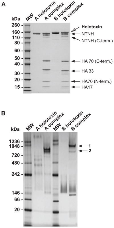
Purified BoNT/A and BoNT/B holotoxins and complexes were analyzed by SDS-PAGE (Fig. 1A). The BoNT/B complex comprises of similarly sized NAP components, NTNH, HA70, HA 33 and HA17. NAPs with similar molecular weights were found in the BoNT/A complex as previously described (Cheng et al., 2008).
Figure 1.
Protein size and content comparisons of BoNT/A and BoNT/B holotoxin and complexes. A. Purified BoNT/A and BoNT/B holotoxins and complexes were size-separated on a 4–12% SDS-PAGE gel and stained with Coomassie Blue. Individual protein components in the toxin complexes were identified by mass spectrometry (arrows). B. The sizes of purified BoNT/A and BoNT/B holotoxins and complexes were compared under native gel conditions. Two different sized BoNT/B complexes were denoted with arrows. Gel bands 1 and 2 were identified by mass spectrometry to contain BoNT/B holotoxin, NTNH, HA70, HA33 and HA17. MW: molecular weight marker (sizes in kDa).
BoNT/A and BoNT/B holotoxins and complexes were size-separated on native PAGE (Fig. 1B). BoNT/B holotoxin separated primarily as monomers, appearing as a smeared band on the gel; BoNT/A holotoxin, on the other hand, separated as multimers of different sizes thus forming a ladder-like pattern. The BoNT/B complex separated primarily as large complexes (bands 1 and 2 of Fig. 1B) that migrated in the region between the 720 and 1048 kDa markers, with band 1 being larger in size than the largest substantial BoNT/A complex. Mass spectrometry analysis of both BoNT/B complex bands confirmed that they contained BoNT/B, HA70, NTNH, HA33, and HA17 proteins. Another prominent BoNT/B complex protein band found about the 146 kDa marker size was identified as BoNT/B.
3.2 Toxin dose response in mouse intraperitoneal and oral intoxication models
The dose responses of mice to ip and oral treatment with BoNT/B holotoxin or complex were determined using Swiss Webster mice that were approximately 5 weeks of age. Toxicity was expressed as mean lethal dose (LD50) values. Thus, the lower the measured unit (weight of toxin/kg), the higher the toxicity, since less toxin was required for lethality. The ip LD50 for BoNT/B holotoxin and complex were 0.33 and 1.1 ng/kg respectively (Table 1). When the amount of BoNT/B complex was adjusted for holotoxin content (average percentage of holotoxin in a purified complex sample was 28.5%), the LD50 value was similar to that of holotoxin alone. The LD50 for BoNT/B holotoxin was slightly lower than the BoNT/A holotoxin LD50 of 0.42 ng/kg (Cheng et al., 2008), however the difference was not statistically significant (p < 0.05).
Table 1.
Mouse intraperitoneal and oral LD50 for purified BoNT/B holotoxin and complex
| BoNT/B | ip LD50 (ng/kg)b | Oral LD50 (μg/kg)b |
|---|---|---|
| Holotoxin (150kDa) | 0.33 ± 0.03 | NDc |
| Complex | 1.1 ± 0.17 | 1.0 ± 0.15 |
| Holotoxin in complexa | 0.33 ± 0.05 | 0.29 ± 0.043 |
LD50 value of BoNT/B complex was normalized to holotoxin content. BoNT/B holotoxin constituted about 28.5% of total protein content.
Mean ± S.E.M, n ≥8.
ND: not determined
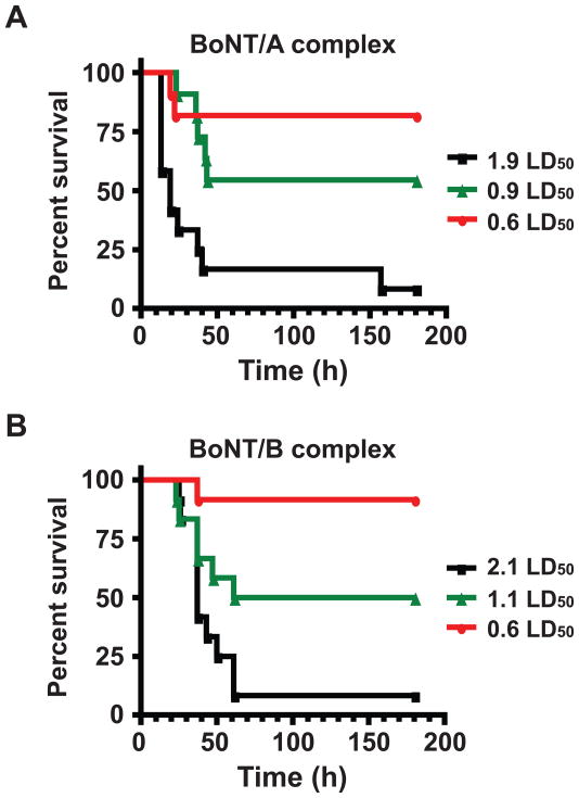
The oral LD50 for BoNT/B complex was 1 ± 0.15 μg/kg in a phosphate gelatin buffer matrix (Table 1). In contrast, BoNT/A complex LD50 was previously determined to be 90±10 μg/kg (Cheng et al., 2008). Thus, BoNT/B complex appears to be about 90 times more toxic than the BoNT/A complex. Because of this large difference in oral toxicity between the two BoNTs, toxin doses were normalized to LD50 units in an effort to compare survival over time. One oral BoNT/A complex LD50 was defined as 1.9 μg per mouse while 1 LD50 of BoNT/B complex was defined as 0.02 μg per mouse (19–20 g each). Mice were monitored over 8 days and the percentage of surviving mice was plotted over time (Fig. 2). Surprisingly, a shorter overall time-to-death was observed in mice that were fed with a high dose of BoNT/A complex as compared to mice fed with a high dose of BoNT/B. The median survival time for mice treated with 1.9 LD50 units of BoNT/A complex was 19 h (Fig. 2A) while the median survival time for mice fed 2.1 LD50 units of BoNT/B complex mice was 37 h (Fig. 2B).
Figure 2.
Survival time comparisons of mice treated orally with BoNTs complexes. Mice were treated with different doses of BoNT/A complex (A) and BoNT/B complex (B) diluted in phosphate gelatin buffer. The percentage of surviving animals was plotted over 180 h. Doses are shown in LD50 units for each toxin for comparison. One BoNT/A LD50 was defined as 1.9 μg/mouse, while 1 LD50 of BoNT/B was defined as 0.02 μg/mouse. N≥12 mice per dose.
3.3 Bioavailability of purified BoNT/B complexes in food matrices
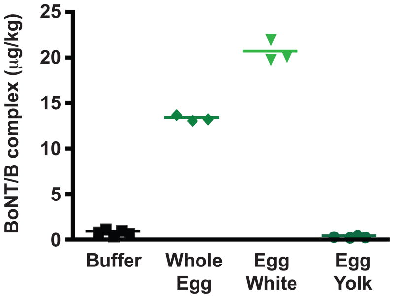
We compared the oral bioavailability of BoNT/B complex in food matrices of buffer, non-fat milk and liquefied whole egg (Table 2 and Fig.3). No statistically significant difference in toxicity was observed between the phosphate gelatin buffer and the non-fat milk matrix. About a 13 fold decrease in toxicity was observed in the whole egg matrix.
Table 2.
Oral LD50 of BoNT/B complex in buffer and egg matrices
| Food Matrix | Oral LD50 (μg/kg)a |
|---|---|
| Buffer | 1.0 ± 0.15 |
| Non-fat milk | 0.88 ± 0.025 |
| Liquid whole egg* | 13.5 ± 0.19 |
| Egg white buffer* | 21.0 ± 0.68 |
| Egg yolk buffer** | 0.50 ± 0.058 |
Mean ± S.E.M. n≥3
p < 0.0001
p = 0.02
Figure 3.
Oral bioavailability of BoNT/B complex in buffer and egg matrices. The oral LD50s of BoNT/B complex in phosphate gelatin buffer and food matrices of liquid whole egg, egg white buffer and egg yolk buffer were plotted for comparison.
We separated egg white from egg yolk, diluted each fraction in buffer, and then determined the BoNT/B oral bioavailability in those two matrices (Table 2 and Fig. 3). An additional 1.5 times decrease in toxicity (as compared to the whole egg matrix) was observed in the egg white matrix (p > 0.0001), while a statistically significant 2 fold increase in toxicity was observed in the egg yolk matrix (p = 0.02).
4. Discussion
Foodborne botulism in the U.S is mainly caused by BoNT serotypes A, B (in the continental U.S.) and E (mainly from outbreaks in Alaska) (Arnon et al., 2001). To date, little has been done to compare the toxin bioavailabilities of different BoNT serotypes in food. Previous studies comparing toxins from different strains revealed increased toxicity of BoNT/B in buffer conditions when compared with other serotypes (Ohishi, 1984; Ohishi, Sugii, and Sakaguchi, 1977). However, the basis for this increased toxicity remains unknown.
In this study, we used mouse intraperitoneal and oral intoxication models to compare the bioavailability of purified holotoxin and complexed toxin versions of BoNT/A and BoNT/B in both buffer and food matrices. Although we observed that the BoNT/B complex contained similar protein components as BoNT/A complex (Fig. 1A), they differed in the size of the toxin complex (Fig.1B). When separated under non-denaturing conditions, BoNT/A and BoNT/B complexes separated mainly as two large protein bands between the range of 720 and 1048 kDa and smaller sized bands of about 150 kDa. The larger of the two BoNT/B complex bands (band 1) was the most abundant (as determined by the intensity of the Coomassie staining) and was, based on its slower mobility on the native gel, larger in size than the BoNT/A complex bands (Fig. 1B). Although mass spectrometry analysis showed that both BoNT/A and BoNT/B contained the same NAP components, the ratios of the holotoxin to individual NAPs in these two complexes remain unknown.
The larger observed size of the BoNT/B complex could explain the overall increase in oral toxicity when compared to BoNT/A complex. In our laboratory, BoNT/B complex (Okra strain) was 90-fold more potent in oral toxicity when compared with the smaller BoNT/A complex (Hall A strain). The difference in toxicity correlated well with previous studies using different strains of serotypes A and B (Ohishi, 1984; Sugii, Ohishi, and Sakaguchi, 1977). A larger BoNT/B complex could presumably better protect the BoNT/B holotoxin from the harsh conditions of the intestinal tract, perhaps by minimizing the amount of holotoxin degradation.
In addition to increased protection from gastric degradation accorded by a larger sized complex, we asked whether other factors, such as toxicity differences between BoNT/A and BoNT/B holotoxins, or the effect of NAPs, especially HA components on intestinal membrane entry, could potentially influence the overall oral toxicity of the BoNT/B complex.
In the absence of the harsh conditions of the gut, such as when toxin is introduced intraperitoneally, the LD50s of BoNT/B and BoNT/A holotoxin or complex were 0.33 ng/kg, and 0.42 ng/kg respectively (Table 1). The slight difference in toxicity was not statistically significant as determined by the unpaired t-test (p < 0.05). Furthermore, other research comparing the neurotoxicity of different BoNTs using a phrenic nerve-hemidiagphragm assay have showed faster paralytic activity of BoNT/A relative to BoNT/B holotoxins (Rasetti-Escargueil et al., 2009). Thus, the increased oral toxicity of BoNT/B complex (compared with BoNT/A complex) was not due to the increased toxicity of the BoNT/B holotoxin.
Previous protection and transmission electron microscopy studies have shown detailed interactions of BoNT/D NTNH and HA70 (Hasegawa et al., 2007; Niwa et al., 2007); HA33 was shown to bind epithelial cells, possibly bringing the toxin in close proximity to its receptors (Matsumura et al., 2008; Zhou et al., 2005). Perhaps the larger BoNT/B complex, with presumably more NAP proteins, can bind intestinal membranes with a higher affinity than the BoNT/A complexes. Our results, however, do not fully support this model. Despite the higher oral toxicity of BoNT/B complex, when mice were fed comparable high LD50 units of BoNT complex toxin, BoNT/B complex dosed mice showed a time-to-death delay of about 18 h when compared to BoNT/A complex dosed mice (Fig. 2). One possibility is that the larger BoNT/B complex requires more time to translocate or cross the intestinal epithelium, leading to the delayed lethality. Lower doses did not produce this time-to-death delay, perhaps because of the longer overall time-to-death, and lower mortality of lower doses would mask these differences. Previous studies have shown that both BoNT/A and BoNT/B HA proteins disrupt the intestinal epithelial barrier at the intercellular tight junctions, with the BoNT/A complex having an increased potency versus the BoNT/B complex (Fujinaga et al., 2009; Jin et al., 2009). The size of BoNT complexes are unknown for these studies. However, our observations of the time-to-death delay correlated well with the research showing decreased membrane disrupting potency of BoNT/B complex. We thus propose that the major contribution to the mouse oral toxicity differences between serotypes A and B lie in the protective properties of the larger BoNT/B complexes. The larger sized BoNT/B complexes may be better protected from the degradative effects of the gastric route. Further studies comparing degradation of different BoNT serotypes and NAPs in different sized complexes are needed for confirmation.
Different BoNT serotypes exerted different toxic effects when prepared in food matrices. BoNT/B complex showed markedly decreased toxicity in egg white buffer matrix, yet slightly increased toxicity in the egg yolk buffer matrix (Table 2 and Fig. 3). Purified BoNT/A complexes did not show any difference in oral bioavailability in these same matrices (Cheng et al., 2008), however there were bioavailability differences in the non-fat milk matrix, underscoring serotype differences. It remains to be assessed which food components in these two matrices contributed to the difference in oral bioavailability. Different sugars have been found to inhibit toxin complex binding to intestinal epithelia (DasGupta and Sugiyama, 1977; Nakamura et al., 2007); it is likely that food components could interact differently with different toxin receptors or to various NAPs, and thus interfere or enhance toxin complex binding.
In addition to serotype, toxicity may be affected by subtype variations (Hill et al., 2009). Different types of NAP proteins are found in other BoNT/A subtypes (such as BoNT/A2 and BoNT/A3) and BoNT/E strains; instead of HA proteins, OrfXs, p21 or p47 proteins are associated with the holotoxin. The toxicity differences of these BoNT serotypes and subtypes and how well mouse oral bioavailability data translates to human oral intoxication remain to be explored.
5. Conclusion
Our studies using the mouse oral intoxication models revealed several different factors that can affect BoNT bioavailability. Size of the toxin complex was the first major difference observed, with BoNT/B complex forming larger-sized complexes that are more lethal in oral intoxication than BoNT/A complexes. Certain food matrices such as the egg white matrix can decrease bioavailability, and others such as the egg yolk matrix can increase bioavailability. Thus, the specific serotype, the presence of associated complex proteins, the size of the toxin complex, or the type of food matrix, can either positively or negatively affect the oral bioavailability of BoNTs. A better understanding of the toxin, its mode of contamination in food, and its bioavailability in complex food matrices would help prepare emergency personnel.
Highlights.
BoNT/B complexes are 90 times more toxic than BoNT/A complexes in a mouse oral intoxication model.
The larger size of BoNT/B complexes contributed to increased oral toxicity.
Different BoNT serotypes have different food bioavailabilities
Acknowledgments
The authors would like to acknowledge Drs. Kirkwood Land, Xiaohua He, and Wallace Yokoyama for their helpful comments; Irina Dynin and Dr. Bruce Onisko for their help with mass spectrometry; Wanless Hatcher, Zeke Martinez and Wentrell Brooks for their help with animal care and handling. The USDA is an equal opportunity provider and employer.
Role of the funding source
This work was funded by the United States Department of Agriculture, Agricultural Research Service, National Program project NP108, CRIS 5325-42000-043-00D. L.W.C. was also funded by the National Institute of Allergy And Infectious Diseases Service Grant U54 AI065359. Funding sources had no role in study design; in the collection, analysis, and interpretation of data; in the writing of the report; and in the decision to submit the paper for publication.
Abbreviations
- BoNTs
Botulinum neurotoxins
- NAPs
neurotoxin associated proteins
- ip
intraperitoneal
- LD50
mean lethal dose
- NTNH
non-toxic, non hemaglutinin protein
- HA
hemaglutinin protein
Footnotes
Publisher's Disclaimer: This is a PDF file of an unedited manuscript that has been accepted for publication. As a service to our customers we are providing this early version of the manuscript. The manuscript will undergo copyediting, typesetting, and review of the resulting proof before it is published in its final citable form. Please note that during the production process errors may be discovered which could affect the content, and all legal disclaimers that apply to the journal pertain.
References
- Ahsan CR, Hajnoczky G, Maksymowych AB, Simpson LL. Visualization of binding and transcytosis of botulinum toxin by human intestinal epithelial cells. J Pharmacol Exp Ther. 2005;315(3):1028–35. doi: 10.1124/jpet.105.092213. [DOI] [PubMed] [Google Scholar]
- Arnon SS, Schechter R, Inglesby TV, Henderson DA, Bartlett JG, Ascher MS, Eitzen E, Fine AD, Hauer J, Layton M, Lillibridge S, Osterholm MT, O’Toole T, Parker G, Perl TM, Russell PK, Swerdlow DL, Tonat K. Botulinum toxin as a biological weapon: medical and public health management. Jama. 2001;285(8):1059–70. doi: 10.1001/jama.285.8.1059. [DOI] [PubMed] [Google Scholar]
- Bigalke H, Rummel A. Medical aspects of toxin weapons. Toxicology. 2005;214(3):210–20. doi: 10.1016/j.tox.2005.06.015. [DOI] [PubMed] [Google Scholar]
- Chen F, Kuziemko GM, Stevens RC. Biophysical characterization of the stability of the 150-kilodalton botulinum toxin, the nontoxic component, and the 900-kilodalton botulinum toxin complex species. Infect Immun. 1998;66(6):2420–5. doi: 10.1128/iai.66.6.2420-2425.1998. [DOI] [PMC free article] [PubMed] [Google Scholar]
- Cheng LW, Onisko B, Johnson EA, Reader JR, Griffey SM, Larson AE, Tepp WH, Stanker LH, Brandon DL, Carter JM. Effects of purification on the bioavailability of botulinum neurotoxin type A. Toxicology. 2008;249(2–3):123–9. doi: 10.1016/j.tox.2008.04.018. [DOI] [PubMed] [Google Scholar]
- DasGupta BR, Sugiyama H. Inhibition of Clostridium botulinum types A and B hemagglutinins by sugars. Can J Microbiol. 1977;23(9):1257–60. doi: 10.1139/m77-188. [DOI] [PubMed] [Google Scholar]
- Eisele KH, Fink K, Vey M, Taylor HV. Studies on the dissociation of botulinum neurtoxin type A complexes. Toxicon. 2011;57(4):555–65. doi: 10.1016/j.toxicon.2010.12.019. [DOI] [PubMed] [Google Scholar]
- Fujinaga Y. Interaction of botulinum toxin with the epithelial barrier. J Biomed Biotechnol. 2010:974943. doi: 10.1155/2010/974943. [DOI] [PMC free article] [PubMed] [Google Scholar]
- Fujinaga Y, Matsumura T, Jin Y, Takegahara Y, Sugawara Y. A novel function of botulinum toxin-associated proteins: HA proteins disrupt intestinal epithelial barrier to increase toxin absorption. Toxicon. 2009;54(5):583–6. doi: 10.1016/j.toxicon.2008.11.014. [DOI] [PubMed] [Google Scholar]
- Hasegawa K, Watanabe T, Suzuki T, Yamano A, Oikawa T, Sato Y, Kouguchi H, Yoneyama T, Niwa K, Ikeda T, Ohyama T. A novel subunit structure of Clostridium botulinum serotype D toxin complex with three extended arms. J Biol Chem. 2007;282(34):24777–83. doi: 10.1074/jbc.M703446200. [DOI] [PubMed] [Google Scholar]
- Hill KK, Smith TJ, Helma CH, Ticknor LO, Foley BT, Svensson RT, Brown JL, Johnson EA, Smith LA, Okinaka RT, Jackson PJ, Marks JD. Genetic diversity among Botulinum Neurotoxin-producing clostridial strains. J Bacteriol. 2007;189(3):818–32. doi: 10.1128/JB.01180-06. [DOI] [PMC free article] [PubMed] [Google Scholar]
- Hill KK, Xie G, Foley BT, Smith TJ, Munk AC, Bruce D, Smith LA, Brettin TS, Detter JC. Recombination and insertion events involving the botulinum neurotoxin complex genes in Clostridium botulinum types A, B, E and F and Clostridium butyricum type E strains. BMC Biol. 2009;7:66. doi: 10.1186/1741-7007-7-66. [DOI] [PMC free article] [PubMed] [Google Scholar]
- Hines HB, Lebeda F, Hale M, Brueggemann EE. Characterization of botulinum progenitor toxins by mass spectrometry. Appl Environ Microbiol. 2005;71(8):4478–86. doi: 10.1128/AEM.71.8.4478-4486.2005. [DOI] [PMC free article] [PubMed] [Google Scholar]
- Inoue K, Fujinaga Y, Watanabe T, Ohyama T, Takeshi K, Moriishi K, Nakajima H, Inoue K, Oguma K. Molecular composition of Clostridium botulinum type A progenitor toxins. Infect Immun. 1996;64(5):1589–94. doi: 10.1128/iai.64.5.1589-1594.1996. [DOI] [PMC free article] [PubMed] [Google Scholar]
- Jin Y, Takegahara Y, Sugawara Y, Matsumura T, Fujinaga Y. Disruption of the epithelial barrier by botulinum haemagglutinin (HA) proteins - differences in cell tropism and the mechanism of action between HA proteins of types A or B, and HA proteins of type C. Microbiology. 2009;155(Pt 1):35–45. doi: 10.1099/mic.0.021246-0. [DOI] [PubMed] [Google Scholar]
- Maksymowych AB, Simpson LL. Binding and transcytosis of botulinum neurotoxin by polarized human colon carcinoma cells. J Biol Chem. 1998;273(34):21950–7. doi: 10.1074/jbc.273.34.21950. [DOI] [PubMed] [Google Scholar]
- Maksymowych AB, Simpson LL. Structural features of the botulinum neurotoxin molecule that govern binding and transcytosis across polarized human intestinal epithelial cells. J Pharmacol Exp Ther. 2004;310(2):633–41. doi: 10.1124/jpet.104.066845. [DOI] [PubMed] [Google Scholar]
- Matsumura T, Jin Y, Kabumoto Y, Takegahara Y, Oguma K, Lencer WI, Fujinaga Y. The HA proteins of botulinum toxin disrupt intestinal epithelial intercellular junctions to increase toxin absorption. Cell Microbiol. 2008;10(2):355–64. doi: 10.1111/j.1462-5822.2007.01048.x. [DOI] [PubMed] [Google Scholar]
- Nakamura T, Takada N, Tonozuka T, Sakano Y, Oguma K, Nishikawa A. Binding properties of Clostridium botulinum type C progenitor toxin to mucins. Biochim Biophys Acta. 2007;1770(4):551–5. doi: 10.1016/j.bbagen.2006.11.006. [DOI] [PubMed] [Google Scholar]
- Niwa K, Koyama K, Inoue S, Suzuki T, Hasegawa K, Watanabe T, Ikeda T, Ohyama T. Role of nontoxic components of serotype D botulinum toxin complex in permeation through a Caco-2 cell monolayer, a model for intestinal epithelium. FEMS Immunol Med Microbiol. 2007;49(3):346–52. doi: 10.1111/j.1574-695X.2006.00205.x. [DOI] [PubMed] [Google Scholar]
- Ohishi I. Oral toxicities of Clostridium botulinum type A and B toxins from different strains. Infect Immun. 1984;43(2):487–90. doi: 10.1128/iai.43.2.487-490.1984. [DOI] [PMC free article] [PubMed] [Google Scholar]
- Ohishi I, Sugii S, Sakaguchi G. Oral toxicities of Clostridium botulinum toxins in response to molecular size. Infect Immun. 1977;16(1):107–9. doi: 10.1128/iai.16.1.107-109.1977. [DOI] [PMC free article] [PubMed] [Google Scholar]
- Rasetti-Escargueil C, Jones RG, Liu Y, Sesardic D. Measurement of botulinum types A, B and E neurotoxicity using the phrenic nerve-hemidiaphragm: improved precision with in-bred mice. Toxicon. 2009;53(5):503–11. doi: 10.1016/j.toxicon.2009.01.019. [DOI] [PubMed] [Google Scholar]
- Reed LJ, Muench H. A simple method of estimating fifty per cent endpoints. Am J Hyg. 1938;27:493. [Google Scholar]
- Scarlatos A, Welt BA, Cooper BY, Archer D, DeMarse T, Chau KV. Methods for detecting botulinum toxin with applicability to screening foods against biological terrorist attacks. Journal of Food Science. 2005;70(8):121–130. [Google Scholar]
- Simpson LL. Identification of the major steps in botulinum toxin action. Annu Rev Pharmacol Toxicol. 2004;44:167–93. doi: 10.1146/annurev.pharmtox.44.101802.121554. [DOI] [PubMed] [Google Scholar]
- Smith TJ, Hill KK, Foley BT, Detter JC, Munk AC, Bruce DC, Doggett NA, Smith LA, Marks JD, Xie G, Brettin TS. Analysis of the neurotoxin complex genes in Clostridium botulinum A1-A4 and B1 strains: BoNT/A3, /Ba4 and /B1 clusters are located within plasmids. PLoS One. 2007;2(12):e1271. doi: 10.1371/journal.pone.0001271. [DOI] [PMC free article] [PubMed] [Google Scholar]
- Sugii S, Ohishi I, Sakaguchi G. Correlation between oral toxicity and in vitro stability of Clostridium botulinum type A and B toxins of different molecular sizes. Infect Immun. 1977;16(3):910–4. doi: 10.1128/iai.16.3.910-914.1977. [DOI] [PMC free article] [PubMed] [Google Scholar]
- Weil CS. Tables for the convenient calculation of median-effective dose (LD50 or ED50) and instructions in their use. Biometrics. 1952;8:249–63. [Google Scholar]
- Zhou Y, Foss S, Lindo P, Sarkar H, Singh BR. Hemagglutinin-33 of type A botulinum neurotoxin complex binds with synaptotagmin II. Febs J. 2005;272(11):2717–26. doi: 10.1111/j.1742-4658.2005.04688.x. [DOI] [PubMed] [Google Scholar]


